Review
. 2025 Jan 27;14(2):150.
doi: 10.3390/antiox14020150.

Do Lifestyle Interventions Mitigate the Oxidative Damage and Inflammation Induced by Obesity in the Testis?

Affiliations
Review

Do Lifestyle Interventions Mitigate the Oxidative Damage and Inflammation Induced by Obesity in the Testis?

Ruben J Moreira et al. Antioxidants (Basel). .

Abstract

Obesity results from a disproportionate accumulation of fat and has become a global health concern. The increase in adipose tissue is responsible for several systemic and testicular changes including hormone levels (leptin, adiponectin, testosterone, estrogen), inflammatory cytokines (increase in TNF-α and IL-6 and decrease in IL-10), and redox state (increase in reactive oxygen species and reduction in antioxidant enzymes). This results in poor sperm quality and compromised fertility in men with obesity. Lifestyle modifications, particularly diet transition to caloric restriction and physical exercise, are reported to reverse these negative effects. Nevertheless, precise mechanisms mediating these benefits, including how they modulate testicular oxidative stress, inflammation, and metabolism, remain to be fully elucidated. The main pathway described by which these lifestyle interventions reverse obesity-induced oxidative damage is the Nrf2-SIRT1 axis, which modulates the overexpression of antioxidant defenses. Of note, some of the detrimental effects of obesity on the testis are inherited by the descendants of individuals with obesity, and while caloric restriction reverses some of these effects, no significant work has been carried out regarding physical exercise. This review discusses the consequences of obesity-induced testicular oxidative stress on adult and pediatric populations, emphasizing the therapeutic potential of lifestyle to mitigate these detrimental effects.

Keywords: caloric restriction; inflammation; obesity; oxidative stress; physical exercise; sperm parameters; testosterone.


Conflict of interest statement

The authors declare no conflicts of interest.

Figures

Figure 1
Figure 1
Mechanisms linking high-fat diet to adipose tissue expansion and systemic effects of inflammation and oxidative stress. A high-fat diet leads to hyperplasia and hypertrophy of adipose tissue. This expansion contributes to hyperleptinemia and leptin resistance. Enlarged adipose tissue experiences hypoxia, which activates hypoxia-inducible factor 1-alpha (HIF-1α) and increases plasminogen activator inhibitor-1 (PAI-1). Additionally, unfolded adiponectin triggers the unfolded protein response (UPR), especially the PERK (protein kinase R-like ER kinase) and IRE1 (inositol-requiring enzyme 1) pathways, responsible for the degradation of misfolded adiponectin and consequent hypoadiponectinemia and insulin resistance. Macrophages infiltrate the adipose tissue Adipose tissue also synthesizes monocyte chemoattractant protein-1 (MCP-1), which disturbs insulin signaling by disrupting Akt phosphorylation. MCP-1 contributes to inflammation by secreting inflammatory cytokines such as Tumor Necrosis Factor-alpha (TNF-α), Interleukin-6 (IL-6), Interleukin-1β (IL-1β), and nitric oxide (NO). Inflammatory and oxidative stress markers, including NADPH oxidase (NOX) activity, exacerbate systemic inflammation and oxidative stress. Together, these pathways contribute to dysfunctions associated with metabolic syndrome. ↑—upregulation.
Figure 2
Figure 2
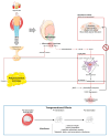
High-fat-diet-induced testicular dysfunction and its transgenerational effects. A high-fat diet induces adipose tissue hypertrophy and aromatase activity, increasing estradiol levels. Nitric oxide (NO) released by macrophages and adipose tissue contributes to nitrosative stress, impairing the hypothalamus–pituitary axis and disrupting LH signaling. Other inflammatory cytokines, such as Tumor Necrosis Factor-alpha (TNF-α), Interleukin-6 (IL-6), Interleukin-1β (IL-1β), exacerbate inflammation, further impairing Leydig cell function. Leydig cells exhibit reduced steroidogenic activity due to the activation of the JNK/ERK/MAPK pathway that culminates in the increased Nuclear Factor Kappa B (NF-κB) and DAX-1, which inhibits Nur77 and steroidogenic factor 1 (SF1), transcription factors involved in the expression of steroidogenic enzymes (steroidogenic acute regulatory protein—StAR; Cytochromes P450 CYP11a and CYP17a; and β-Hydroxysteroid dehydrogenases—β-HSD, resulting in decreased testosterone production. The seminiferous tubules show increased apoptosis, disrupted Sertoli cell activity, and compromised blood–testis barrier integrity, contributing to poor sperm quality. Elevated oxidative stress, driven by reduced nuclear factor erythroid 2-related factor 2 (Nrf2)-mediated antioxidant defenses and increased NADPH oxidase (NOX) activity, exacerbates lipid peroxidation and testicular damage. Transgenerational effects observed in the F1 and F2 generations include reduced testicular weight, decreased sperm parameters, and impaired antioxidant capacity, accompanied by metabolic, lipidic, and transcriptomic inheritance. Red arrows represent the negative effects induced (regular arrow—effect induced; inhibition arrow—process inhibition). ↓—downregulation. ↑—upregulation. Red box—negative effects. Blue box—transgenerational effects.
Figure 3
Figure 3
Effects of dietary transition from a high-fat diet to caloric restriction on male reproductive health and transgenerational outcomes. Transitioning from a high-fat diet to caloric restriction leads to improvements in serum markers, including increased testosterone and adiponectin levels, and reduced insulin and leptin levels. In the testes, enhanced expression of key genes—fat mass and obesity-associated protein (FTO), melanocortin 4 receptor (MC4R), sirtuin 1 (SIRT1), glucosamine-6-phosphate deaminase 2 (GNPDA2), transmembrane protein 18 (TMEM18)—promotes antioxidant enzyme activity (via Nrf2), sperm viability, and insulin sensitization. However, caloric restriction is associated with an increase in abnormal sperm head morphology. Transgenerational benefits observed in the F1 and F2 generations include restored testicular weight, improved sperm quality, increased testicular antioxidant capacity, and enhanced metabolic, lipidic, and transcriptomic profiles, suggesting intergenerational inheritance of positive dietary effects. ↓—decrease. ↑—increase. Green box—positive effects. Red box—negative effects. Blue box—transgenerational effects.
Figure 4
Figure 4
Effects of physical exercise on male reproductive health and potential transgenerational effects. Moderate-intensity physical exercise decreases inflammatory cytokines (Tumor Necrosis Factor-alpha—TNF-α; Interleukin-6—IL-6; Interleukin-1β—IL-1β; nitric oxide—NO; and monocyte chemoattractant protein-1—MCP-1), insulin, and leptin levels, while increasing testosterone levels, improving sperm motility, count, and normal morphology, and resulting in insufficient apoptosis. The positive effects of moderate exercise loads are associated with decrease in microRNA mir34a, an increase in sirtuin 1 (SIRT1) and nuclear factor erythroid 2-related factor 2 (Nrf2), which is associated with higher testicular antioxidant capacity. High-intensity physical exercise shows no changes in inflammatory cytokines, testosterone production, or sperm motility, count, and normal morphology, but increases Sertoli cell activity. Transgenerational effects of physical exercise on damaged testicles remain unknown. ↓—downregulation. ↑—upregulation. Green box—positive effects. Red box—negative effects. Blue box—transgenerational effects.

Similar articles

Cited by

References

    1. Bowman-Busato J., Schreurs L., Halford J.C., Yumuk V., O’Malley G., Woodward E., De Cock D., Baker J.L. Providing a common language for obesity: The European Association for the Study of Obesity obesity taxonomy. Int. J. Obes. 2024:1–10. doi: 10.1038/s41366-024-01565-9. - DOI - PMC - PubMed
    1. World Health Organization . World Health Statistics 2024: Monitoring Health for the SDGs, Sustainable Development Goals. World Health Organization; Geneva, Switzerland: 2024.
    1. Carrageta D.F., Oliveira P.F., Alves M.G., Monteiro M.P. Obesity and male hypogonadism: Tales of a vicious cycle. Obes. Rev. 2019;20:1148–1158. doi: 10.1111/obr.12863. - DOI - PubMed
    1. Guo D., Wu W., Tang Q., Qiao S., Chen Y., Chen M., Teng M., Lu C., Ding H., Xia Y. The impact of BMI on sperm parameters and the metabolite changes of seminal plasma concomitantly. Oncotarget. 2017;8:48619. doi: 10.18632/oncotarget.14950. - DOI - PMC - PubMed
    1. Hammoud A.O., Wilde N., Gibson M., Parks A., Carrell D.T., Meikle A.W. Male obesity and alteration in sperm parameters. Fertil. Steril. 2008;90:2222–2225. doi: 10.1016/j.fertnstert.2007.10.011. - DOI - PubMed

Grants and funding

LinkOut - more resources