. 2025 Feb 19;14(2):241.
doi: 10.3390/antiox14020241.

Antioxidant and Photoprotective Activities of 3,4-Dihydroxybenzoic Acid and (+)-Catechin, Identified from Schima argentea Extract, in UVB-Irradiated HaCaT Cells

Affiliations

Antioxidant and Photoprotective Activities of 3,4-Dihydroxybenzoic Acid and (+)-Catechin, Identified from Schima argentea Extract, in UVB-Irradiated HaCaT Cells

Qi He et al. Antioxidants (Basel). .

Abstract

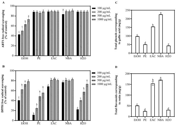
In traditional Chinese medicine, the root bark and leaves of Schima argentea are utilized to treat dysentery, parasitic infections, and digestive disorders. In this study, the n-butanol extract of S. argentea (NBA) exhibited potent antioxidant properties, protecting HaCaT cells from UVB-induced damage, and was abundant in phenolic and flavonoid compounds. Using UPLC-QTOF-MS analysis, several antioxidants within NBA were identified. Among these, 3,4-dihydroxybenzoic acid, (+)-catechin, and procyanidin B2 effectively reduced ROS levels after 1 h post-UVB treatment (225 mJ/cm2). Notably, all three compounds significantly decreased the phosphorylation of p38 and JNK in a dose-dependent manner. Additionally, the cell survival rate of these compounds was assessed after 12 h post-UVB treatment (225 mJ/cm2). Both 3,4-dihydroxybenzoic acid and (+)-catechin significantly prevented UVB-induced apoptosis in HaCaT cells, as evidenced by MTT, Hoechst, Calcein/PI staining, and flow cytometry analyses. Proteomic analysis revealed that 3,4-dihydroxybenzoic acid achieved photoprotection by downregulating c-Fos and Jun and modulating cell cycle proteins, while (+)-catechin promoted cell repair through the PI3K-Akt and Wnt signaling pathways. These results demonstrated that both compounds can directly absorb UVB, scavenge ROS, and provide cell photoprotection by modulating multiple signaling pathways. The n-butanol extract of S. argentea holds promising potential for future medical applications.

Keywords: (+)-Catechin; 3,4-Dihydroxybenzoic acid; Schima argentea; UVB-induced apoptosis; proteomic analysis.


Conflict of interest statement

The authors declare no conflicts of interest.

Figures

Figure 1
Figure 1
Antioxidant capacity and phytochemical composition of S. argentea extracts. (A) ABTS free radical scavenging of S. argentea extracts. (B) DPPH free radical scavenging of S. argentea extracts. (C) Total phenols of S. argentea extracts (expressed as gallic acid equivalents). (D) Total flavonoids of S. argentea extracts (expressed as rutin equivalents). EtOH, PE, EAC, NBA, and H2O indicate S. argentea extracts obtained using ethanol, petroleum ether, ethyl acetate, n-butanol, and distilled water, respectively. Different letters above the columns indicate statistically significant differences (p < 0.05). Results are presented as mean ± S.D. (n = 3).
Figure 2
Figure 2
Effect of NBA on HaCaT cell viability and ROS generation. Cell viability of HaCaT cells treated with NBA in the (A) absence and (B) presence of UVB irradiation (225 mJ/cm2) after 1 h of incubation. (C) ROS levels in HaCaT cells treated with NBA after 1 h post-UVB treatment (225 mJ/cm2). The control was not exposed to UVB irradiation. Cell viability of HaCaT cells treated with NBA in the (D) absence and (E) presence of UVB irradiation (225 mJ/cm2) after 12 h of incubation. Different letters above the columns indicate statistically significant differences (p < 0.05). Results are presented as mean ± S.D. (n = 3).
Figure 3
Figure 3
Antioxidant capacity of 3,4-dihydroxybenzoic acid, (+)-catechin, and procyanidin B2. (A) ABTS free radical scavenging of 3,4-dihydroxybenzoic acid, (+)-catechin, and procyanidin B2. (B) DPPH free radical scavenging of 3,4-dihydroxybenzoic acid, (+)-catechin, and procyanidin B2. The control was treated with H2O. DA, Cat, and PB2 indicate 3,4-dihydroxybenzoic acid, (+)-catechin, and procyanidin B2, respectively. Different letters above the columns indicate statistically significant differences (p < 0.05). Results are presented as mean ± S.D. (n = 3).
Figure 4
Figure 4
Effect of 3,4-dihydroxybenzoic acid, (+)-catechin, and procyanidin B2 on HaCaT cell viability and ROS generation. Cell viability of HaCaT cells treated with 3,4-dihydroxybenzoic acid, (+)-catechin, and procyanidin B2 in the (A) absence and (B) presence of UVB irradiation (225 mJ/cm2) after 1 h of incubation. (C) ROS levels in HaCaT cells treated with 3,4-dihydroxybenzoic acid, (+)-catechin, and procyanidin B2 after 1 h post-UVB treatment (225 mJ/cm2). Control groups were not exposed to UVB irradiation. Different letters above the columns indicate statistically significant differences (p < 0.05). Results are presented as mean ± S.D. (n = 3).
Figure 5
Figure 5
ROS assays of HaCaT cells treated with (A) 3,4-dihydroxybenzoic acid, (B) (+)-catechin, and (C) procyanidin B2 were assessed using a fluorescence microscope after 1 h post-UVB treatment (225 mJ/cm2).
Figure 6
Figure 6
Western blot analysis of p-p38 and p-JNK expression in HaCaT cells treated with (A) 3,4-dihydroxybenzoic acid, (B) (+)-catechin, and (C) procyanidin B2 after 1 h post-UVB treatment (225 mJ/cm2). The relative intensities of proteins treated with (D) 3,4-dihydroxybenzoic acid, (E) (+)-catechin, and (F) procyanidin B2 were analyzed using ImageJ 1.53e software. Different letters above the columns indicate statistically significant differences (p < 0.05). Results are presented as mean ± S.D. (n = 3).
Figure 7
Figure 7
Viability of HaCaT cells treated with 3,4-dihydroxybenzoic acid, (+)-catechin, and procyanidin B2 was assessed after 12 h post-UVB treatment (225 mJ/cm2). Control groups were not exposed to UVB irradiation. DA, Cat, and PB2 indicate 3,4-dihydroxybenzoic acid, (+)-catechin, and procyanidin B2, respectively. Different letters above the columns indicate statistically significant differences (p < 0.05). Results are presented as mean ± S.D. (n = 3).
Figure 8
Figure 8
Hoechst staining of HaCaT cells treated with (A) 3,4-dihydroxybenzoic acid, (B) (+)-catechin, and (C) procyanidin B2 was assessed using fluorescence microscope after 12 h post-UVB treatment (225 mJ/cm2).
Figure 9
Figure 9
Calcein/PI staining of HaCaT cells treated with (A) 3,4-dihydroxybenzoic acid, (B) (+)-catechin, and (C) procyanidin B2 was assessed using fluorescence microscope after 12 h post-UVB treatment (225 mJ/cm2). Calcein and PI indicate viable and non-viable cells, respectively.
Figure 10
Figure 10
Flow cytometry analysis of HaCaT cells was assessed in the (A) absence and (B) presence of UVB irradiation (225 mJ/cm2) after 12 h of cultivation. Apoptosis of HaCaT cells treated with (C) 3,4-dihydroxybenzoic acid, (D) (+)-catechin, and (E) procyanidin B2 was assessed using flow cytometry after 12 h post-UVB treatment (225 mJ/cm2). Representative images are displayed. (F) The apoptotic cells were counted in quadrants Q2 and Q4. Different letters above the columns indicate statistically significant differences (p < 0.05). Results are presented as mean ± S.D. (n = 3).

Similar articles

Cited by

References

    1. Wei M., He X., Liu N., Deng H. Role of reactive oxygen species in ultraviolet-induced photodamage of the skin. Cell Div. 2024;19:1. doi: 10.1186/s13008-024-00107-z. - DOI - PMC - PubMed
    1. Gromkowska-Kępka K.J., Puścion-Jakubik A., Markiewicz-Żukowska R., Socha K. The impact of ultraviolet radiation on skin photoaging—review of in vitro studies. J. Cosmet. Dermatol. 2021;20:3427–3431. doi: 10.1111/jocd.14033. - DOI - PMC - PubMed
    1. Schuch A.P., Moreno N.C., Schuch N.J., Menck C.F.M., Garcia C.C.M. Sunlight damage to cellular DNA: Focus on oxidatively generated lesions. Free Radic. Biol. Med. 2017;107:110–124. doi: 10.1016/j.freeradbiomed.2017.01.029. - DOI - PubMed
    1. Kim D.J., Iwasaki A., Chien A.L., Kang S. UVB-mediated DNA damage induces matrix metalloproteinases to promote photoaging in an AhR- and SP1-dependent manner. JCI Insight. 2022;7:e156344. doi: 10.1172/jci.insight.156344. - DOI - PMC - PubMed
    1. She Q.B., Ma W.Y., Dong Z. Role of MAP kinases in UVB-induced phosphorylation of p53 at serine 20. Oncogene. 2002;21:1580–1589. doi: 10.1038/sj.onc.1205239. - DOI - PubMed

LinkOut - more resources