Case Reports
. 2025 Feb 13;15(4):459.
doi: 10.3390/diagnostics15040459.

A Case Report of Madelung's Disease in Romania

Affiliations
Case Reports

A Case Report of Madelung's Disease in Romania

Andrei Ionut Cucu et al. Diagnostics (Basel). .

Abstract

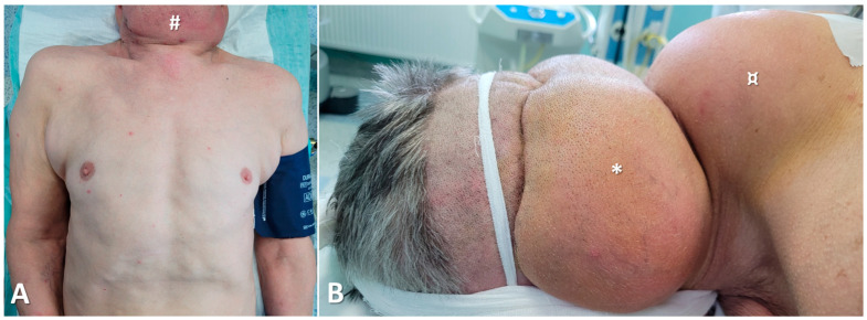
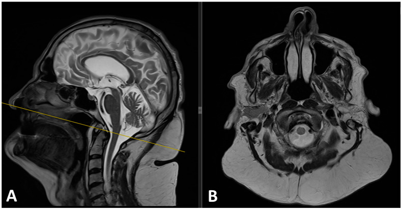
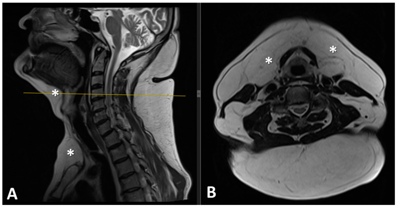
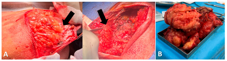
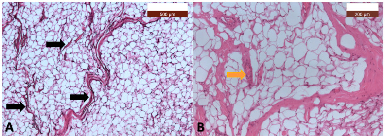
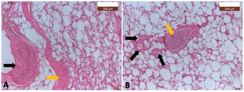
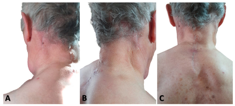
Background: Madelung's disease is a rare lipid metabolic disorder characterized by diffuse and symmetrical adipose tissue deposits in the subcutaneous fascial spaces, presenting with multiple painless masses throughout the body. The disease is more common in middle-aged adults with a history of chronic alcohol consumption. Case Report: This article reports a case of Madelung's disease from Romania in a 67-year-old man admitted to our department for multiple adipose masses located in the neck and upper back. MRI examination of the head and neck revealed symmetrical and non-encapsulated fat deposition. Surgical intervention was performed to resect the adipose masses. The article also discusses the etiology, clinical manifestations, diagnosis, and surgical treatment of large adipose lesions. Conclusions: This case report provides insights for the diagnosis and treatment of Madelung's syndrome.

Keywords: Launois–Bensaude syndrome; Madelung syndrome; benign symmetric lipomatosis.


Conflict of interest statement

The authors declare no conflicts of interest.

Figures

Figure 1
Figure 1
(A) Left lateral cervical view highlighting the lipomatous masses: a bulky mass located in the anterior neck region (#), the posterior occipito-cervical region (*), and the cervico-dorsal region (¤); (B) Posterior view showing two masses in the occipito-cervical region (*) and one mass in the cervico-dorsal region (¤); (C) Posterior cervico-dorsal view (detailed).
Figure 2
Figure 2
(A) Anterior view showing the lipomatous mass in the anterior neck region (#) and trunk lipomatosis; (B) Lateral view highlighting the masses located in the occipito-cervical region (*) and the cervico-dorsal region (“buffalo hump”) (¤).
Figure 3
Figure 3
(A) Preoperative sagittal T2 and (B) axial T2 MRI images revealed significant hypertrophic fat tissue around the neck and upper back, consistent with the diagnosis of Madelung disease.
Figure 4
Figure 4
(A) Preoperative sagittal T2 and (B) axial T2 MRI images revealed significant soft-tissue mass of high-signal intensity with low-signal intensity fibrous septa at the level of the chin, submandibular, and suprasternal region (*).
Figure 5
Figure 5
(A) Surgical findings: it was observed that the lipoma lacks a capsule, with no cleavage plane between the tumor and the adjacent anatomical structures (black arrows); (B) Postoperative specimen: yellow adipose tissue, soft and solid, with an envelope in some areas, and lobulated.
Figure 6
Figure 6
(A) Adipose tissue not delimited by a capsule, intersected by numerous connective septa of varying thicknesses, lengths, and orientations (black arrows). The adipose tissue consists of mature adipocytes, but with varying shapes and sizes (HE, ×5); (B) With a higher magnification (HE, ×10), it is observed that the connective septa are numerous and contain blood vessels of the arteriole type (yellow arrow) (HE, ×10).
Figure 7
Figure 7
(A) The adipose tissue, composed of mature adipocytes with varying shapes and sizes, is intersected by simple, densely cellularized connective septa (orange arrow) and connective septa containing muscular-type arteries (black arrow) (HE, ×10); (B) The adipose tissue, composed of mature adipocytes with varying shapes and sizes, is intersected by numerous short connective septa containing capillaries or arterioles (black arrows), which appear to separate the cells from one another. Additionally, there is a longer and thicker septum within which a nerve fiber is observed (orange arrow) (HE, ×10).
Figure 8
Figure 8
(A) Left lateral cervical view; (B) right lateral cervical view; (C) posterior view, showing the surgical outcomes one year after liposuction. No recurrence was observed.

Similar articles

References

    1. Wanke J., Yongjing F. A Case Report of Madelung’s Disease in China. Radiol. Case Rep. 2022;17:320–325. doi: 10.1016/j.radcr.2021.10.035. - DOI - PMC - PubMed
    1. Liu Q., Lyu H., Xu B., Lee J.H. Madelung Disease Epidemiology and Clinical Characteristics: A Systemic Review. Aesthetic Plast. Surg. 2021;45:977–986. doi: 10.1007/s00266-020-02083-5. - DOI - PubMed
    1. Zielińska-Kaźmierska B., Lewicki M., Manowska B. Madelung Disease. Adv. Dermatol. Allergol. 2015;32:400–403. doi: 10.5114/pdia.2015.54750. - DOI - PMC - PubMed
    1. Jimenez Aragon F., Morales Puebla J.M., Corzón Pereira T. Madelung’s Disease. Acta Otorrinolaringol. Esp. 2013;64:166–167. doi: 10.1016/j.otoeng.2013.04.015. - DOI - PubMed
    1. Ruzicka T., Vieluf D., Landthaler M., Braun-Falco O. Benign Symmetric Lipomatosis Launois-Bensaude. Report of Ten Cases and Review of the Literature. J. Am. Acad. Dermatol. 1987;17:663–674. doi: 10.1016/S0190-9622(87)70253-9. - DOI - PubMed

Publication types

LinkOut - more resources