Review
. 2025 Jan 31;13(2):327.
doi: 10.3390/biomedicines13020327.

Mitochondrial Dysfunction in Neurodegenerative Diseases: Mechanisms and Corresponding Therapeutic Strategies

Affiliations
Review

Mitochondrial Dysfunction in Neurodegenerative Diseases: Mechanisms and Corresponding Therapeutic Strategies

Kai Meng et al. Biomedicines. .

Abstract

Neurodegenerative disease (ND) refers to the progressive loss and morphological abnormalities of neurons in the central nervous system (CNS) or peripheral nervous system (PNS). Examples of neurodegenerative diseases include Alzheimer's disease (AD), Parkinson's disease (PD), and amyotrophic lateral sclerosis (ALS). Recent studies have shown that mitochondria play a broad role in cell signaling, immune response, and metabolic regulation. For example, mitochondrial dysfunction is closely associated with the onset and progression of a variety of diseases, including ND, cardiovascular diseases, diabetes, and cancer. The dysfunction of energy metabolism, imbalance of mitochondrial dynamics, or abnormal mitophagy can lead to the imbalance of mitochondrial homeostasis, which can induce pathological reactions such as oxidative stress, apoptosis, and inflammation, damage the nervous system, and participate in the occurrence and development of degenerative nervous system diseases such as AD, PD, and ALS. In this paper, the latest research progress of this subject is detailed. The mechanisms of oxidative stress, mitochondrial homeostasis, and mitophagy-mediated ND are reviewed from the perspectives of β-amyloid (Aβ) accumulation, dopamine neuron damage, and superoxide dismutase 1 (SOD1) mutation. Based on the mechanism research, new ideas and methods for the treatment and prevention of ND are proposed.

Keywords: mitochondrial dynamics; mitochondrial energy metabolism; mitochondrial homeostasis imbalance; mitophagy; neurodegenerative diseases.


Conflict of interest statement

The authors declare that the research was conducted in the absence of any commercial or financial relationships that could be construed as a potential conflict of interest.

Figures

Figure 1
Figure 1
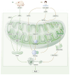
Aβ-Induced mitochondrial dysfunction triggers neuronal degeneration in AD. (a) Aβ protein, which is an AD marker, acts on mitochondria and causes a variety of mitochondrial abnormalities by inhibiting complex III/IV, OGDC, and PDH activity, which in turn inhibits ATP synthesis and induces the onset of mitophagy. The release of Cyt-c from the mitochondria into the cytoplasm is an important factor in mitophagy. The most important aspect is that Aβ causes the rise in ROS, while a large level of ROS inhibits PP2A and activates GSK 3β, leading to Tau over-phosphorylation, causing neuronal fiber tangles. At the same time, ROS promotes APP expression, APP further promotes ROS and Aβ production, and APP and Aβ block APP and Aβ production. This leads to tau hyperphosphorylation, which causes neuronal fiber tangles. Meanwhile, ROS promotes APP expression, while APP further promotes ROS and Aβ generation, and APP and Aβ block protein transport in mitochondria, causing neuronal damage. (b) Treatment of rat pericytes with low doses of Aβ significantly upregulated the expression of PINK1/Parkin and inhibited the phosphorylation of Tau, which in turn promoted mitophagy; however, the accumulation of Aβ in the neurons of AD patients, which is usually observed in pathological states, has been shown to inhibit mitophagy, leading to a decrease in the production of ROS, which causes neuronal apoptosis. (c) In neurons of the cerebral cortex of AD patients, Aβ protein causes S-nitrosylation of DRP1, which increases GTPase activity and accelerates mitochondrial fission; at the same time, Aβ interacts with DRP1 and activates DRP1 and FIS1, which results in mitochondrial fission, and both pathways contribute to apoptotic neuronal death. (d) High levels of phosphorylated tau have been observed in patients with AD or in 3XTg mice. Their presence promoted the interaction of Aβ with DRP1 in AD mouse models and triggered neuronal apoptosis by increasing GTPase activity. Aβ: Amyloid-β; OGDC: oxoglutarate dehydrogenase complex; PDH: pyruvate dehydrogenase; ROS: Reactive oxygen species; PP2A: proteinphosphatase 2A; GSK 3β: glycogen synthase kinase-3; APP: amyloid precursor protein; DRP1: Dynamic-related protein 1; FIS1: Mitochondrial fission protein 1.
Figure 2
Figure 2
Mechanisms of mitochondrial homeostasis affecting PD. (a) In patients with PD, the excessive accumulation of ROS in the CNS leads to an imbalance in glutamate metabolism, neuronal excitotoxicity, and the release of Cyt-c. This results in an imbalance in mitochondrial homeostasis, secretion of the toxic lipid particles APOE and APOJ by astrocytes, and apoptosis of dopaminergic neurons, ultimately inducing PD. (b) In PD model mice, the knockout of PINK1 in dopaminergic neurons leads to mitochondrial calcium accumulation, resulting in mitochondrial calcium overload, increased ROS production, the promotion of mitochondrial fission, and the mediation of neuronal apoptosis. In the brains of these mice, dopaminergic cells are exposed to oxidative stress, with translocated DRP1 binding to P53 on the OMM, exacerbating oxidative stress and inhibiting GTP and ATP synthesis. Alternatively, DRP1-dependent mitochondrial fission is indirectly promoted by miR-499 transcription, thus mediating neuronal apoptosis and PD. In the neurons of PD patients, α-syn co-localizes with cardiolipin and rapidly oligomerizes, inhibiting complex I synthesis, reducing mitochondrial membrane potential difference, promoting the opening of the mPTP, and generating mROS. This accelerates mitochondrial oxidative stress, inhibits mitophagy, and induces neuronal apoptosis. In PD model rats, dopaminergic neurons exposed to neurotoxins, such as 6-OHDA, rotenone, and MPP+, induce Bax translocation to the OMM, resulting in the opening of mPTP, gradual loss of mitochondrial membrane potential, excessive release of Cyt-c and AIF, activation of caspase-9, and formation of apoptosomes, resulting in cell apoptosis and inducing PD. (c) In the microglia of patients with PD, the LRRK2 G2019S mutation inhibits the removal of MIRO, causing mitochondrial oxidative stress, suppressing mitophagy, and leading to excessive cell activation. In the brain tissue of PD model mice exposed to copper, activated microglia secrete inflammatory products, upregulate NLRP3/caspase-1 axis proteins, downregulate Parkin and PINK1, promote ROS generation, and activate the NF-κB pathway. This inhibits mitophagy and activates microglia to synthesize and release pro-NGF, which binds to the sortilin receptor, upregulates JNK and c-Jun proteins, and activates the JNK-JUN signaling pathway. This sequence initiates neuronal apoptosis, induces dopaminergic neuronal apoptosis, and triggers PD. Cyt-c: cytochrome C; APOE: apolipoprotein E.; APOJ: apolipoprotein E; PINK1: PTEN-induced putative kinase 1. OMM: outer mitochondrial membrane. GTP: Guanosine triphosphate. ATP: Adenosine triphosphate. α-syn: alpha-Synuclein. mPTP: mitochondrial permeability transition pore. mROS: Mitochondrial reactive oxygen species. 6-OHDA: 6-hydroxydopamine. MPP+: 1-methyl-4-phenylpyridinium. AIF: Apoptosis-inducing factor. Caspase 9: Cysteine-requiring Aspartate Protease 9. MIRO: mitochondrial Rho. NLRP3: Nucleotide-binding oligomerization domain receptor 3. Caspase1: Cysteine-requiring Aspartate Protease 1. ProNGF: pro-nerve growth factor. JNK: c-Jun N-Terminal Kinase.
Figure 3
Figure 3
Mechanisms of mitochondrial dysfunction and neuronal apoptosis in ALS. (a) DRP1 is overexpressed in the cytoplasm of ganglion cells of ALS model rats, where it binds to APAF1, recruiting and activating the precursor of caspase-9. This upregulates the expression of the apoptotic execution protein caspase-3, promotes mitochondrial fission, and induces neuronal apoptosis. DRP1, by translocating and binding to the mitochondrial membrane, opens the mPTP, releasing ROS and Cyt-c, leading to mitochondrial oxidative stress, mitochondrial fission dysfunction, and neuronal apoptosis. (b) In patients with ALS, C9orf72 expansion upregulates TDP-43, promoting the transcription of ND3/6 mRNA, which inhibits mitochondrial complex I. The activation of mPTP promotes mtDNA release, activates the cGAS-STING signaling pathway, and induces an inflammatory response. Following treatment with CCCP, the overexpression of TDP-43 downregulated caspase-3 and promoted autophagosome formation, leading to sustained mitochondrial fission, disruption of mitochondrial homeostasis, and the induction of neuronal apoptosis. In patients with ALS, OPTN mutations cause failure to bind to LC3B in damaged mitochondria and exhibit aberrant localization. The clearance of damaged mitochondria by OPTN is inhibited, leading to abnormal mitophagy and ALS induction. APAF 1: apoptotic protease-activating factor 1. Caspase 3: Cysteine-requiring Aspartate Protease 3. C9orf72: chromosome 9 open reading frame 72. TDP-43: TAR DNA-binding protein, 43 kDa. mtDNA: Mitochondrial DNA. cGAS-STING: Cyclic GMP-AMP synthase stimulator of interferon genes. CCCP: carbonyl cyanide, m-chlorophenyl hydrazone. OPTN: optineurin. LC3B: microtubule-associated protein light chain 3 beta.

References

    1. Seifan A., Ganzer C.A., Ryon K., Lin M., Mahmudur R., Adolfo H., Shih C., Jacobs A.R., Greenwald M., Isaacson R.S. Detecting Non-cognitive Features of Prodromal Neurodegenerative Diseases. Curr. Aging Sci. 2019;11:242–249. doi: 10.2174/1874609812666190204094233. - DOI - PMC - PubMed
    1. Brody J.A., Stanhope J.M., Kurland L.T. Patterns of Amyotrophic Lateral Sclerosis and Parkinsonism-Dementia on Guam. Contemp. Neurol. Ser. 1975;12:45–70. - PubMed
    1. de Haan P., Klein H.C., ’t Hart B.A. Autoimmune Aspects of Neurodegenerative and Psychiatric Diseases: A Template for Innovative Therapy. Front. Psychiatry. 2017;8:46. doi: 10.3389/fpsyt.2017.00046. - DOI - PMC - PubMed
    1. Messner D.A., Rabins P., Downing A.C., Irizarry M., Foster N.L., Al Naber J., Dabbous O., Fillit H., Gabler S., Krakauer R., et al. Designing Trials of Disease Modifying Agents for Early and Preclinical Alzheimer’s Disease Intervention: What Evidence is Meaningful to Patients, Providers, and Payers? J. Prev. Alzheimers Dis. 2019;6:20–26. doi: 10.14283/jpad.2018.42. - DOI - PubMed
    1. Jethwa K.D. Antipsychotics for the management of Parkinson’s disease psychosis. Int. J. Geriatr. Psychiatry. 2017;32:464–465. doi: 10.1002/gps.4640. - DOI - PubMed

LinkOut - more resources