. 2025 Feb 17;13(2):501.
doi: 10.3390/biomedicines13020501.

Dupilumab in the Treatment of Severe Uncontrolled Chronic Rhinosinusitis with Nasal Polyps (CRSwNP) and Comorbid Asthma-A Multidisciplinary Monocentric Real-Life Study

Affiliations

Dupilumab in the Treatment of Severe Uncontrolled Chronic Rhinosinusitis with Nasal Polyps (CRSwNP) and Comorbid Asthma-A Multidisciplinary Monocentric Real-Life Study

Gian Luca Fadda et al. Biomedicines. .

Abstract

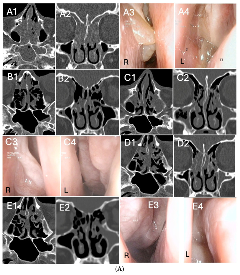
Background: Chronic rhinosinusitis with nasal polyps (CRSwNP) and asthma are mutually correlated with Type-2 inflammation. Dupilumab is effective in uncontrolled and relapsing CRSwNP. However, the precise characterization of Type-2 inflammation and the impact of previous surgery on clinical outcomes need clarification. Methods: We present a prospective observational study on a 38 CRSwNP-patient cohort, whose Type-2 endotype was confirmed after a multidisciplinary approach shared among ENTs, pneumologists and allergologists. Patients were treated with dupilumab and evaluated at 15 days and 1-3-6-12-18-24-30 months, focusing on clinical (VAS, nasal polyp score-NPS), radiological (Lund-Mackay) and quality of life (SNOT-22) parameters, as well olfactory function, asthma control, variation of Type-2 markers and number and extent (ACCESS score) of previous surgeries. Results: We confirmed the efficacy of dupilumab in total and sub-items VAS, NPS, SNOT-22 and sniffing score, as well as Lund-Mackay score improvements, observable and significant after 2 weeks of treatment (p < 0.0001) and long-lasting over 30 months. Good to excellent response criteria to biologic treatment at 6 months was observed in 30/32 patients. Comorbid asthma reached rapid control (p < 0.0001) and exhaled nitric oxide normalization was achieved. One single "not adequate" surgery showed a trend to milder improvement, as well as a higher ACCESS score to better olfactory outcome. Conclusions: The accurate selection of uncontrolled relapsing CRSwNP in terms of Type-2 endotyping by multidisciplinary approach can maximize dupilumab efficacy. The number and extent of previous surgeries may differentiate the response, although this effect is difficult to catch in real life. "Adequate" ESS surgery before dupilumab may drive mostly effective disease control.

Keywords: asthma; biologic treatment; biomarkers; chronic rhinosinusitis; dupilumab; endoscopic sinus surgery; nasal polyps; type-2 inflammation.


Conflict of interest statement

G.L.F. reports a fee as a speaker for GSK, Sanofi, all outside of the submitted work G.G. reports a fee as a speaker for AstraZeneca, Menarini, all outside of the submitted work; F.L.M.R. reports grants, personal fees, and other compensation from AstraZeneca, Boehringer Ingelheim, Chiesi, GSK, and Novartis, and personal fees and grants to support scientific research from Sanofi, all outside of the submitted work. All the other authors declare no conflicts of interest.

Figures

Figure 1
Figure 1
Clinical, endoscopic and radiologic scores evaluated before and after dupilumab treatment. From V1 to V9. Total nasal symptoms visual analogue scale (total VAS) (A), SNOT-22 (B), nasal polyps score (NPS) (C); Sniffin’s Sticks test (SSIT-16); (D), Lund–Mackay score (LMS) (E). * p < 0.0001 (paired-sample t-test).
Figure 2
Figure 2
Absolute blood eosinophil count (B-EOS, ×109/L) in total CRSwNP population (A), a patient subgroup according to at least 50% increase from basal (B) and Total IgE (KU/L) (C) evaluated before and after dupilumab treatment. From V1 to V9. * p < 0.0001; ** p < 0.001; *** p < 0.01, # p < 0.05 (paired-sample t-test).
Figure 3
Figure 3
(A). Radiological and endoscopic (R = right, L = left) images in relation to dupilumab therapy without surgery in CRSwNP (eosinophils>30/hpf). (A1A4): start of dupilumab. Axial (A1) and coronal (A2) computed tomography (CT) images show pansinusitis. Endoscopic examination (A3,A4) of both nasal cavities shows polypoid formation completely occupying the nasal fossa (total NPS = 7) S:sinus; TI:turbinate. (B1,B2): 6 months after initiation of dupilumab. Sphenoidal (B1), frontal and maxillary sinuses (B2) completely disease-free. Pictures (C1C4): 12 months after initiation of dupilumab. Radiologic (C1,C2) and endoscopic examination (C3,C4) of both nasal cavities show significant reduction of polypoid mass. (D1,D2): axial (D1) and coronal (D2) computed tomography (CT) images 18 months after initiation of dupilumab. Outcomes are maintained in the long run. (E1E4): 24 months after initiation of dupilumab. Maintenance of the radiological (E1,E2) and endoscopic (E3,E4) outcomes is evident, even at long term follow-up visit. (B). Radiological and endoscopic (R = right, L =left) images in relation to dupilumab therapy after “adequate” Full House FESS of revision for CRSwNP. (A1A4): CT (A1,A2) and MRI (A3,A4) images showing CRSwNP recurrence after two “not adequate” FESS (ACCESS score 20/24). Intra-operatory images (RESS): (A5) right nasal fossa NPS = 4; (A6) debridement of nasal polyps; (A7) left maxillary sinus empyema; (A8) frontal sinusotomy. (B1B4): start of dupilumab 18 months after “adequate” Full House FESS, following CRSwNP relapse (eosinophils>50/hpf). Endoscopic (B1,B2) and radiological (B3,B4) images, ACCESS score 2/24. (C): 6 months after initiation of dupilumab. Radiologic (C1,C2) and endoscopic examination (C3,C4) of both nasal cavities shows partial reduction of polypoid mass. (D1D4): 12 months after initiation of dupilumab, a local control of the disease is evidenced in both axial (D1) and coronal (D2) computed tomography (CT) and endoscopic (D3,D4) images (E1E4): 24 months after initiation of dupilumab, images highlight the maintenance of the radiological (E1,E2) and endoscopic (E3,E4) results even at long term follow-up visits. Inferior turbinates are preserved, while middle turbinate was remodeled for correct sinus ventilation and disease control. “ss”: sphenoid sinus; “sm”: maxillary sinus. (C). Radiological and endoscopic (R = right, L = left) images in relation to dupilumab therapy after “adequate” salvage Full House FESS in patient who underwent two previous “not adequate” surgeries (last surgery performed 18 months before starting dupilumab). (A1A4): The patient started biologic treatment with dupilumab. Coronal (A1) and axial (A2) CT scans evidenced ethmoidal cells still not opened and hyperostosis of the anterior and posterior compartment. Endoscopic images (A3,A4): NP relapse and tissue remodeling. (B1B4): 6 months after initiation of dupilumab, persistence of nasal polyps seen radiologically (B1,B2) and endoscopically (B3,B4). (C1C4): 18 months after initiation of dupilumab, ACCESS score 22/24 (C1,C2). Endoscopic sinus surgery (C3,C4) was performed in this patient, keeping on treatment with dupilumab. (D1D4): 1 year after “adequate” Full House FESS and 3 years after dupilumab. TC scans (D1,D2) evidenced paranasal sinuses fully opened. Corresponding endoscopic image (D3,D4).
Figure 3
Figure 3
(A). Radiological and endoscopic (R = right, L = left) images in relation to dupilumab therapy without surgery in CRSwNP (eosinophils>30/hpf). (A1A4): start of dupilumab. Axial (A1) and coronal (A2) computed tomography (CT) images show pansinusitis. Endoscopic examination (A3,A4) of both nasal cavities shows polypoid formation completely occupying the nasal fossa (total NPS = 7) S:sinus; TI:turbinate. (B1,B2): 6 months after initiation of dupilumab. Sphenoidal (B1), frontal and maxillary sinuses (B2) completely disease-free. Pictures (C1C4): 12 months after initiation of dupilumab. Radiologic (C1,C2) and endoscopic examination (C3,C4) of both nasal cavities show significant reduction of polypoid mass. (D1,D2): axial (D1) and coronal (D2) computed tomography (CT) images 18 months after initiation of dupilumab. Outcomes are maintained in the long run. (E1E4): 24 months after initiation of dupilumab. Maintenance of the radiological (E1,E2) and endoscopic (E3,E4) outcomes is evident, even at long term follow-up visit. (B). Radiological and endoscopic (R = right, L =left) images in relation to dupilumab therapy after “adequate” Full House FESS of revision for CRSwNP. (A1A4): CT (A1,A2) and MRI (A3,A4) images showing CRSwNP recurrence after two “not adequate” FESS (ACCESS score 20/24). Intra-operatory images (RESS): (A5) right nasal fossa NPS = 4; (A6) debridement of nasal polyps; (A7) left maxillary sinus empyema; (A8) frontal sinusotomy. (B1B4): start of dupilumab 18 months after “adequate” Full House FESS, following CRSwNP relapse (eosinophils>50/hpf). Endoscopic (B1,B2) and radiological (B3,B4) images, ACCESS score 2/24. (C): 6 months after initiation of dupilumab. Radiologic (C1,C2) and endoscopic examination (C3,C4) of both nasal cavities shows partial reduction of polypoid mass. (D1D4): 12 months after initiation of dupilumab, a local control of the disease is evidenced in both axial (D1) and coronal (D2) computed tomography (CT) and endoscopic (D3,D4) images (E1E4): 24 months after initiation of dupilumab, images highlight the maintenance of the radiological (E1,E2) and endoscopic (E3,E4) results even at long term follow-up visits. Inferior turbinates are preserved, while middle turbinate was remodeled for correct sinus ventilation and disease control. “ss”: sphenoid sinus; “sm”: maxillary sinus. (C). Radiological and endoscopic (R = right, L = left) images in relation to dupilumab therapy after “adequate” salvage Full House FESS in patient who underwent two previous “not adequate” surgeries (last surgery performed 18 months before starting dupilumab). (A1A4): The patient started biologic treatment with dupilumab. Coronal (A1) and axial (A2) CT scans evidenced ethmoidal cells still not opened and hyperostosis of the anterior and posterior compartment. Endoscopic images (A3,A4): NP relapse and tissue remodeling. (B1B4): 6 months after initiation of dupilumab, persistence of nasal polyps seen radiologically (B1,B2) and endoscopically (B3,B4). (C1C4): 18 months after initiation of dupilumab, ACCESS score 22/24 (C1,C2). Endoscopic sinus surgery (C3,C4) was performed in this patient, keeping on treatment with dupilumab. (D1D4): 1 year after “adequate” Full House FESS and 3 years after dupilumab. TC scans (D1,D2) evidenced paranasal sinuses fully opened. Corresponding endoscopic image (D3,D4).
Figure 4
Figure 4
Asthma control test (ACT, score) (A) and exhaled nitric oxide (FENO, parts per billion, PPB) (B) evaluated in patient with concomitant asthma and CRSwNP before and after dupilumab treatment from V0 to V9 and before and after treatment (at least 6 months of follow-up); * p < 0.0001 (paired-sample t-test).

Similar articles

References

    1. Fokkens W.J., Lund V.J., Hopkins C., Hellings P.W., Kern R., Reitsma S., Toppila-Salmi S., Bernal-Sprekelsen M., Mullol J., Alobid I., et al. European Position Paper on Rhinosinusitis and Nasal Polyps 2020. Rhinology. 2020;58((Suppl. S29)):1–464. doi: 10.4193/Rhin20.401. - DOI - PubMed
    1. Kato A., Peters A.T., Stevens W.W., Schleimer R.P., Tan B.K., Kern R.C. Endotypes of chronic rhinosinusitis: Relationships to disease phenotypes, pathogenesis, clinical findings, and treatment approaches. Allergy. 2022;77:812–826. doi: 10.1111/all.15074. - DOI - PMC - PubMed
    1. Bachert C., Han J.K., Desrosiers M., Hellings P.W., Amin N., Leeet S.E., Mullol J., Greos L.S., Bosso J.V., Laidlaw T.M., et al. Efficacy and safety of dupilumab in patients with severe chronic rhinosinusitis with nasal polyps (LIBERTY NP SINUS-24 and LIBERTY NP SINUS-52): Results from two multicentre, randomised, double-blind, placebo-controlled, parallel-group phase 3 trials. Lancet. 2019;394:1638–1650. doi: 10.1016/S0140-6736(19)31881-1. Erratum in Lancet 2019, 394, 1618. - DOI - PubMed
    1. De Corso E., Pasquini E., Trimarchi M., La Mantia I., Pagella F., Ottaviano G., Garzaro M., Pipolo C., Torretta S., Seccia V., et al. Dupilumab in the treatment of severe uncontrolled chronic rhinosinusitis with nasal polyps (CRSwNP): A multicentric observational Phase IV real-life study (DUPIREAL) Allergy. 2023;78:2669–2683. doi: 10.1111/all.15772. - DOI - PubMed
    1. Laidlaw T.M., Mullol J., Woessner K.M., Amin N., Mannent L.P. Chronic Rhinosinusitis with Nasal Polyps and Asthma. J. Allergy Clin. Immunol. Pract. 2021;9:1133–1141. doi: 10.1016/j.jaip.2020.09.063. - DOI - PubMed

LinkOut - more resources