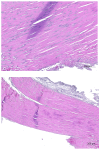
Histological Alterations and Interferon-Gamma and AKT-mTOR Expression in an Experimental Model of Achilles Tendinopathy-A Comparison of Stem Cell and Amniotic Membrane Treatment
- PMID: 40002938
- PMCID: PMC11852843
- DOI: 10.3390/biomedicines13020525
Histological Alterations and Interferon-Gamma and AKT-mTOR Expression in an Experimental Model of Achilles Tendinopathy-A Comparison of Stem Cell and Amniotic Membrane Treatment
Abstract
Achilles tendon injuries are extremely common and have a significant impact on the physical and mental health of individuals. Both conservative and surgical treatments have unsatisfactory results. The search for new therapeutic tools, using cell therapies with stem cells (SC) and biological tissues, such as amniotic membranes (AM), has proved useful for the regeneration of injured tendons. Background/Objectives: This research was carried out to assess the capacity of tissue repair in animal models of Achilles tendinopathy, in which rats were submitted to complete sections of the tendon, and the effects of using bone marrow SC and/or AM graft are evaluated. Methods: Thirty-seven Wistar rats, submitted to complete surgical section of the Achilles tendon and subsequent tenorrhaphy, were randomized into four groups: Control Group (CG), received saline solution; SC Group (SCG) received an injection of SC infiltrated directly into the tendon; AM Group (AMG), the tendon was covered with an AM graft; SC + AM Group (SC+AMG), has been treated with an AM graft and SC local injection. Six weeks later, the Achilles tendons were evaluated using a histological score and immunohistochemical pro-healing markers such as Interferon-γ, AKT, and mTOR. Results: There were no differences between morphometric histological when evaluating the Achilles tendons of the samples. No significant differences were found regarding the expression of AKT-2 and mTOR markers between the study groups. The main finding was the presence of a higher concentration of Interferon-γ in the group treated with SC and AM. Conclusions: The isolated use of SC, AM, or the combination of SC-AM did not produce significant changes in tendon healing when the histological score was evaluated. Similarly, no difference was observed in the expression of AKT-2 and mTOR markers. An increase in the expression of Interferon-γ was observed in SC+AMG. This suggests that such therapies may be potentially beneficial for the regeneration of injured tendons. However, as tendon repair mechanisms are very complex, further studies should be carried out to verify the benefits of the tendon structure and function.
Keywords: AKT-mTOR; Achilles tendon; amniotic membrane; interferon-γ; mononuclear stem cells; tendinopathy.
Conflict of interest statement
The authors declare that there is no conflict of interest.
Figures


References
LinkOut - more resources
Full Text Sources
Miscellaneous

