Review
. 2025 Feb 5;15(2):239.
doi: 10.3390/life15020239.

The Perception of the Diaphragm with Ultrasound: Always There Yet Overlooked?

Affiliations
Review

The Perception of the Diaphragm with Ultrasound: Always There Yet Overlooked?

Kathleen Möller et al. Life (Basel). .

Abstract

Diaphragm ultrasound makes it possible to diagnose diaphragmatic atrophy and dysfunction. Important indications include unclear dyspnea; diaphragmatic elevation; assessment of diaphragm dysfunction in pulmonary, neuromuscular and neurovascular diseases; and in critically ill patients before noninvasive and mechanical ventilation and follow-up of diaphragm thickness and function during mechanical ventilation with potential prediction of prolonged weaning. In patients with respiratory insufficiency and potential diaphragm dysfunction, it is possible to objectify the contribution of diaphragm dysfunction. In addition, assessment of diaphragmatic hernias, tumors and diaphragmatic dysfunction in COVID-19 and diaphragmatic ultrasound in sports medicine have been described. This narrative review includes the sonomorphology of the diaphragm, standardization of ultrasonographic investigation with transducer positions and ultrasound techniques, normal findings and diagnostic criteria for pathological findings. The correct sonographic measurement, calculation and evaluation can ultimately influence further therapeutic procedures for the patient suffering from diaphragm dysfunction in various diseases.

Keywords: amplitude; diaphragm dysfunction; diaphragm thickening fraction; diaphragm thickness; ultrasonographic diagnosis.


Conflict of interest statement

The authors declare that they have no financial conflict of interest with regard to the content of this report.

Figures

Figure 1
Figure 1
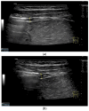
Diaphragm on the right (a) and left (b). Hypoechoic muscle, covered to the pleural cavity by the hyperechoic parietal pleura and to the abdomen by the hyperechoic peritoneum. In the center of the muscle there is another echogenic layer, which corresponds to narrow collagenous fibers. The diaphragm is particularly visible here, as it is not overlaid by lung artifacts in a pleural effusion.
Figure 2
Figure 2
With abdominal sector transducers (a) and in elderly people (b), the diaphragm can appear hyperechoic. D—diaphragm.
Figure 2
Figure 2
With abdominal sector transducers (a) and in elderly people (b), the diaphragm can appear hyperechoic. D—diaphragm.
Figure 3
Figure 3
Transducer positions for visualization of the apposition zone on the right. The transducer is positioned longitudinally and laterally in the area of the mid-axillary line or slightly ventrally between the anterior and mid-axillary line, approximately in the 8th or 10th intercostal space (a,b). The diaphragm is located caudal to the pulmonary glide (typical A-lines/reverberation artifacts) (c). It is located on the inside of the intercostal spaces.
Figure 3
Figure 3
Transducer positions for visualization of the apposition zone on the right. The transducer is positioned longitudinally and laterally in the area of the mid-axillary line or slightly ventrally between the anterior and mid-axillary line, approximately in the 8th or 10th intercostal space (a,b). The diaphragm is located caudal to the pulmonary glide (typical A-lines/reverberation artifacts) (c). It is located on the inside of the intercostal spaces.
Figure 4
Figure 4
Measurement of the diaphragm thickness on the right and left in the apposition zone (9 MHz linear transducer). The markers are positioned within the peritoneum and the pleura. They are located on the outer contour of the hypoechoic muscle (a,b). The delicate hypoechoic collagenous central fiber is also visible. The diaphragm is thinner in the end-expiratory phase (a) than in the contracted end-inspiratory phase (b). The same measurements are performed on the left in the apposition zone. The diaphragm between the intercostal spaces and the spleen can be seen below the pulmonary glide, thinner at end-expiration phase (c) and during end-inspiratory contraction (d). D—diaphragm.
Figure 4
Figure 4
Measurement of the diaphragm thickness on the right and left in the apposition zone (9 MHz linear transducer). The markers are positioned within the peritoneum and the pleura. They are located on the outer contour of the hypoechoic muscle (a,b). The delicate hypoechoic collagenous central fiber is also visible. The diaphragm is thinner in the end-expiratory phase (a) than in the contracted end-inspiratory phase (b). The same measurements are performed on the left in the apposition zone. The diaphragm between the intercostal spaces and the spleen can be seen below the pulmonary glide, thinner at end-expiration phase (c) and during end-inspiratory contraction (d). D—diaphragm.
Figure 5
Figure 5
Subcostal transducer positions. The transducer is placed between the medioclavicular line and the anterior axillary line (a,b). The diaphragm excursion can then be visualized and measured in M-mode during breath holding and deep inspiration (c,d) and voluntary “sniffing” or coughing (e).
Figure 5
Figure 5
Subcostal transducer positions. The transducer is placed between the medioclavicular line and the anterior axillary line (a,b). The diaphragm excursion can then be visualized and measured in M-mode during breath holding and deep inspiration (c,d) and voluntary “sniffing” or coughing (e).
Figure 6
Figure 6
Subxiphoid transducer positions. The right diaphragmatic fat limb runs between the inferior vena cava and the aorta to the right of the spine. The left diaphragmatic limb is shown to the left of and dorsal to the aorta. Diaphragm between the markings (a). The diaphragm in the color Doppler imaging (CDI) between the vessels. The incisions should not be confused with lymphomas (b). The diaphragm on the right between the inferior vena cava and the spine (between the markings) and in front of the abdominal aorta (c). The thickened diaphragm during inspiration (between the markings) in the same position. Under optimal examination conditions, the contraction can also be visible in this position (d). The diaphragm between the aorta and liver is visible in longitudinal section (e). Endosonography shows the diaphragmatic limb ventral to the aorta (arrow) (f). Ams—superior mesenteric artery, Ao—aorta, Ard—right renal artery, D—diaphragm; IVC—vena cava inferior, PV—portal vein, TC—celiac trunk, Vl—splenic vein.
Figure 6
Figure 6
Subxiphoid transducer positions. The right diaphragmatic fat limb runs between the inferior vena cava and the aorta to the right of the spine. The left diaphragmatic limb is shown to the left of and dorsal to the aorta. Diaphragm between the markings (a). The diaphragm in the color Doppler imaging (CDI) between the vessels. The incisions should not be confused with lymphomas (b). The diaphragm on the right between the inferior vena cava and the spine (between the markings) and in front of the abdominal aorta (c). The thickened diaphragm during inspiration (between the markings) in the same position. Under optimal examination conditions, the contraction can also be visible in this position (d). The diaphragm between the aorta and liver is visible in longitudinal section (e). Endosonography shows the diaphragmatic limb ventral to the aorta (arrow) (f). Ams—superior mesenteric artery, Ao—aorta, Ard—right renal artery, D—diaphragm; IVC—vena cava inferior, PV—portal vein, TC—celiac trunk, Vl—splenic vein.
Figure 7
Figure 7
When imaging the right adrenal gland, the diaphragm is in one orientation. The diaphragmatic limb passes dorsal to the right adrenal gland and ventral to the vertebral column (a). The diaphragm in expiration ventral to the vertebral column with transducer position in the right flank longitudinal (arrows) (b) and thickened in inspiration (c). Diaphragm with typical triple layering in the subcostal section in the medioclavicular line, well delineated in pleural effusion (d). The transducer is directed medially in the MCL (e). In the case of pleural effusion and lack of total lung reflection, the diaphragm can also be seen in a longer extension on the left side (f). Mirror reflection of the diaphragm. Hyperechoic peritoneum, hypoechoic diaphragm muscle, hyperechoic total reflection of the lung. The hyperechoic parietal pleura cannot be differentiated from the total reflection of the lung. This also applies to the mirrored side. In the area of the mirror artifact are the hypoechoic diaphragmatic muscle and the hyperechoic parietal peritoneum. The diaphragm is not thickened, and the total reflection of the lung should not be confused with the collagenous layer (g). Diaphragmatic incisions (h).
Figure 7
Figure 7
When imaging the right adrenal gland, the diaphragm is in one orientation. The diaphragmatic limb passes dorsal to the right adrenal gland and ventral to the vertebral column (a). The diaphragm in expiration ventral to the vertebral column with transducer position in the right flank longitudinal (arrows) (b) and thickened in inspiration (c). Diaphragm with typical triple layering in the subcostal section in the medioclavicular line, well delineated in pleural effusion (d). The transducer is directed medially in the MCL (e). In the case of pleural effusion and lack of total lung reflection, the diaphragm can also be seen in a longer extension on the left side (f). Mirror reflection of the diaphragm. Hyperechoic peritoneum, hypoechoic diaphragm muscle, hyperechoic total reflection of the lung. The hyperechoic parietal pleura cannot be differentiated from the total reflection of the lung. This also applies to the mirrored side. In the area of the mirror artifact are the hypoechoic diaphragmatic muscle and the hyperechoic parietal peritoneum. The diaphragm is not thickened, and the total reflection of the lung should not be confused with the collagenous layer (g). Diaphragmatic incisions (h).
Figure 8
Figure 8
In pleural effusion and ascites, diaphragmatic movements can be seen. In expiration (a) and during deep inspiration, the diaphragm moves towards the abdominal cavity (b). Intracavitary contrast administration with SonoVue (off-label use) was performed for ascites and right pleural effusion under the suspicion of a fistula between the pleura and abdomen with recurrent right-sided pleural effusion. However, there was no transfer of the contrast medium from the pleural effusion into the ascites (c).
Figure 8
Figure 8
In pleural effusion and ascites, diaphragmatic movements can be seen. In expiration (a) and during deep inspiration, the diaphragm moves towards the abdominal cavity (b). Intracavitary contrast administration with SonoVue (off-label use) was performed for ascites and right pleural effusion under the suspicion of a fistula between the pleura and abdomen with recurrent right-sided pleural effusion. However, there was no transfer of the contrast medium from the pleural effusion into the ascites (c).

Similar articles

Cited by

References

    1. van Doorn J.L.M., Wijntjes J., Saris C.G.J., Ottenheijm C.A.C., van Alfen N., Doorduin J. Association of diaphragm thickness and echogenicity with age, sex, and body mass index in healthy subjects. Muscle Nerve. 2022;66:197–202. doi: 10.1002/mus.27639. - DOI - PMC - PubMed
    1. Ueki J., De Bruin P.F., Pride N.B. In vivo assessment of diaphragm contraction by ultrasound in normal subjects. Thorax. 1995;50:1157–1161. doi: 10.1136/thx.50.11.1157. - DOI - PMC - PubMed
    1. Spiesshoefer J., Herkenrath S., Henke C., Langenbruch L., Schneppe M., Randerath W., Young P., Brix T., Boentert M. Evaluation of Respiratory Muscle Strength and Diaphragm Ultrasound: Normative Values, Theoretical Considerations, and Practical Recommendations. Respiration. 2020;99:369–381. doi: 10.1159/000506016. - DOI - PubMed
    1. Haaksma M.E., Smit J.M., Boussuges A., Demoule A., Dres M., Ferrari G., Formenti P., Goligher E.C., Heunks L., Lim E.H.T., et al. EXpert consensus On Diaphragm UltraSonography in the critically ill (EXODUS): A Delphi consensus statement on the measurement of diaphragm ultrasound-derived parameters in a critical care setting. Crit. Care. 2022;26:99. doi: 10.1186/s13054-022-03975-5. - DOI - PMC - PubMed
    1. Laghi F.A., Jr., Saad M., Shaikh H. Ultrasound and non-ultrasound imaging techniques in the assessment of diaphragmatic dysfunction. BMC Pulm. Med. 2021;21:85. doi: 10.1186/s12890-021-01441-6. - DOI - PMC - PubMed