Review
. 2025 Feb 15;15(2):304.
doi: 10.3390/life15020304.

Anatomy-Based Filler Injection: Treatment Techniques for Supraorbital Hollowness and Charming Roll

Affiliations
Review

Anatomy-Based Filler Injection: Treatment Techniques for Supraorbital Hollowness and Charming Roll

Gi-Woong Hong et al. Life (Basel). .

Abstract

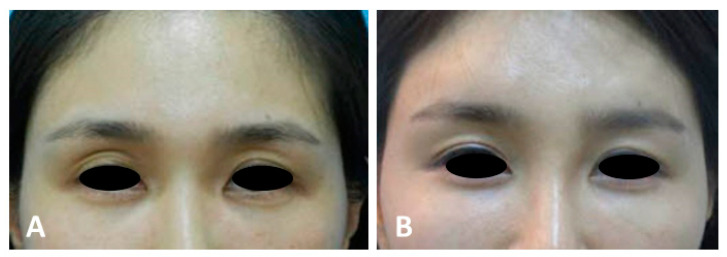
Supraorbital hollowness and pretarsal fullness, commonly known as the sunken eyelid and charming roll, respectively, are significant anatomical features that impact the aesthetic appearance of the periorbital region. Supraorbital hollowness is characterized by a recessed appearance of the upper eyelid, often attributed to genetic factors, aging, or surgical alterations, such as excessive fat removal during blepharoplasty. This condition is particularly prevalent among East Asians due to anatomical differences, such as weaker levator muscles and unique fat distribution patterns. Pretarsal fullness, also known as aegyo-sal, enhances the youthful and expressive appearance of the lower eyelid, forming a roll above the lash line that is considered aesthetically desirable in East Asian culture. Anatomical-based filler injection techniques are critical for correcting these features, involving precise placement within the correct tissue planes to avoid complications and achieve natural-looking results. This approach not only improves the aesthetic appeal of the eyelid but also enhances the overall facial harmony, emphasizing the importance of tailored procedures based on individual anatomy and cultural preferences.

Keywords: eyebrow anatomy; hyaluronic acid fillers; periorbital rejuvenation; pretarsal fullness; supraorbital hollowness.


Conflict of interest statement

Author Jovian Wan was employed by the Medical Research Inc. The remaining authors declare that the research was conducted in the absence of any commercial or financial relationships that could be construed as a potential conflict of interest.

Figures

Figure 1
Figure 1
Before (A) and after (B) treatment of supraorbital hollowness.
Figure 2
Figure 2
Anatomical layers of the supraorbital region.
Figure 3
Figure 3
Vascular structures of the orbital region.
Figure 4
Figure 4
Injection entry point and technique for the cannula. Injection entry point: Vertical line drawn above or outside the lateral canthus, around the lower margin of the superior orbital rim. Focus on the medial and middle parts of the periorbital rim, under the brow, to avoid the supraorbital and supratrochlear main arteries. Above the supratarsal lid crease and below the orbicularis retaining ligament. Injection technique: Patient in vertical sitting position with voluntarily opened eyes. Retrograde linear tiny injection technique with very slow release.
Figure 5
Figure 5
Anatomy of the preseptal space.
Figure 6
Figure 6
Injection planes: Supraperiosteal and submuscular injections around the orbital rim over the orbital septum to fill the hollowness. Subdermal injection of very soft HA filler to smooth the surface and remove unnecessary multiple eyelid lines.
Figure 7
Figure 7
Ideal position and shape of the eyebrow.
Figure 8
Figure 8
Ratio difference between the size of the eye and eyebrow.
Figure 9
Figure 9
Common classification of eyebrow shapes around the world.
Figure 10
Figure 10
Retro-orbicularis oculi fat (ROOF) in the eyebrow region.
Figure 11
Figure 11
Injection plane for the cannula. Submuscular injection into ROOF (retro-orbicularis oculi fat) for eyebrow augmentation. Subdermal injection of very soft filler to even out the surface and remove unnecessary multiple eyelid lines.
Figure 12
Figure 12
Structure of the lower eyelid roll muscle.
Figure 13
Figure 13
Injection techniques for the cannula or needle. Linear threading, retrograde tiny injection, very slow release, serial puncture, and tenting technique.
Figure 14
Figure 14
Injection planes: Deep subdermal or supramuscular injections. Subdermal injection to smooth the surface, close to the eyelash.
Figure 15
Figure 15
Anatomy of the superior and inferior palpebral arteries.

References

    1. Spada J. Treating Sunken Upper Eyelid With Hyaluronic Acid: Recommendations and Results. J. Drugs Dermatol. 2022;21:1002–1008. doi: 10.36849/JDD.6745. - DOI - PubMed
    1. Wong C.H., Mendelson B. Newer Understanding of Specific Anatomic Targets in the Aging Face as Applied to Injectables: Aging Changes in the Craniofacial Skeleton and Facial Ligaments. Plast. Reconstr. Surg. 2015;136:44s–48s. doi: 10.1097/PRS.0000000000001752. - DOI - PubMed
    1. Wan D., Amirlak B., Rohrich R., Davis K. The clinical importance of the fat compartments in midfacial aging. Plast. Reconstr. Surg. Glob. Open. 2013;1:e92. doi: 10.1097/GOX.0000000000000035. - DOI - PMC - PubMed
    1. Wakade D.V., Nayak C.S., Bhatt K.D. A Study Comparing the Efficacy of Monopolar Radiofrequency and Glycolic Acid Peels in Facial Rejuvenation of Aging Skin Using Histopathology and Ultrabiomicroscopic Sonography (UBM)—An Evidence Based Study. Acta Medica. 2016;59:14–17. doi: 10.14712/18059694.2016.49. - DOI - PubMed
    1. Swift A., Liew S., Weinkle S., Garcia J.K., Silberberg M.B. The Facial Aging Process From the “Inside Out”. Aesthet. Surg. J. 2021;41:1107–1119. doi: 10.1093/asj/sjaa339. - DOI - PMC - PubMed

LinkOut - more resources