. 2025 Feb 8;26(4):1434.
doi: 10.3390/ijms26041434.

Iron Accumulation and Lipid Peroxidation in Cellular Models of Nemaline Myopathies

Affiliations

Iron Accumulation and Lipid Peroxidation in Cellular Models of Nemaline Myopathies

Alejandra López-Cabrera et al. Int J Mol Sci. .

Abstract

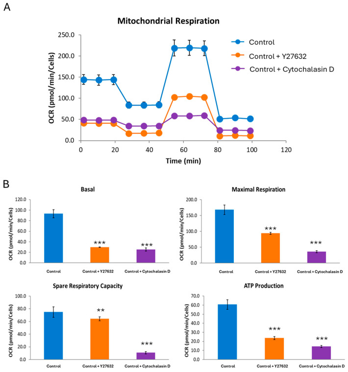
One of the most prevalent types of congenital myopathy is nemaline myopathy (NM), which is recognized by histopathological examination of muscle fibers for the presence of "nemaline bodies" (rods). Mutations in the actin alpha 1 (ACTA1) and nebulin (NEB) genes result in the most prevalent types of NM. Muscle weakness and hypotonia are the main clinical characteristics of this disease. Unfortunately, the pathogenetic mechanisms are still unknown, and there is no cure. In previous work, we showed that actin filament polymerization defects in patient-derived fibroblasts were associated with mitochondrial dysfunction. In this manuscript, we examined the pathophysiological consequences of mitochondrial dysfunction in patient-derived fibroblasts. We analyzed iron and lipofuscin accumulation and lipid peroxidation both at the cellular and mitochondrial level. We found that fibroblasts derived from patients harboring ACTA1 and NEB mutations showed intracellular iron and lipofuscin accumulation, increased lipid peroxidation, and altered expression levels of proteins involved in iron metabolism. Furthermore, we showed that actin polymerization inhibition in control cells recapitulates the main pathological alterations of mutant nemaline cells. Our results indicate that mitochondrial dysfunction is associated with iron metabolism dysregulation, leading to iron/lipofuscin accumulation and increased lipid peroxidation.

Keywords: iron accumulation; lipid peroxidation; nemaline myopathy.


Conflict of interest statement

The authors declare no conflicts of interest.

Figures

Figure 1
Figure 1
Iron accumulation in NM cells. (A) Prussian Blue staining of control cells (C1) and NM fibroblasts (P1, P2, P3, and P4) was carried out as described in Section 4, Materials and Methods. P1 fibroblasts were exposed to 100 µM deferiprone (Def), an iron chelating agent, for 24 h as a negative control. Images were made in brightfield by an Axio Vert A1 inverted optical microscope (Zeiss, Oberkochen, Germany) with a 40× objective and were analyzed using Fiji-ImageJ software (version 2.9.0/1.53t) (National Institute of Health, Bethesda, MD, USA). Scale bar = 20 µm. (B) Quantification of Prussian Blue staining images was performed by the Image J software (version 1.54f). (C) Iron content determined by ICP-MS in NM fibroblast. ICP-MS was used to measure the total iron content of control and NM patients as detailed in the Material and Methods. Data represent the mean ± SD of three separate experiments. * p < 0.05, ** p < 0.01, *** p < 0.001 between NM fibroblasts and controls; aa p < 0.01 between untreated and treated NM cells between the presence and the absence of deferiprone (Def). A.U., arbitrary units.
Figure 2
Figure 2
Lipofuscin accumulation in NM fibroblasts. (A) Sudan black staining of control cells (C1) and NM fibroblasts (P1, P2, P3, and P4) was performed as described in Section 4, Materials and Methods. P1 fibroblasts were exposed to 100 µM deferiprone (Def), an iron chelating agent, for 24 h, as a negative control. Images were made in brightfield by an Axio Vert A1 inverted optical microscope (Zeiss, Oberkochen, Germany) with a 40× objective and were analyzed using Fiji-ImageJ software (version 2.9.0/1.53t) (National Institute of Health, Bethesda, MD, USA). Scale bar = 20 µm. (B) Quantification of Sudan Black in control and NM fibroblasts was performed by ImageJ software (version 1.54f). Data represent the mean ± SD of three separate experiments. ** p < 0.01, *** p < 0.001 between NM fibroblasts and controls; aa p < 0.01 between untreated and treated NM cells between the presence and the absence of deferiprone (Def). A.U., arbitrary units.
Figure 3
Figure 3
Lipofuscin-like aggregates in NM fibroblasts. (A) Representative autofluorescence and bright field (BF) images of control (C1) and NM fibroblasts (P1, P2, P3, and P4). Control cell and P1 were treated with 100 μM deferiprone (Def) for 24 h as a negative control. Scale bar = 20 μm. (B) Quantification of cell autofluorescence was determined by image analysis using the Fiji software (version 2.9.0/1.53t). (C) The autofluorescence spectra of lipofuscin granules were measured by confocal laser scanning microscopy (Nikon A1R, Shinagawa, Tokyo, Japan) in control, NM fibroblasts (P1, P2, P3, and P4), and negative controls (control and P1 treated with 100 μM deferiprone (Def)). Excitation laser source: 405 nm. The emission spectra were recorded in 20 large lipofuscin granules in 20 cells. Results are expressed as mean ± SD of autofluorescence intensity. ** p < 0.01, *** p < 0.001 between NM fibroblasts and controls; aa p < 0.01 between the presence and the absence of DEF in controls and Patient 1 cells. A.U., arbitrary units.
Figure 4
Figure 4
Electron microscopy images of control and P2 (ACTA1) and P3 (NEB) fibroblasts. (A) Representative electron microscopy images of control (C1) and NM fibroblasts (P2 and P3). Red arrows were used to highlight the lipofuscin-like granules. (B) Quantification of lipofuscin-like aggregates. (C) P2 cells showed mitochondrial vacuolization, and condensation/lateralization of mitochondrial membranes (orange arrow). Scale bar = 2 μm. Data represent the mean ± SD of the examination of 50 cells per condition. *** p < 0.001 between NM cells and controls.
Figure 5
Figure 5
Mitochondrial ferrous iron (Fe2+) in control and NM cells. (A) Levels of mitochondrial ferrous iron in control (C1) and NM fibroblasts (P1, P2, P3, and P4) were assayed by MitoFerroGreen staining as described in Materials and Methods. Cellular models of pantothenate kinase-associated neurodegeneration (PKAN) were used as a positive control. PKAN cells were exposed to 100 µM deferiprone (Def), an iron chelating agent, for 24 h as a negative control. (B) MitoFerroGreen staining quantification was conducted by using Fiji software (version 2.9.0/1.53t). Cells were incubated with MitoTrackerTM Deep Red FM to demonstrate that MitoFerroGreen signal colocalizes with a mitochondrial marker. The colocalization of both markers was assessed by the DeltaVision software (version softWoRx 7.0; Applied Precision; Issaquah, Washington (WA), United states (USA)) calculating the Pearson correlation coefficient. Pearson correlation coefficient was >0.75 in NM fibroblasts and control. Scale bar = 20 µm. *** p < 0.001 between control and NM fibroblasts; aaa p < 0.001 between the presence and the absence of DEF between treated and untreated PKAN patient cells. Data represent the mean ± SD of four separate experiments. A.U., arbitrary units.
Figure 6
Figure 6
Cellular lipid peroxidation in control and NM cells. (A) Levels of cellular lipid peroxidation in control (C1) and NM fibroblasts (P1, P2, P3, and P4) were measured using BODIPY® staining as detailed in Section 4, Material and Methods. (B) BODIPY® staining quantification was performed by using the Fiji software (version 2.9.0/1.53t). Images were made in by an Axio Vert A1 inverted optical microscope (Zeiss, Oberkochen, Germany) with a 40× objective. Control fibroblasts were treated with 500 µM Luperox (Tert-butyl hydroperoxide) for 15 min as positive control of lipid peroxidation. Scale bar = 20 µm. *** p < 0.001 between control and NM fibroblasts; aaa p < 0.001 between the presence and the absence of Luperox between treated and untreated Control cells. Data represent the mean ± SD of four separate experiments. A.U., arbitrary units.
Figure 7
Figure 7
Mitochondria lipid peroxidation in control and NM cells. (A) Levels of mitochondrial lipid peroxidation in control (C1) and NM fibroblasts (P1, P2, P3, and P4) were assayed by MitoPeDPP staining as detailed in Section 4, Material and Methods. (B) MitoPeDPP staining quantification was performed by using the Fiji software (version 2.9.0/1.53t). Cells were incubated with MitoTrackerTM Deep Red FM to demonstrate that MitoPeDPP signal colocalizes with a mitochondrial marker. Control fibroblasts were treated with 500 µM Luperox (Tert-butyl hydroperoxide) for 15 min as positive control of lipid peroxidation. Colocalization of both markers MitoPeDPP and MitoTrackerTM was assessed by the DeltaVision software (version softWoRx 7.0Applied Precision; Issaquah, Washington (WA), United states (USA)). Scale bar = 20 µm. ** p < 0.01, *** p < 0.001 between control and NM fibroblasts; aaa p < 0.001 between the presence and the absence of Luperox between treated and untreated control cells. Data represent the mean ± SD of four separate experiments. A.U., arbitrary units.
Figure 8
Figure 8
Iron metabolism-related protein expression levels in NM fibroblast. (A) Immunoblotting analysis of cellular extracts from controls (C1 and C2) and NM patient cell lines P1, P2, P3, and P4. Protein extracts (50 μg) were separated on a SDS polyacrylamide gel and immunostained with antibodies against TFR, DMT1, IRP1, Ferritin, Mitoferritin, Mitoferrin2, FXN, ISCU, Mt-ACP, and PANK2. Tubulin was used as a loading control. (B) Densitometry of Western blotting. For control cells (C1 and C2), data are the mean ± SD of the two control cell lines. Data represent the mean ± SD of three separate experiments. * p < 0.05, ** p < 0.01, *** p < 0.001 between NM cells and controls. A.U., arbitrary units.
Figure 9
Figure 9
Labile iron pool (LIP). LIP in control (C1) and NM fibroblasts (P1, P2, P3, and P4) were measured as described in Section 4, Materials and Methods. As a negative control, control cells were exposed to 100 μM deferiprone (Def) for 24 h. For control cells, data are mean ± SD of three control cell lines. * p < 0.05 between control and NM fibroblasts. a p < 0.05 between the presence and the absence of DEF between treated and untreated Control cells. Data represent the mean ± SD of six separate experiments.
Figure 10
Figure 10
Actin staining by Rhodamine–Phalloidin and MitotrackerTM DeepRed of control fibroblasts treated with Y27632 or Cytochalasin D inhibitors. (A) Control fibroblasts (C1) were treated with 10 µM Y27632 for 3 h or with 20 µM Cytochalasin D for 24 h. Control fibroblasts were stained with Rhodamine–Phalloidin and MitotrackerTM DeepRed and visualized under a widefield fluorescence microscope. Nuclei were revealed by DAPI staining. Treated control fibroblasts presented smaller and unstructured actin filaments compared to untreated control fibroblasts. Images were taken using the 40× lens and processed by the ImageJ software (version 1.54f). (B) Measurement of the length of actin filaments (µm). The length of the actin filaments was measured in triplicate with the ImageJ software (version 1.54f) in 30 images. (C) Quantification of tubular and rounded percentage of mitochondria in control cells. Data represent the mean ± SD of three separate experiments (at least 100 cells for each condition and experiment were analyzed). *** p < 0.001 between treated and untreated control cells. Scale bar = 20 µm.
Figure 11
Figure 11
Bioenergetic analysis of control cells untreated and treated with Y27632 inhibitor or Cytochalasin D. (A) Respiratory profile of untreated and treated control cells (C1). (B) Basal, maximal, and spare respiratory capacity and ATP production were determined in untreated and treated control fibroblast using the Seahorse analyzer. Control fibroblasts were treated with 10 µM of Y27632 for 24 h or with 20 µM of Cytochalasin D for 3 h. ** p < 0.01, *** p < 0.001 between untreated and treated control cells.
Figure 12
Figure 12
Expression levels of iron metabolism-related proteins in control fibroblasts treated with acting depolymerizing agents. (A) Immunoblotting analysis of cellular extracts from control cells (C1) treated with 10 µM of Y27632 for 24 h or 20 µM Cytochalasin D for 3 h. Protein extracts (50 μg) were separated on a SDS polyacrylamide gel and immunostained with antibodies against TFR, DMT1, IRP1, Ferritin, Mitoferritin, Mitoferrin2, FXN, ISCU, Mt-ACP, and PANK2. Tubulin was used as a loading control. (B) Densitometry of Western blotting. Data represent the mean ± SD of three separate experiments. * p < 0.05, ** p < 0.01, *** p < 0.001 between untreated and treated controls. A.U., arbitrary units.
Figure 13
Figure 13
Iron accumulation in control cells treated with Y27632 or Cytochalasin D inhibitors. (A) Prussian Blue staining of control (C1) (−) (−) and control cells treated with 10 µM Y27632 for 24 h, or with 20 µM Cytochalasin D for 3 h. Images were made in brightfield by an Axio Vert A1 inverted optical microscope (Zeiss, Oberkochen, Germany) with a 40× objective and were analyzed using Fiji-ImageJ software (version 2.9.0/1.53t) (National Institute of Health, Bethesda, MD, USA). Scale bar = 20 µm. (B) Quantification of Prussian Blue staining images was performed by the Image J software (version 1.54f). Data represent the mean ± SD of three separate experiments. ** p < 0.01, *** p < 0.001 between untreated and treated control cells. A.U., arbitrary units.
Figure 14
Figure 14
Lipofuscin accumulation in control cells treated with Y27632 or Cytochalasin D inhibitor. (A) Sudan Black staining of control cells (C1) treated with 10 µM Y27632 for 24 h, or with 20 µM Cytochalasin D for 3 h. Images were made in brightfield by an Axio Vert A1 inverted optical microscope (Zeiss, Oberkochen, Germany) with a 40× objective and were analyzed using Fiji-ImageJ software(version 2.9.0/1.53t) (National Institute of Health, Bethesda, MD, USA). Scale bar = 20 µm. (B) Quantification of Sudan Black staining images was performed by the Image J software (version 1.54f). Data represent the mean ± SD of three separate experiments. *** p < 0.001 between untreated and treated control cells. A.U., arbitrary units.
Figure 15
Figure 15
Cellular lipid peroxidation in control cells treated with Y27632 or Cytochalasin D inhibitors. (A) Control cells (C1) were treated with 10 µM Y27632 for 24 h, or with 20 µM Cytochalasin D for 3 h. The levels of cellular lipid peroxidation were measured using BODIPY® staining as detailed in the Material and Methods. (B) BODIPY® staining quantification was performed by using the Fiji software (version 2.9.0/1.53t). Control fibroblasts were treated with 500 µM Luperox (LUP, Tert-butyl hydroperoxide) for 15 min as positive control of lipid peroxidation. Scale bar = 20 µm. ** p < 0.01, *** p < 0.001 between treated and untreated control fibroblasts. Data represent the mean ± SD of four separate experiments. A.U., arbitrary units.

References

    1. North K.N., Wang C.H., Clarke N., Jungbluth H., Vainzof M., Dowling J.J., Amburgey K., Quijano-Roy S., Beggs A.H., Sewry C., et al. Approach to the diagnosis of congenital myopathies. Neuromuscul. Disord. 2014;24:97–116. doi: 10.1016/j.nmd.2013.11.003. - DOI - PMC - PubMed
    1. Ogasawara M., Nishino I. A review of major causative genes in congenital myopathies. J. Hum. Genet. 2023;68:215–225. doi: 10.1038/s10038-022-01045-w. - DOI - PubMed
    1. Cassandrini D., Trovato R., Rubegni A., Lenzi S., Fiorillo C., Baldacci J., Minetti C., Astrea G., Bruno C., Santorelli F.M., et al. Congenital myopathies: Clinical phenotypes and new diagnostic tools. Ital. J. Pediatr. 2017;43:101. doi: 10.1186/s13052-017-0419-z. - DOI - PMC - PubMed
    1. Christophers B., Lopez M.A., Gupta V.A., Vogel H., Baylies M. Pediatric Nemaline Myopathy: A Systematic Review Using Individual Patient Data. J. Child. Neurol. 2022;37:652–663. doi: 10.1177/08830738221096316. - DOI - PMC - PubMed
    1. Conen P.E., Murphy E.G., Donohue W.L. Light and Electron Microscopic Studies of “Myogranules” in a Child with Hypotonia and Muscle Weakness. Can. Med. Assoc. J. 1963;89:983–986. - PMC - PubMed

LinkOut - more resources