Review
. 2025 Feb 10;26(4):1457.
doi: 10.3390/ijms26041457.

The Crosstalk Between Endothelial Cells, Smooth Muscle Cells, and Macrophages in Atherosclerosis

Affiliations
Review

The Crosstalk Between Endothelial Cells, Smooth Muscle Cells, and Macrophages in Atherosclerosis

Sihe Gong et al. Int J Mol Sci. .

Abstract

Atherosclerosis (AS) is a chronic inflammatory vascular disease closely tied to cellular metabolism. Recent genome-wide association study data have suggested the significant roles of endothelial cells, smooth muscle cells, and macrophages in the regression and exacerbation of AS. However, the impact of cellular crosstalk and cellular metabolic derangements on disease progression in AS is vaguely understood. In this review, we analyze the roles of the three cell types in AS. We also summarize the crosstalk between the two of them, and the associated molecules and consequences involved. In addition, we emphasize potential therapeutic targets and highlight the importance of the three-cell co-culture model and extracellular vesicles in AS-related research, providing ideas for future studies.

Keywords: atherosclerosis; endothelial cells; macrophages; smooth muscle cells.


Conflict of interest statement

All authors declare that they have no conflicts of interest.

Figures
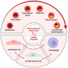
Figure 1
Figure 1
Intercellular communication affects the progression of atherosclerosis. In atherosclerosis, initial changes at the cellular and molecular levels lead to endothelial injury, which may not immediately cause luminal narrowing or impairment of tissues and organs. The foam cells then gather below the endothelium cells and appear as yellow fat splotches. This is followed by the accumulation of lipids within and outside the cells, generating lipid pools that disrupt the intimal structure and deform the arterial wall. Smooth muscle cells migrate to participate in the formation of a fibrous cap, leading to the emergence of white plaques that protrude into the arterial lumen, causing narrowing and the formation of atherosclerotic and fibrous plaques. On the basis of these plaques, secondary lesions such as hemorrhage, necrosis, ulceration, calcification, and mural thrombosis can occur, potentially triggering acute cardiovascular events. In this process, endothelial cells, smooth muscle cells, and macrophages all play important roles.
Figure 2
Figure 2
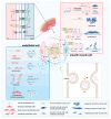
Crosstalk between endothelial cells and smooth muscle cells and the extracellular vesicle transfer process. Endothelial cells (ECs) and smooth muscle cells (SMCs) communicate directly through gap junctions and adhesion junctions, involving proteins such as Connexin (Cx) 37, Cx40, Cx43, and N-cadherin. The two cells can also influence each other through various vasoactive substances and extracellular vesicles. Extracellular vesicles are formed from the plasma membrane and transport molecules such as miRNAs and peptides. These chemicals regulate EC proliferation, dysfunction, migration, and expression of inflammatory factors. They can also alter SMC phenotypic transition, apoptosis, proliferation, migration, and expression of inflammatory factors. Furthermore, the extracellular matrix can mediate communication between ECs and SMCs through its protein or carbohydrate components and mechanical transmission.
Figure 3
Figure 3
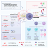
Crosstalk between endothelial cells and macrophages and different phenotypes of macrophages. Different modes of direct contact between endothelial cells and macrophages provide avenues for communication between these two cell types. Vasoactive substances as well as miRNAs contained within extracellular vesicles can lead to proliferation, dysfunction, changes in permeability, and expression of inflammatory factors in endothelial cells. Moreover, those can also induce phenotypic transformation and migration in macrophages. Additionally, components of the extracellular matrix can influence their communication. Macrophages are classified into two main phenotypes: M1, which secretes pro-inflammatory factors, and M2, which secretes anti-inflammatory cytokines. Beyond these, other phenotypes such as M(Hb), Mhem, HA-mac, Mox, and M4 also exist.
Figure 4
Figure 4
Crosstalk between smooth muscle cells and macrophages and atherosclerotic scenarios in the vasculature. Membrane surface proteins like ICAM-1, VCAM-1, and CX3CL1 can mediate the interaction between smooth muscle cells and macrophages, or they can interact directly through signaling pathways like Dll4/Notch. Vasoactive substances such as IL, TGF, TNF, IFN, and PDGF play crucial roles in the crosstalk between smooth muscle cells and macrophages. Factors secreted by both types of cells can influence smooth muscle cell phenotypic switching, apoptosis, proliferation, migration, and changes in inflammatory factor expression, as well as affect macrophage phenotypic conversion and the expression of inflammatory factors in atherosclerosis. miRNAs such as miR-503-5p and miR-145-5p in extracellular vesicles also play a significant role in this process. The extracellular matrix, which constitutes the cellular environment, can be altered by the communication between macrophages and smooth muscle cells.

Similar articles

Cited by

References

    1. Libby P., Buring J.E., Badimon L., Hansson G.K., Deanfield J., Bittencourt M.S., Tokgözoğlu L., Lewis E.F. Atherosclerosis. Nat. Rev. Dis. Primers. 2019;5:56. doi: 10.1038/s41572-019-0106-z. - DOI - PubMed
    1. Soehnlein O., Libby P. Targeting inflammation in atherosclerosis—From experimental insights to the clinic. Nat. Rev. Drug Discov. 2021;20:589–610. doi: 10.1038/s41573-021-00198-1. - DOI - PMC - PubMed
    1. Mayerl C., Lukasser M., Sedivy R., Niederegger H., Seiler R., Wick G. Atherosclerosis research from past to present—On the track of two pathologists with opposing views, Carl von Rokitansky and Rudolf Virchow. Virchows Arch. 2006;449:96–103. doi: 10.1007/s00428-006-0176-7. - DOI - PubMed
    1. Duff G.L. The pathogenesis of atherosclerosis. Can. Med. Assoc. J. 1951;64:387–394. - PMC - PubMed
    1. Ross R. Atherosclerosis—An Inflammatory Disease. N. Engl. J. Med. 1999;340:115–126. doi: 10.1056/NEJM199901143400207. - DOI - PubMed

LinkOut - more resources