. 2025 Feb 11;26(4):1493.
doi: 10.3390/ijms26041493.

Paeoniflorin Attenuates APAP-Induced Liver Injury via Intervening the Crosstalk Between Hepatocyte Pyroptosis and NETs

Affiliations

Paeoniflorin Attenuates APAP-Induced Liver Injury via Intervening the Crosstalk Between Hepatocyte Pyroptosis and NETs

Yu-Ru Zhu et al. Int J Mol Sci. .

Abstract

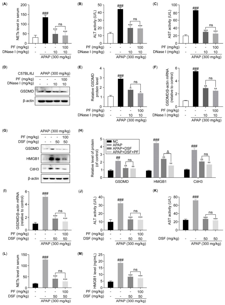
(1) Liver injury caused by an overdose of acetaminophen (APAP) represents a major public health concern. Paeoniflorin (PF) has been reported to have anti-inflammatory and liver-protective effects, but the underlying mechanisms remain unclear. This study aimed to investigate the effect of PF on the crosstalk between pyroptosis and NETs in AILI. (2) APAP-treated C57BL/6J mice were used to demonstrate the protective effect of PF on liver injury. HepG2 and dHL-60 cells were cultured to study the effects of PF on hepatocyte pyroptosis and neutrophil extracellular traps (NETs) in vitro. Moreover, cell co-culture experiments were performed, and mice were treated with a NETs-depleting agent and hepatocyte pyroptosis inhibitor to investigate the improvement of AILI induced by PF through regulating the crosstalk between hepatocyte pyroptosis and NETs. (3) PF significantly alleviated AILI. Additionally, PF inhibited the expression of pyroptosis-related proteins, high-mobility group box 1 (HMGB1), and NETs-associated proteins in vitro and in vivo. The co-culture experiments demonstrated that PF not only inhibited the NETs triggered by hepatocyte pyroptosis, but also suppressed the hepatocyte pyroptosis induced by NETs. In mice with depleted neutrophils, the level of hepatocyte pyroptosis notably decreased, indicating a diminished impact of PF. Similarly, NETs formation was reduced in mice receiving a pyroptosis inhibitor compared to the APAP group. Compared with DNase I alone, the reduction effect of PF combined with DNase I on serum ALT and AST levels decreased from 46.857% and 39.927% to 44.347% and 33.419%, respectively. Compared with DSF alone, PF combined with DSF reduced the ALT and AST levels from 46.857% and 39.927% to 45.347% and 36.419%, respectively. (4) PF demonstrated therapeutic effects on AILI. Its mechanism involves the regulation of the crosstalk between hepatocyte pyroptosis and NETs. This research substantiates the pharmacological promise of PF as a therapeutic intervention for acute AILI.

Keywords: acetaminophen; liver injury; neutrophil extracellular traps; paeoniflorin; pyroptosis.


Conflict of interest statement

The authors declare no conflicts of interest.

Figures

Figure 1
Figure 1
PF attenuates AILI in mice. (A) Schematic diagram of drug administration to animals (n = 6); (B) body weights of mice during administration; (C) liver index measurements; (DG) serum concentrations of ALT, AST, LDH, and MDA; (H) MPO levels in liver tissues of mice; (I) H&E staining images. The magnification used for the H&E staining images was ×100, with the scale bars representing 100 μm. All experimental data are presented as the mean ± SD. ## p < 0.01 and ### p < 0.001 vs. the NC group; * p < 0.05, ** p < 0.01, and *** p < 0.001 vs. the APAP group.
Figure 2
Figure 2
PF attenuates APAP-induced hepatocyte inflammation and pyroptosis. (A) The mRNA expression levels of IL-1β, TNF-α, IL-6, and IL-18 in the liver of the mice were quantified using the quantitative real-time polymerase chain reaction (qRT-PCR). (B) The mRNA expression levels of NLRP3, caspase-1, GSDMD, and HMGB1 in the livers of mice were measured via qRT-PCR. (C,D) The protein levels of NLRP3, caspase-1, GSDMD, and HMGB1 in the livers of mice were assessed using Western blotting (WB) analysis and are reported relative to β-actin levels. (E) The levels of HMGB1 in the serum of mice were quantified using enzyme-linked immunosorbent assays (ELISAs). (F) HepG2 cells were treated with various concentrations of PF (0, 10, 20, and 40 μM) for 24 h, and then the cell viability was assessed using the thiazolyl blue tetrazolium bromide (MTT) assay (n = 3). (G) HepG2 cells were pretreated with various concentrations of PF (0, 10, 20, and 40 μM) for 24 h and subsequently treated with APAP (10 mM) for 6 h, and then the cell viability was assessed using the MTT assay (n = 3). (H) The mRNA expression levels of IL-1β, TNF-α, IL-6, and IL-18 in HepG2 cells were quantified using qRT-PCR. (I) The mRNA expression levels of NLRP3, caspase-1, GSDMD, and HMGB1 in the HepG2 cells were quantified using qRT-PCR. (J,K) The protein levels of NLRP3, caspase-1, GSDMD, and HMGB1 in the HepG2 cells were assessed using WB analysis, and are reported relative to β-actin levels. (L) The concentration of HMGB1 in the culture medium of the HepG2 cells was quantified using ELISA. All experimental data are presented as the mean ± SD. ## p < 0.01 and ### p < 0.001 vs. the NC group; * p< 0.05, ** p< 0.01, and *** p< 0.001 vs. the APAP group.
Figure 3
Figure 3
PF inhibits the APAP-induced formation of NETs. (A) NETs in the serum were quantified using an ELISA for NE–DNA complexes. (B,C) The protein levels of MPO, PADI4, NE, and CitH3 in the mouse liver were assessed using WB analysis and are reported relative to β-actin levels. (D) To evaluate the distribution of MPO and CitH3, the positive areas for MPO and CitH3 in liver sections were assessed using IHC staining. The magnification in the images is ×200. (E) The surface characterization of the dHL-60 cells was performed using FESEM. The magnification used was ×2500, with the scale bars representing 20 μm. (F,G) A cellular immunofluorescence analysis was conducted to assess the protein expression of MPO and CitH3 in the dHL-60 cells (n = 3). The magnification used was ×400, with the scale bars representing 20 μm. (H,I) The protein levels of MPO, PADI4, NE, and CitH3 in the dHL-60 cells were assessed using WB analysis, and are reported relative to β-actin levels. All experimental data are presented as the mean ± SD. ## p < 0.01 and ### p < 0.001 vs. the NC group; * p < 0.05, ** p < 0.01, and *** p < 0.001 vs. the APAP group or PMA group.
Figure 4
Figure 4
PF intervenes the crosstalk between hepatocyte pyroptosis and NETs. (A) HepG2 cells in the co-culture were treated with PMA (100 nM) for 4 h, followed by treatment with various concentrations of PF (0, 10, 20, and 40 μM) for 24 h; subsequently, cell viability was assessed using the MTT assay (n = 3). (B) The mRNA expression level of GSDMD in the HepG2 cells in the co-culture was quantified using qRT-PCR. (C,D) The protein level of GSDMD in the HepG2 cells in the co-culture was assessed by WB analysis and reported relative to β-actin levels. (E,F) The protein levels of CitH3 in the dHL-60 cells in the co-culture were assessed using WB analysis and are reported relative to β-actin levels. (G) A cellular immunofluorescence analysis was performed to evaluate the expression of MPO and CitH3 in the dHL-60 cells in the co-culture (n = 3). The magnification used was ×400. All experimental data are presented as the mean ± SD. ## p < 0.01 and ### p < 0.001 vs. the NC group; * p < 0.05, ** p < 0.01, and *** p < 0.001 vs. the APAP group or PMA group.
Figure 5
Figure 5
PF attenuates AILI in mice by intervening the crosstalk between hepatocyte pyroptosis and NETs. (A) The levels of NETs in the serum of the neutropenic mice were quantified using ELISAs. (B,C) The ALT and AST levels in the serum of the neutropenic mice. (D,E) The protein levels of GSDMD in the liver of the neutropenic mice were determined using WB analysis and are reported relative to β-actin levels. (F) The mRNA expression level of GSDMD in the liver of the neutropenic mice was quantified via qRT-PCR. (G,H) The protein levels of GSDMD, HMGB1, and CitH3 in the liver of the pyroptosis inhibitor-treated mice were assessed using WB analysis and are reported relative to β-actin levels. (I) The mRNA expression level of GSDMD in the liver of the pyroptosis inhibitor-treated mice was quantified via qRT-PCR. (J,K) The serum levels of ALT and AST in the pyroptosis inhibitor-treated mice. (L,M) The levels of NETs and HMGB1 in the serum of the pyroptosis inhibitor-treated mice were measured using ELISA. All experimental data are presented as the mean ± SD. ## p < 0.01 and ### p < 0.001 vs. the NC group, * p < 0.05, ** p < 0.01, and *** p < 0.001 vs. the APAP or PMA group; Δ p < 0.05, ns p > 0.05 vs. the APAP + DNase I or APAP + DSF group.

References

    1. Allison R., Guraka A., Shawa I.T., Tripathi G., Moritz W., Kermanizadeh A. Drug induced liver injury—A 2023 update. J. Toxicol. Environ. Health B Crit. Rev. 2023;26:442–467. doi: 10.1080/10937404.2023.2261848. - DOI - PubMed
    1. Jaeschke H., Xie Y., McGill M.R. Acetaminophen-induced Liver Injury: From Animal Models to Humans. J. Clin. Transl. Hepatol. 2014;2:153–161. - PMC - PubMed
    1. Gil-Pitarch C., Serrano-Maciá M., Simon J., Mosca L., Conter C., Rejano-Gordillo C.M., Zapata-Pavas L.E., Peña-Sanfélix P., Azkargorta M., Rodríguez-Agudo R., et al. Neddylation inhibition prevents acetaminophen-induced liver damage by enhancing the anabolic cardiolipin pathway. Cell Rep. Med. 2024;5:101653. doi: 10.1016/j.xcrm.2024.101653. - DOI - PMC - PubMed
    1. Inagaki N.F., Nakanishi H., Ohto T., Shindou H., Shimizu T. LPCAT3/LPLAT12 deficiency in the liver ameliorates acetaminophen-induced acute liver injury. FASEB J. 2024;38:e23328. doi: 10.1096/fj.202301744R. - DOI - PubMed
    1. Pettie J.M., Caparrotta T.M., Hunter R.W., Morrison E.E., Wood D.M., Dargan P.I., Thanacoody R.H., Thomas S.H.L., Elamin M., Francis B., et al. Safety and Efficacy of the SNAP 12-hour Acetylcysteine Regimen for the Treatment of Paracetamol Overdose. EClinicalMedicine. 2019;11:11–17. doi: 10.1016/j.eclinm.2019.04.005. - DOI - PMC - PubMed

MeSH terms

LinkOut - more resources