. 2025 Feb 12;26(4):1537.
doi: 10.3390/ijms26041537.

Early Intervention in Herpes Simplex Virus-1 Replication in Vitro with Allenic Macrolide Archangiumide

Affiliations

Early Intervention in Herpes Simplex Virus-1 Replication in Vitro with Allenic Macrolide Archangiumide

You Li et al. Int J Mol Sci. .

Abstract

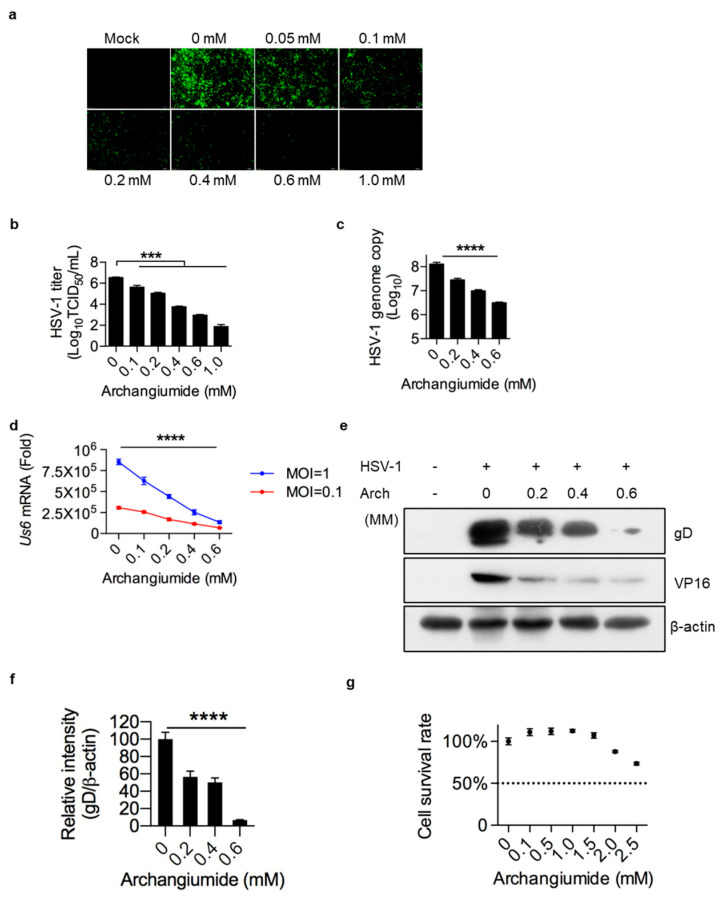
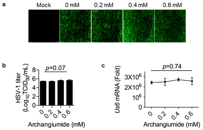
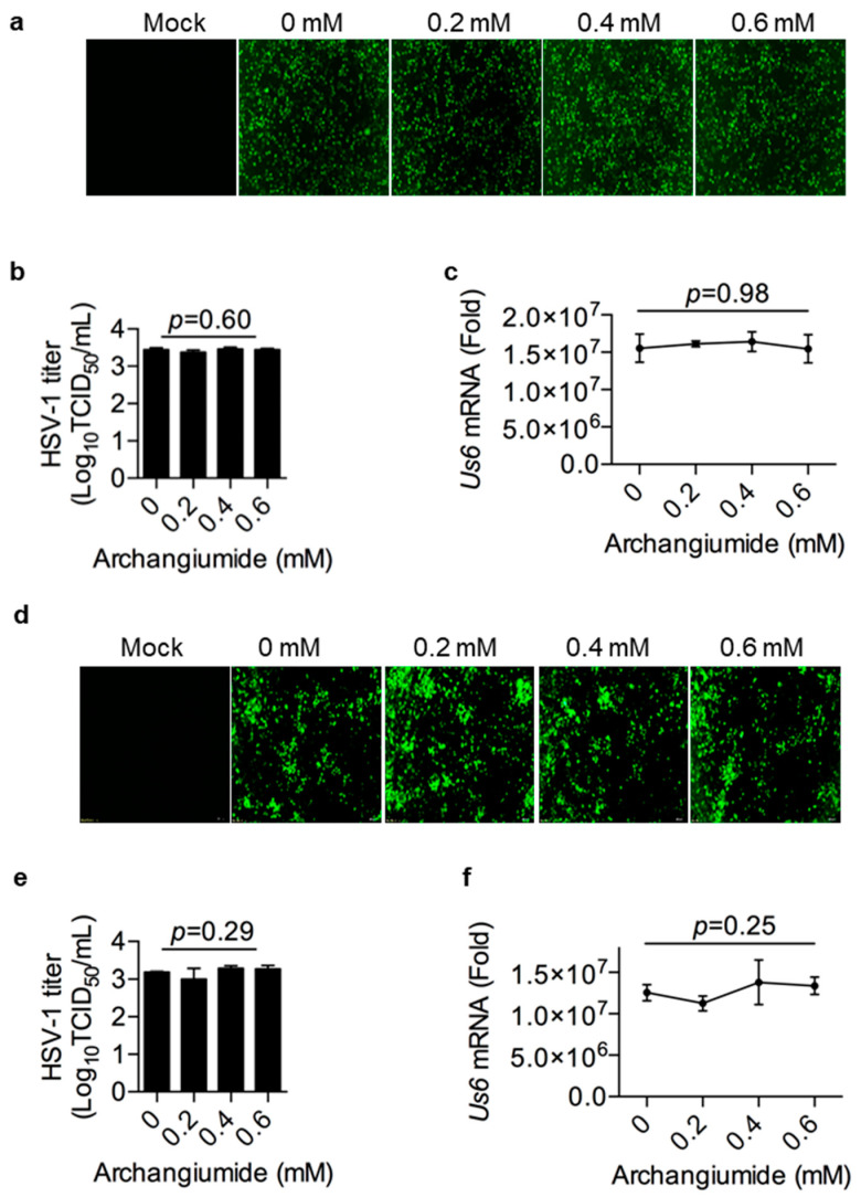
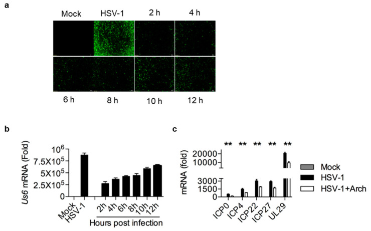
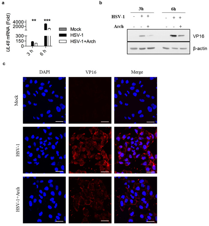
Archangiumide is a unique macrolide natural product that features an endocyclic allene functionality, rendering it a prototype of a new class of secondary metabolites of microbial origin. However, its biological and/or pharmaceutical roles remain obscure. In this study, we have unveiled an antiviral potency of archangiumide that was effective against herpes simplex virus (HSV-1) replication. We found that archangiumide did not affect host cell viability, nor pathogen infectivity, but suppressed HSV-1 early replication, in terms of early replication genes, such as ICP0, ICP4, etc. Further scrutinizing the underlined master regulator, we found that HSV-1 VP16 protein expression was inhibited by archangiumide, as well as VP16 nuclear translocation. As VP16 is a coactivator of transcription, archangiumide harnessed the master regulator of HSV-1 early replication. Together, here we provide evidence that allene macrolide archangiumide possesses robust antiviral functions that may be valuable for a novel viral infection intervention, as macrolides are generally safe drugs for prolonged treatments.

Keywords: HSV-1; antiviral; archangiumide.


Conflict of interest statement

The authors declare no conflicts of interest.

Figures

Figure 1
Figure 1
Archangiumide suppressed HSV-1 replication. (a,b) Archangiumide was added to HSV-1-eGFP virus-infected HeLa cells (MOI = 0.5) and cultured for 24 h. Sham infection was performed as control (Mock). GFP signal was captured (a). Fluorescent images were in 200× magnification. HSV-1 titer (TCID50/mL) was analyzed in culture supernatant after 24 h (b). Statistics were analyzed with non-parametric one-way ANOVA (Kruskl–Wallis test). ***, p < 0.001. n = 3. (c) Archangiumide was added to infected cells (MOI = 0.1). After 24 h culture, genomice DNA was extracted and the HSV-1 genome was analyzed by qPCR. Kruskl–Wallis one-way ANOVA. ****, p < 0.0001. n = 3. (d) gD transcription (Us6 mRNA) was analyzed by qPCR under high and low dose infections (MOI = 1 and MOI = 0.1). Kruskl–Wallis one-way ANOVA for each MOI group. ****, p < 0.0001. n = 3. (e) Western of gD and VP14 protein under archangiumide. β-actin as control. (f) Quantification of e.****, p < 0.0001. n = 3. (g) Cell survival rate under different doses of archangiumide. Dashed line represents CC50. n = 3. Data shown represent three independent experiments.
Figure 2
Figure 2
Archangiumide showed no effect on virus inactivation. Archangiumide was pre-incubated with HSV-1-eGFP virus stock for 2 h, then the stock was diluted into MOI = 0.1 to infect cells. (a) GFP signal was captured at 24 h. Fluorescent images were in 200× magnification. (b) HSV-1 titer (TCID50/mL) was analyzed in culture supernatant after 24 h. Kruskl–Wallis one-way ANOVA. n = 3. (c) gD transcription (Us6 mRNA) was analyzed by qPCR after 24 h. Kruskl–Wallis one-way ANOVA. n = 3. Data shown represent three independent experiments.
Figure 3
Figure 3
Archangiumide showed no effects on viral attachment and penetration. (ac) With or without archangiumide, HSV-1 (MOI = 0.01) was loaded onto cells at 4 °C for 2 h for attachment, then removed. Infected cells were cultured in fresh medium and the GFP signal was captured at 24 h (a). Fluorescent images were in 200× magnification. (b) HSV-1 titer (TCID50/mL) was analyzed in culture supernatant after 24 h. Kruskl–Wallis one-way ANOVA. n = 3. (c) gD transcription (Us6 mRNA) was analyzed by qPCR after 24 h. Kruskl–Wallis one-way ANOVA. n = 3. (df) HSV-1 (MOI = 0.01) was loaded onto cells at 4 °C for 2 h for attachment, then removed. With or without archangiumide, attached HSV-1 was stimulated to penetrate at 37 °C for 15 min. After using an acidic PBS wash (pH = 3) to remove non-penetrated virus then neutralizing cells with a basal PBS wash (pH = 11), infected cells were cultured 24 h for analysis. (d) GFP signal was captured at 24 h. Fluorescent images were in 200× magnification. (e) HSV-1 titer (TCID50/mL) was analyzed in culture supernatant after 24 h. Kruskl–Wallis one-way ANOVA. n = 3. (f) gD transcription (Us6 mRNA) was analyzed by qPCR after 24 h. Kruskl–Wallis one-way ANOVA. n = 3. Data shown represent three independent experiments.
Figure 4
Figure 4
Archangiumide suppressed early viral replication. At different times after HSV-1 infection (MOI = 0.1), 0.4 mM archangiumide was added to culture systems to suppress replication. (a) GFP signal was captured at 24 h. Fluorescent images were in 200× magnification. (b) gD transcription (Us6 mRNA) was analyzed by qPCR after 24 h. Unpaired test, two-tailed. **, p < 0.01, n = 3. (c) Early-replication-related gene transcriptions (ICP0, ICP4, ICP22, ICP27, UL29 mRNA) were analyzed by qPCR before 6 h post infection. Kruskl–Wallis one-way ANOVA for each gene. n = 3. Data shown represent three independent experiments.
Figure 5
Figure 5
Archangiumide down-regulated VP16 expression and blocked its nuclear translocation. 0.4 mM archangiumide was added to HSV-1-infected cells (MOI = 4). (a) At 3 h and 6 h, VP16 (UL48 mRNA) was analyzed by qPCR. Kruskl–Wallis one-way ANOVA for each timepoint. ***, p < 0.001, **, p < 0.01, n = 3. (b) Western blot of VP16 protein under archangiumide. β-actin as control. (c) Intracellular distribution of VP16. DAPI as nuclear indicator. Bars indicate 10 μm. Data shown represent three independent experiments.

Similar articles

References

    1. Dinos G.P. The macrolide antibiotic renaissance. Br. J. Pharmacol. 2017;174:2967–2983. doi: 10.1111/bph.13936. - DOI - PMC - PubMed
    1. Fernandes P., Martens E., Pereira D. Nature nurtures the design of new semi-synthetic macrolide antibiotics. J. Antibiot. 2017;70:527–533. doi: 10.1038/ja.2016.137. - DOI - PMC - PubMed
    1. Myers A.G., Clark R.B. Discovery of Macrolide Antibiotics Effective against Multi-Drug Resistant Gram-Negative Pathogens. Acc. Chem. Res. 2021;54:1635–1645. doi: 10.1021/acs.accounts.1c00020. - DOI - PubMed
    1. Kim K., Jung S., Kim M., Park S., Yang H.J., Lee E. Global Trends in the Proportion of Macrolide-Resistant Mycoplasma pneumoniae Infections: A Systematic Review and Meta-analysis. JAMA Netw. Open. 2022;5:e2220949. doi: 10.1001/jamanetworkopen.2022.20949. - DOI - PMC - PubMed
    1. Hu J.-Q., Wang J.-J., Li Y.-L., Zhuo L., Zhang A., Sui H.-Y., Li X.-J., Shen T., Yin Y., Wu Z.-H., et al. Combining NMR-Based Metabolic Profiling and Genome Mining for the Accelerated Discovery of Archangiumide, an Allenic Macrolide from the Myxobacterium Archangium violaceum SDU8. Org. Lett. 2021;23:2114–2119. doi: 10.1021/acs.orglett.1c00265. - DOI - PubMed

LinkOut - more resources