. 2025 Feb 12;26(4):1552.
doi: 10.3390/ijms26041552.

The Simultaneous Deletion of pH-Sensing Receptors GPR4 and OGR1 (GPR68) Ameliorates Colitis with Additive Effects on Multiple Parameters of Inflammation

Affiliations

The Simultaneous Deletion of pH-Sensing Receptors GPR4 and OGR1 (GPR68) Ameliorates Colitis with Additive Effects on Multiple Parameters of Inflammation

Federica Foti et al. Int J Mol Sci. .

Abstract

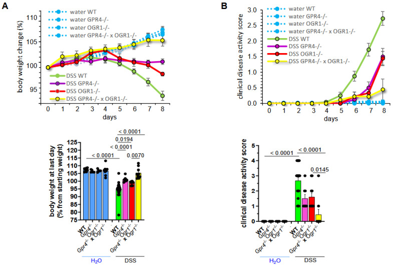
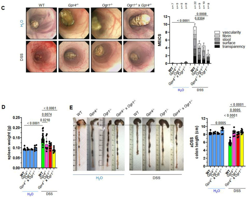
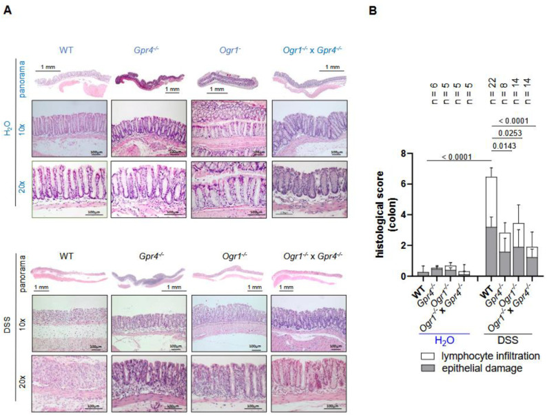
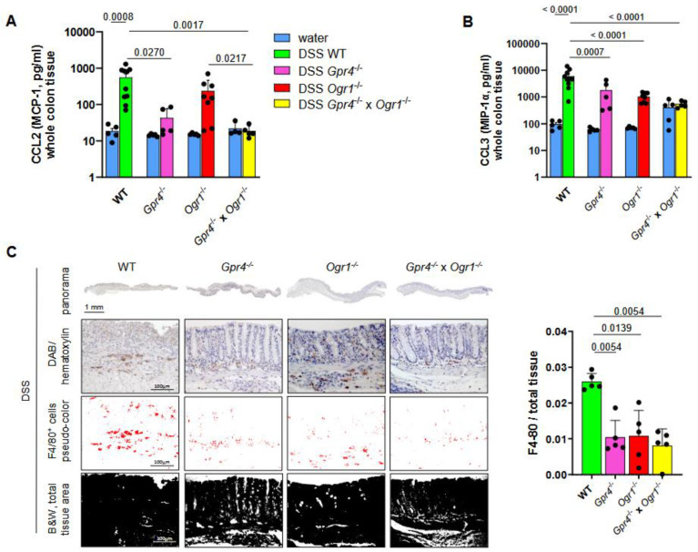
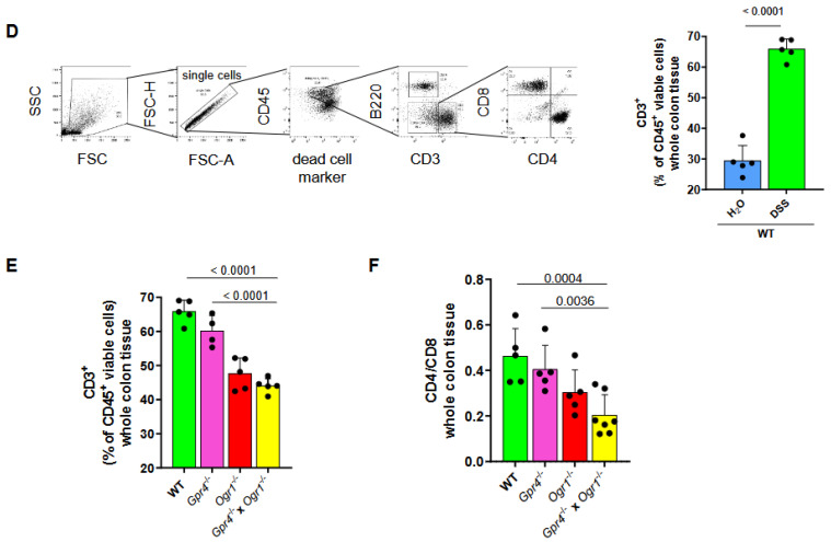
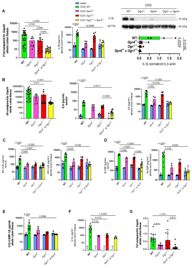
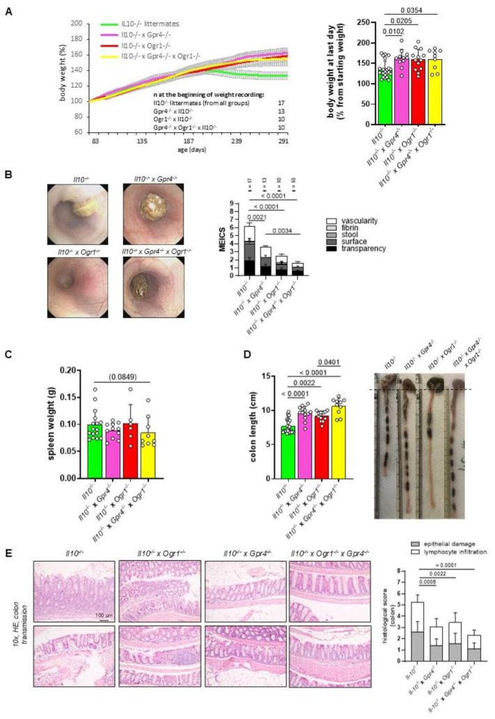
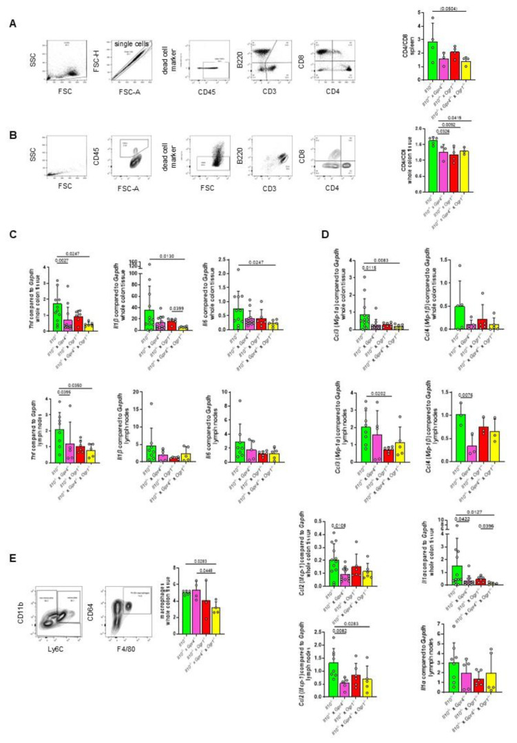
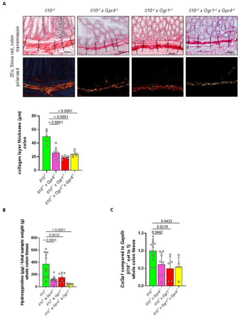
G protein-coupled receptors (GPRs), including pro-inflammatory GPR4 and ovarian cancer GPR1 (OGR1/GPR68), are involved in the pH sensing of the extracellular space and have been implicated in inflammatory bowel disease (IBD). Previous data show that a loss of GPR4 or OGR1 independently is associated with reduced intestinal inflammation in mouse models of experimental colitis. In the present manuscript, we investigated the impact of the simultaneous loss of GPR4 and OGR1 in animal models of IBD. To study the effects of combined loss of Gpr4 Ogr1 in IBD we used the well-established acute dextran sodium sulfate (DSS) and spontaneous Il10-/- murine colitis models. Disease severity was assessed using multiple clinical scores (e.g., body weight loss, disease activity score, murine endoscopic index of colitis severity (MEICS) and histological analyses). Real-time quantitative polymerase chain reaction (qPCR), Western blot, and flow cytometry were used to investigate changes in pro-inflammatory cytokines expression and immune cells infiltration. We found that a combined loss of GPR4 and OGR1 significantly reduces colon inflammation in IBD relative to single deficiencies as evidenced by reduced body weight loss, disease score, CD4/CD8 ratio, and Il1β, Il6, and Tnf in the colon. Similarly, in the II10 deficiency model, the inflammation was significantly ameliorated upon the simultaneous deletion of GPR4 and OGR1, evidenced by a reduction in the MEICS score, colon length, Tnf and Il1β measurements, and a decrease in the number of macrophages in the colon, as compared to single deletions. Importantly, hydroxyproline levels were decreased close to baseline in Il10-/- × Gpr4-/- × Ogr1-/- mice. Our findings demonstrate that the simultaneous loss of GRP4 and OGR1 functions exerts an additive effect on multiple parameters associated with colonic inflammation. These results further reinforce the hypothesis that chronic inflammatory acidosis is a driver of fibrosis and is dependent on GPR4 and OGR1 signaling. The inhibition of both GPR4 and OGR1 by pH-sensing receptor modulators may constitute as a potential therapeutic option for IBD, as both pH-sensing receptors appear to sustain inflammation by acting on complementary pro-inflammatory pathways.

Keywords: GPR4; OGR1; inflammatory bowel disease; pH-sensing G protein-coupled receptors.


Conflict of interest statement

The authors declare no competing interests. G.R. discloses grant support from AbbVie, Ardeypharm, MSD, FALK, Flamentera, Novartis, Roche, Tillots, UCB, and Zeller. All other authors have nothing to disclose.

Figures

Figure 1
Figure 1
The absence of both GPR4 and OGR1 additively reduces inflammation in DSS-induced colitis. (A) Body weight, ±SEM. (B) Clinical disease activity score, ±SEM. (C) MEICS, ±SD, and exemplary pictures of colonoscopy from each group. (D) Spleen weight, ±SD. (E) Colon length, ±SD, and exemplary pictures of colons from each group. Non-parametric distribution (Shapiro–Wilk test). One-way ANOVA, multiple comparisons test, Kruskal–Wallis test, two-stage linear step-up procedure of Benjamini, Krieger, and Yekutieli. p-values and n as indicated.
Figure 1
Figure 1
The absence of both GPR4 and OGR1 additively reduces inflammation in DSS-induced colitis. (A) Body weight, ±SEM. (B) Clinical disease activity score, ±SEM. (C) MEICS, ±SD, and exemplary pictures of colonoscopy from each group. (D) Spleen weight, ±SD. (E) Colon length, ±SD, and exemplary pictures of colons from each group. Non-parametric distribution (Shapiro–Wilk test). One-way ANOVA, multiple comparisons test, Kruskal–Wallis test, two-stage linear step-up procedure of Benjamini, Krieger, and Yekutieli. p-values and n as indicated.
Figure 2
Figure 2
Decreased histological score in Gpr4−/− × Ogr1−/− compared with WT mice upon acute DSS-induced colitis. (A) Exemplary microscopic pictures of HE-stained colons. (B) Histological score, non-parametric distribution (Shapiro–Wilk test). One-way ANOVA, multiple comparisons test, Kruskal–Wallis test, two-stage linear step-up procedure of Benjamini, Krieger, and Yekutieli. ±SD, p-values, and n as indicated.
Figure 3
Figure 3
Decreased F4/80+ and CD4/CD8 in Gpr4−/− × Ogr1−/− compared with WT mice upon acute DSS-induced colitis. Immunoassay, (A) MCP-1, and (B) CCL3 in whole colon tissue, ±SEM each. (C) IHC, F4/80, ±SD. Flow cytometry for (D,E) CD3, ±SD, and (F) CD4/CD8, ±SD. (D) Unpaired t-test. (A,C) Non-parametric distribution (Shapiro–Wilk test). (B,DF) Normal distribution (Shapiro–Wilk test). (AC,E,F) One-way ANOVA, multiple comparisons test, Kruskal–Wallis test, two-stage linear step-up procedure of Benjamini, Krieger, and Yekutieli. p-values and n as indicated.
Figure 3
Figure 3
Decreased F4/80+ and CD4/CD8 in Gpr4−/− × Ogr1−/− compared with WT mice upon acute DSS-induced colitis. Immunoassay, (A) MCP-1, and (B) CCL3 in whole colon tissue, ±SEM each. (C) IHC, F4/80, ±SD. Flow cytometry for (D,E) CD3, ±SD, and (F) CD4/CD8, ±SD. (D) Unpaired t-test. (A,C) Non-parametric distribution (Shapiro–Wilk test). (B,DF) Normal distribution (Shapiro–Wilk test). (AC,E,F) One-way ANOVA, multiple comparisons test, Kruskal–Wallis test, two-stage linear step-up procedure of Benjamini, Krieger, and Yekutieli. p-values and n as indicated.
Figure 4
Figure 4
GPR4 and OGR1 deficiency additively reduces pro-inflammatory cytokines in acute DSS-induced colitis. (A) Il1β. qPCR, ±SD; immunoassay, ±SEM; and WB, ±SD, in whole colon tissue each. (B) Il6. qPCR, ±SD and immunoassay, ±SEM, in whole colon tissue and serum as indicated. (C) KC, (D) G-CSF, (E) CCL4, and (F) IL1α in serum or whole colon tissue as indicated, ±SEM. (G) qPCR, Tnf, ±SD. (AG) One-way ANOVA, multiple comparisons test, Kruskal–Wallis test, two-stage linear step-up procedure of Benjamini, Krieger, and Yekutieli. p-values and n as indicated. Non-parametric distribution, except IL-6 whole colon tissue, KC serum, and G-CSF serum, which showed normal distribution (Shapiro–Wilk test).
Figure 5
Figure 5
The absence of both GPR4 and OGR1 reduces inflammation upon spontaneous colitis. (A) Body weight, ±SEM. (B) MEICS, ±SD. (C) Spleen weight. (D) Colon length, ±SD, and exemplary pictures of colons from each group. (E) Exemplary microscopic pictures of HE-stained colons and histological score. Non-parametric distribution (Shapiro–Wilk test). One-way ANOVA, multiple comparisons test, Kruskal–Wallis test, two-stage linear step-up procedure of Benjamini, Krieger, and Yekutieli. p-values and n as indicated.
Figure 6
Figure 6
The absence of both GPR4 and OGR1 reduces CD4/CD8 ratio and macrophages upon spontaneous colitis. Flow cytometry for CD4/CD8 in (A) spleen, and (B) whole colon tissue. (C) qPCR, pro-inflammatory Tnf, Il1β, and Il6 in whole colon tissue and lymph nodes as indicated. (D) qPCR, monocyte-attracting Ccl3, and Ccl4. (E) Flow cytometry for F4/80+ in whole colon tissue and qPCR, Ccl2, and Il1α mainly produced by monocytes. Non-parametric distribution (Shapiro–Wilk test) except for (B), (D) lymph nodes, (E) Ccl2 lymph nodes. ±SD, one-way ANOVA, multiple comparisons test, Kruskal–Wallis test, two-stage linear step-up procedure of Benjamini, Krieger, and Yekutieli. p-values and n as indicated.
Figure 7
Figure 7
Decreased fibrosis in Gpr4−/− × Ogr1−/− compared with WT mice upon spontaneous colitis. (A) Sirius red staining and collagen layer thickness. (B) Hydroxyproline assay. (C) qPCR, Col3a1. Normal distribution (Shapiro–Wilk test), one-way ANOVA, multiple comparisons test, Kruskal–Wallis test, two-stage linear step-up procedure of Benjamini, Krieger, and Yekutieli, ±SD, p-values, and n as indicated.

Similar articles

Cited by

References

    1. Ludwig M.G., Vanek M., Guerini D., Gasser J.A., Jones C.E., Junker U., Hofstetter H., Wolf R.M., Seuwen K. Proton-sensing G-protein-coupled receptors. Nature. 2003;425:93–98. doi: 10.1038/nature01905. - DOI - PubMed
    1. Wyder L., Suply T., Ricoux B., Billy E., Schnell C., Baumgarten B.U., Maira S.M., Koelbing C., Ferretti M., Kinzel B., et al. Reduced pathological angiogenesis and tumor growth in mice lacking GPR4, a proton sensing receptor. Angiogenesis. 2011;14:533–544. doi: 10.1007/s10456-011-9238-9. - DOI - PubMed
    1. Dong L., Li Z., Leffler N.R., Asch A.S., Chi J.T., Yang L.V. Acidosis activation of the proton-sensing GPR4 receptor stimulates vascular endothelial cell inflammatory responses revealed by transcriptome analysis. PLoS ONE. 2013;8:e61991. doi: 10.1371/journal.pone.0061991. - DOI - PMC - PubMed
    1. Miltz W., Velcicky J., Dawson J., Littlewood-Evans A., Ludwig M.G., Seuwen K., Feifel R., Oberhauser B., Meyer A., Gabriel D., et al. Design and synthesis of potent and orally active GPR4 antagonists with modulatory effects on nociception, inflammation, and angiogenesis. Bioorg. Med. Chem. 2017;25:4512–4525. doi: 10.1016/j.bmc.2017.06.050. - DOI - PubMed
    1. Jasso G.J., Jaiswal A., Varma M., Laszewski T., Grauel A., Omar A., Silva N., Dranoff G., Porter J.A., Mansfield K., et al. Colon stroma mediates an inflammation-driven fibroblastic response controlling matrix remodeling and healing. PLoS Biol. 2022;20:e3001532. doi: 10.1371/journal.pbio.3001532. - DOI - PMC - PubMed

MeSH terms

LinkOut - more resources