

Influence of Super-Low-Intensity Microwave Radiation on Mesenchymal Stem Cells
- PMID: 40004170
- PMCID: PMC11855362
- DOI: 10.3390/ijms26041705
Influence of Super-Low-Intensity Microwave Radiation on Mesenchymal Stem Cells
Abstract
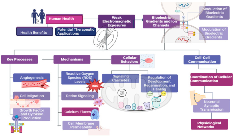
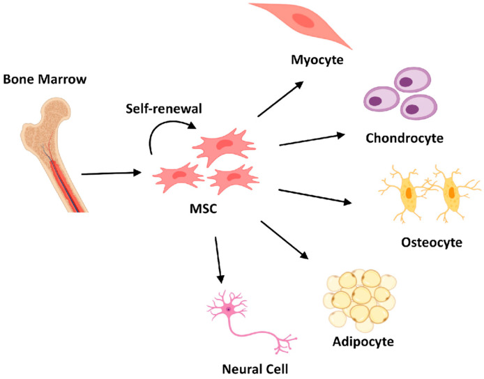
Mesenchymal stem cells (MSCs) have emerged as a promising tool for regenerative medicine due to their multipotency and immunomodulatory properties. According to recent research, exposing MSCs to super-low-intensity microwave radiation can have a significant impact on how they behave and operate. This review provides an overview of the most recent studies on the effects of microwave radiation on MSCs with power densities that are much below thermal values. Studies repeatedly show that non-thermal mechanisms affecting calcium signaling, membrane transport, mitochondrial activity, along ion channel activation may increase MSC proliferation, differentiation along mesodermal lineages, paracrine factor secretion, and immunomodulatory capabilities during brief, regulated microwave exposures. These bioeffects greatly enhance MSC regeneration capability in preclinical models of myocardial infarction, osteoarthritis, brain damage, and other diseases. Additional study to understand microwave treatment settings, biological processes, and safety assessments will aid in the translation of this unique, non-invasive strategy of activating MSCs with microwave radiation to improve cell engraftment, survival, and tissue healing results. Microwave-enhanced MSC treatment, if shown safe and successful, might have broad relevance as a novel cell-based approach for a variety of regenerative medicine applications.
Keywords: mesenchymal stem cells; regenerative medicine; super-low-intensity microwave field; tissue regeneration; weak electromagnetic field.
Conflict of interest statement
The authors declare no conflicts of interest.
Figures
Similar articles
-
Effects of pulsed 2.856 GHz microwave exposure on BM-MSCs isolated from C57BL/6 mice.PLoS One. 2015 Feb 6;10(2):e0117550. doi: 10.1371/journal.pone.0117550. eCollection 2015. PLoS One. 2015. PMID: 25658708 Free PMC article.
-
Therapeutic Properties of Mesenchymal Stromal/Stem Cells: The Need of Cell Priming for Cell-Free Therapies in Regenerative Medicine.Int J Mol Sci. 2021 Jan 14;22(2):763. doi: 10.3390/ijms22020763. Int J Mol Sci. 2021. PMID: 33466583 Free PMC article. Review.
-
The Application of Photobiomodulation on Mesenchymal Stem Cells and its Potential Use for Tenocyte Differentiation.Curr Stem Cell Res Ther. 2025;20(3):232-245. doi: 10.2174/011574888X295488240319111911. Curr Stem Cell Res Ther. 2025. PMID: 38847377 Review.
-
The Therapeutic Use and Potential of MSCs: Advances in Regenerative Medicine.Int J Mol Sci. 2025 Mar 27;26(7):3084. doi: 10.3390/ijms26073084. Int J Mol Sci. 2025. PMID: 40243782 Free PMC article. Review.
-
Mesenchymal stem cell secretome for regenerative medicine: Where do we stand?J Adv Res. 2025 Apr;70:103-124. doi: 10.1016/j.jare.2024.05.004. Epub 2024 May 9. J Adv Res. 2025. PMID: 38729561 Free PMC article. Review.
References
-
- Surkov V., Hayakawa M. Ultra and Extremely Low Frequency Electromagnetic Fields. Volume 486 Springer; Berlin/Heidelberg, Germany: 2014.
-
- Barnes F., Greenenbaum B. Some Effects of Weak Magnetic Fields on Biological Systems: RF fields can change radical concentrations and cancer cell growth rates. IEEE Power Electron. Mag. 2016;3:60–68. doi: 10.1109/MPEL.2015.2508699. - DOI
Publication types
MeSH terms
LinkOut - more resources
Full Text Sources
