. 2025 Feb 18;26(4):1728.
doi: 10.3390/ijms26041728.

Inhibitory Effects of Heat-Processed Gynostemma pentaphyllum Extract (Actiponin®) and Its Components on Cartilage Breakdown in Osteoarthritis

Affiliations

Inhibitory Effects of Heat-Processed Gynostemma pentaphyllum Extract (Actiponin®) and Its Components on Cartilage Breakdown in Osteoarthritis

Seul Ah Lee et al. Int J Mol Sci. .

Abstract

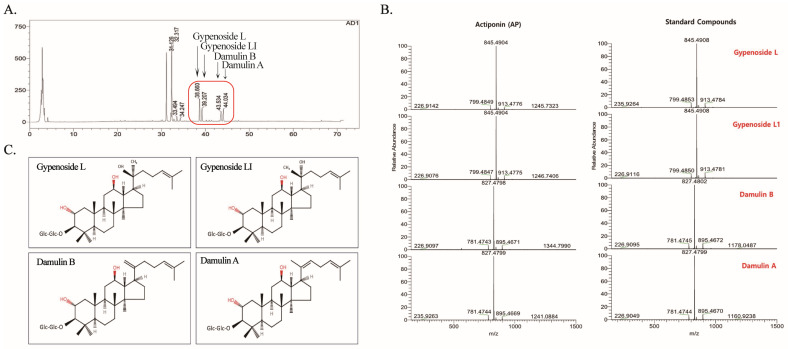
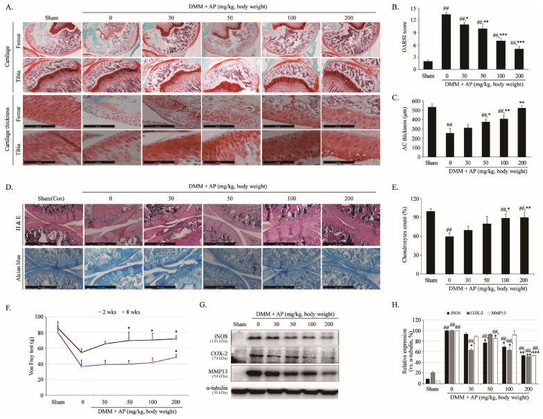
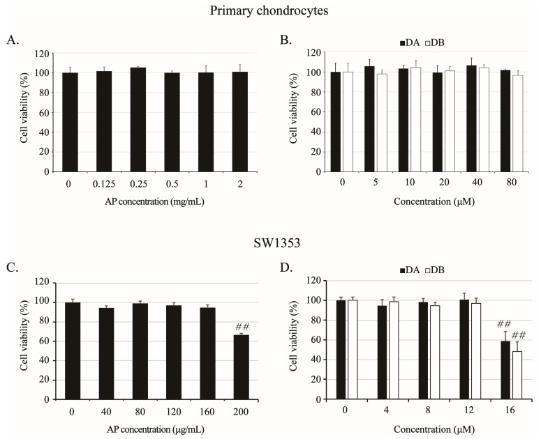
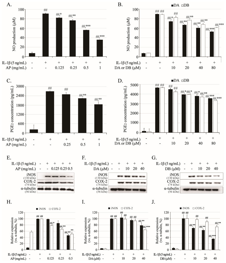
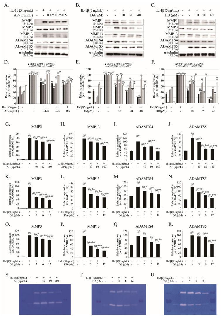
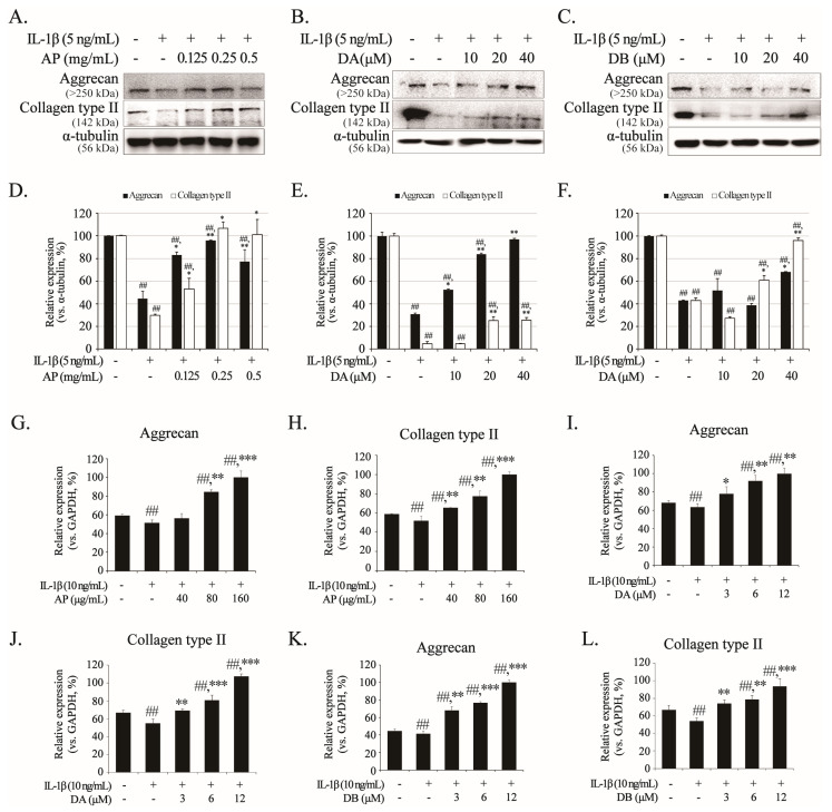
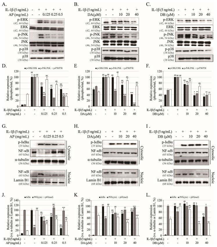
Osteoarthritis (OA), caused by the long-term use of joints, is a representative degenerative disease in the elderly. However, recently, the age of onset has been decreasing owing to excessive activities among young people in their 20s and 30s. Gynostemma pentaphyllum (Thunb.) Makino (GP), a perennial herb of the Cucurbitaceae family, has been used since the Ming dynasty as a medicinal material to treat various ailments, such as rheumatism, liver disease, and diabetes. In this study, we investigated the anti-arthritic effects of heat-processed Gynostemma pentaphyllum extract (Actiponin (AP)) and its derivatives, damulin A (DA) and damulin B (DB), using in vitro (primary rat chondrocytes and SW1353 cells) and in vivo (destabilization of the medial meniscus (DMM)-induced OA model) systems. Histological analysis results from the in vivo study showed that the group that underwent DMM surgery induced degeneration by the loss of proteoglycan and the destruction of cartilage (OARSI score 14 ± 0.57), whereas the group that received AP daily for 8 weeks maintained an intact condition (OARSI score 5 ± 0.28 at 200 mg/kg, p < 0.001). In addition, cartilage thickness and chondrocytes were reduced in the DMM group, but were restored in the AP-administered group. Furthermore, the von Frey analysis results showed that the pain threshold of the DMM group was considerably low (54.5 g at 8 weeks), whereas that of the AP group was dose-dependently increased (65.5, 69.5, 70.3, and 71.8 at 8 weeks for 30, 50, 100, and 200 mg/kg, respectively). In vitro studies showed that AP, DA, and DB reduced the expression of interleukin-1β alone-induced nitrite; inducible nitric oxide synthase; cyclooxygenase-2; matrix metallopeptidase 1/3/13; and a disintegrin and metalloproteinase with thrombospondin motifs 4/5. They also restored the expression of collagen type II and aggrecan, which are components of the extracellular matrix. The anti-arthritic effects of AP, DA, and DB were confirmed to be mediated by the mitogen-activated protein kinase and nuclear factor kappa-light-chain-enhancer of activated B cell signaling pathways. Collectively, these results suggest that AP is a potential therapeutic agent for mitigating OA progression and chondroprotection.

Keywords: Gynostemma pentaphyllum; actiponin; anti-arthritic; chondrocytes; osteoarthritis.


Conflict of interest statement

The authors declare no conflicts of interest.

Figures

Figure 1
Figure 1
HPLC/MS analysis of Actiponin (AP). (A) HPLC chromatogram of Actiponin. Red rectangle represents major gypenosides present in AP. (B) Major gypenoside compounds identified by HPLC/MS analysis of AP and standard gypenoside compounds. (C) Chemical structure of gypenoside L, gypenoside LI, damulin B and damulin A.
Figure 2
Figure 2
Histological evaluation of cartilage-protective effect of AP against cartilage degradation in DMM model. After sham or DMM surgery, rats received gavage of distilled water and AP every day for 8 weeks. Histological analysis of cartilage destruction was evaluated by safranin O/Fast Green, scale bar = 20 μm (A) and H&E and Alcian blue, scale bar = 541.1 μm (D). (B) Osteoarthritis Research Society International (OARSI) advanced Osteoarthritis Cartilage Histopathology Assessment System. (C) Articular cartilage (AC) thickness was evaluated by staining safranin O thickness using Leica application suite X program. (E) Number of chondrocytes in cartilage tissue was counted through a three-person blind test using H&E staining. (F) Measuring plantar pain using von Frey tests. (G) Protein levels of iNOS, COX-2, and MMP13 determined using Western blot. (H) Quantitative data of (G) were analyzed using ImageJ software (version 1.53). α-tubulin served as internal control. In vivo studies were performed three independent times, and Western blot analyses were performed at least five times. ANOVA and Dunnett tests were used to evaluate significance of results. ## p < 0.05 compared with sham group; * p < 0.05, ** p < 0.01, *** p < 0.001 compared with DMM group.
Figure 3
Figure 3
Effects of AP, DA, and DB on viability of rat primary chondrocytes and SW1353 cells. Cells were treated with AP (primary chondrocytes, 0.125, 0.25, 0.5, 1, and 2 mg/mL; SW1353, 40, 80, 120, 160, and 200 μg/mL), DA (primary chondrocytes, 5, 10, 20, 40, and 80 μM; SW1353, 4, 8, 12, and 16 μM), and DB (primary chondrocytes, 5, 10, 20, 40, and 80 μM; SW1353, 4, 8, 12, and 16 μM) for 24 h, and viability was determined by MTT assay. Cytotoxicity of AP (A), DA and DB (B) on primary chondrocytes; AP (C), DA and DB (D) on SW1353 cells. Untreated cells served as controls and were considered 100% viable. Data represented as mean ± SD of five independent experiments. ## p < 0.05 compared with control (0) group.
Figure 4
Figure 4
Inhibitory effects of AP, DA, and DB on IL-1β-induced nitrite, PGE2, iNOS, and COX-2. Primary chondrocytes were pre-treated with AP (0.125, 0.25, 0.5, and 1 mg/mL), DA (10, 20, 40, and 80 μM) and DB (5, 10, 20, 40, and 80 μM) for 1 h, followed by IL-1β (5 ng/mL) stimulation for 24 h. Nitrite production of AP (A), DA, and DB (B) was determined in cultured medium using Griess reagent. PGE2 production AP (C), DA and DB (D) was determined in cultured medium using ELISA kit. Expression of iNOS and COX-2 was determined using Western blot analysis; AP (E), DA (F) and DB (G). (HJ) Quantitative data of (EG) were analyzed using ImageJ software (version 1.53). α-Tubulin served as internal control. n = 5 per group. Data are represented as mean ± SD of five independent experiments. ## p < 0.05 vs. control group; * p <0.05, ** p <0.01, and *** p < 0.001 compared with IL-1β-treated group.
Figure 5
Figure 5
Inhibitory effects of AP, DA and DB on IL-1β-induced MMP1, MMP3, MMP13, ADAMTNS4, and ADAMTS5. Primary chondrocytes and SW1353 cells were pre-treated with AP, DA, and DB for 1 h, followed by IL-1β stimulation for 24 h. Protein levels of MMP1, MMP3, MMP13, ADAMTS-4, and ADAMTS5 in primary chondrocytes were determined using Western blot analysis; AP (A), DA (B), and DB (C). (DF) Quantitative data of (AC) were analyzed using ImageJ software (version 1.53). α-tubulin served as internal control. mRNA levels of MMP3, MMP13, ADAMTS4, and ADAMTS5 in SW1353 cells were determined using real-time PCR; AP (GJ), DA (KN), and DB (OR). Activities of secreted cartilage-degrading enzymes were analyzed using gelatin zymography; AP (S), DA (T) and DB (U). n = 5 per group. Data are represented as mean ± SD of five independent experiments. ## p < 0.05 vs. control group; * p < 0.05, ** p < 0.01, and *** p < 0.001 compared with the IL-1β-treated group.
Figure 6
Figure 6
Inhibitory effects of AP, DA, and DB on IL-1β-induced aggrecan and collagen type II degradation. Primary chondrocytes and SW1353 cells were pre-treated with AP, DA and DB for 1 h, followed by IL-1β stimulation for 24 h. Protein levels of aggrecan and collagen type II in primary chondrocytes were determined using Western blot analysis; AP (A), DA (B), and DB (C). (DF) Quantitative data of (AC) were analyzed using ImageJ software (version 1.53). α-tubulin served as an internal control. mRNA levels of aggrecan and collagen type II in SW1353 cells were determined using real-time PCR; AP (G,H), DA (I,J), and DB (K,L). n = 5 per group. Data are represented as mean ± SD of five independent experiments. ## p < 0.05 vs. control group; * p < 0.05, ** p < 0.01, and *** p < 0.001 compared with the IL-1β-treated group.
Figure 7
Figure 7
Effects of AP, DA, and DB on IL-1β-induced phosphorylation of MAPKs (ERK, JNK, and p38) and activation of NF-κB signaling. Primary chondrocytes were pre-treated with AP (0.125, 0.25, and 0.5 mg/mL), DA (10, 20, and 40 μM), and DB (10, 20, and 40 μM) for 1 h, followed by IL-1β (5 ng/mL) stimulation for 3 h. Protein expression levels of MAPK (ERK, JNK, and p38) and NF-κB (IκBα and p65) were determined using Western blot analysis. MAPKs: AP (A), DA (B), and DB (C); NF-κB: AP (G), DA (H), DB (I). (DF,JL) Quantitative data of (AC,GI) were analyzed using ImageJ software (version 1.53). n = 5 per group. Data are represented as mean ± SD of five independent experiments. ## p < 0.05 vs. control group; * p < 0.05, ** p < 0.01, and *** p < 0.001 compared with IL-1β-treated group.

References

    1. Chen J., Shen W., Zhao T., Wang L., Han J.L., Hamilton H.J., Im H.J. Osteoarthritis: Toward a comprehensive understanding of pathological mechanism. Bone Res. 2017;5:16044. doi: 10.1038/boneres.2016.44. - DOI - PMC - PubMed
    1. Krakowski P., Rejniak A., Sobczyk J., Karpiński R. Cartilage integrity: A review of mechanical and frictional properties and repair approaches in osteoarthritis. Healthcare. 2024;12:1648. doi: 10.3390/healthcare12161648. - DOI - PMC - PubMed
    1. Jenei-Lanzl Z., Meurer A., Zaucke F. Interleukin-1β signaling in osteoarthritis-chondrocytes in focus. Cell Signal. 2019;53:212–223. doi: 10.1016/j.cellsig.2018.10.005. - DOI - PubMed
    1. Hunziker E.B., Quinn T.M., Häuselmann H.J. Quantitative structural organization of normal adult human articular cartilage. Osteoarthr. Cartil. 2002;10:564–572. doi: 10.1053/joca.2002.0814. - DOI - PubMed
    1. Maldonado M., Nam J. The role of changes in extracellular matrix of cartilage in the presence of inflammation on the pathology of osteoarthritis. Biomed. Res. Int. 2013;2013:284873. doi: 10.1155/2013/284873. - DOI - PMC - PubMed