

Fabrication and Characterization of a Stretchable Sodium Alginate Hydrogel Patch Combined with Silicon Nitride and Metalized Halloysite Nanotubes to Develop a Chronic Wound Healing Treatment
- PMID: 40004197
- PMCID: PMC11855668
- DOI: 10.3390/ijms26041734
Fabrication and Characterization of a Stretchable Sodium Alginate Hydrogel Patch Combined with Silicon Nitride and Metalized Halloysite Nanotubes to Develop a Chronic Wound Healing Treatment
Abstract
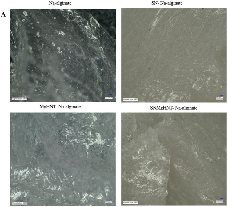
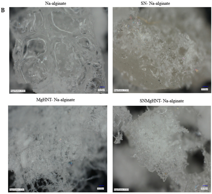
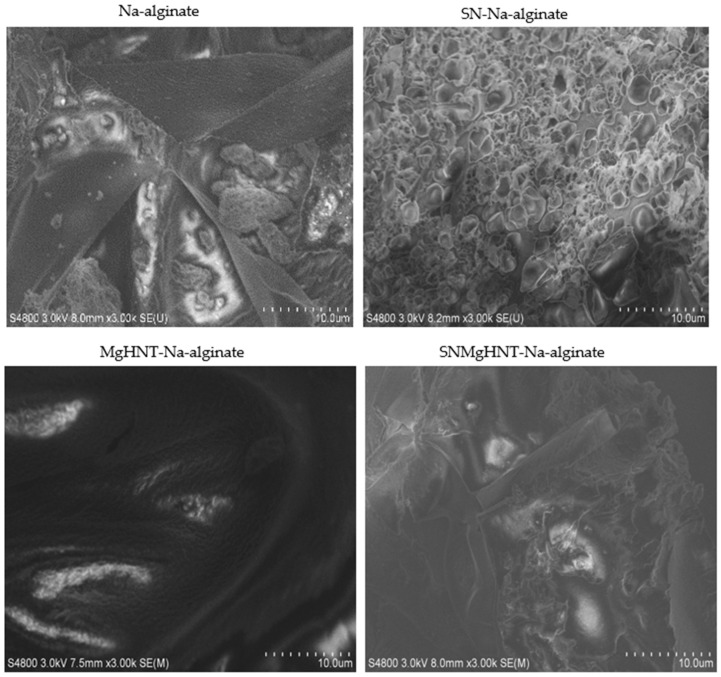
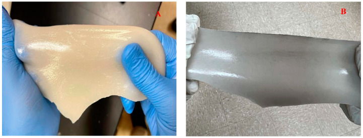
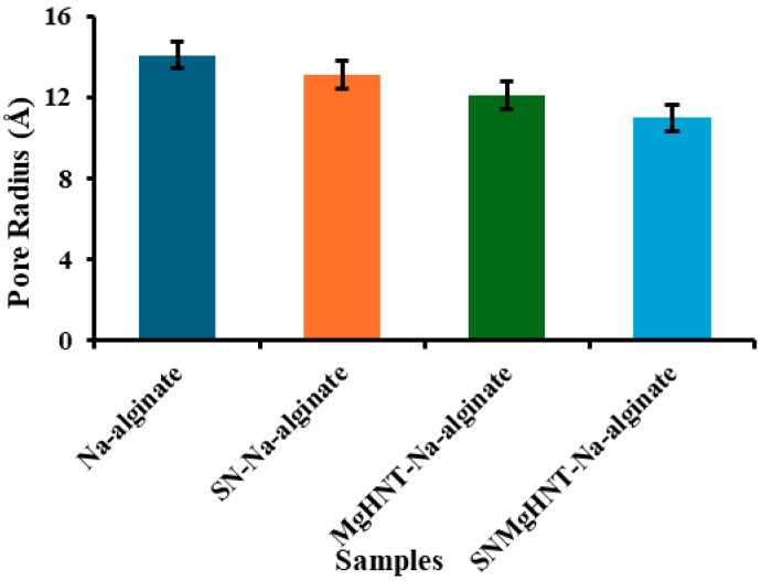
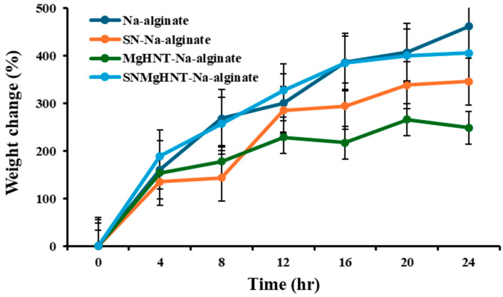
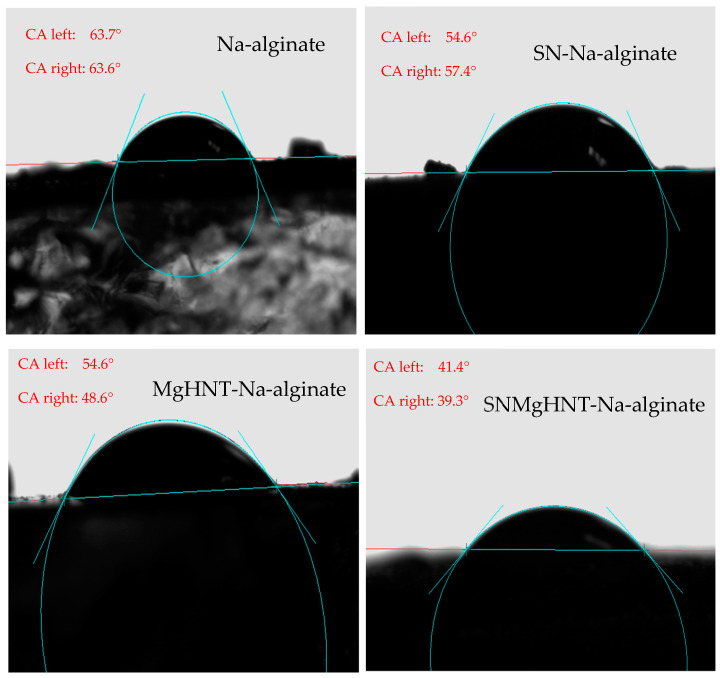
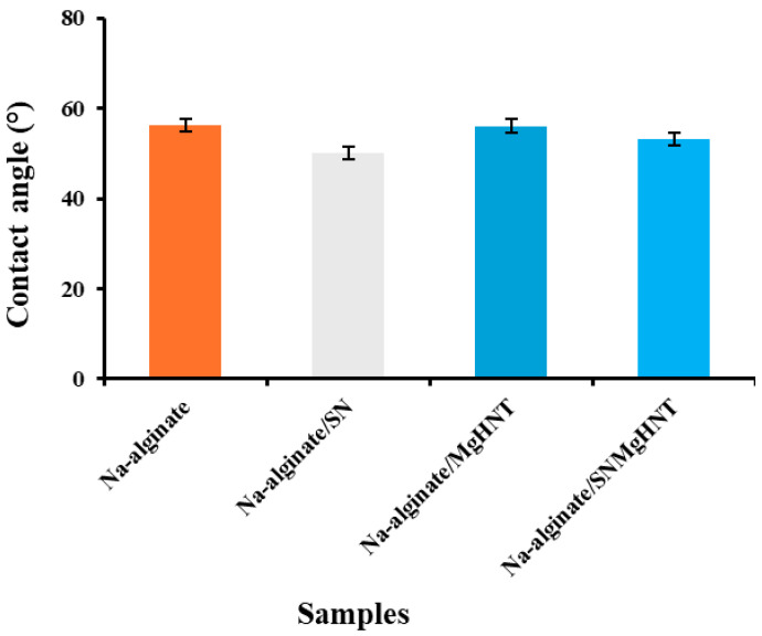
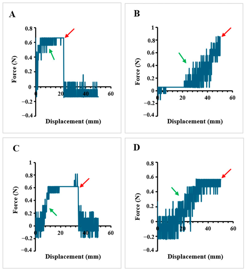
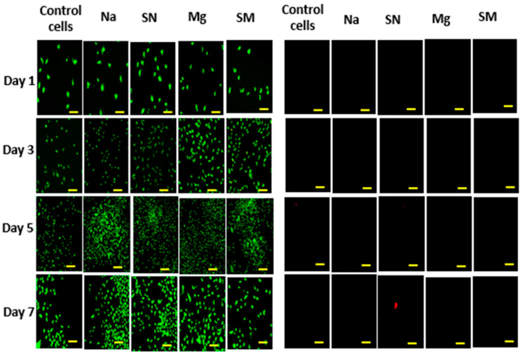
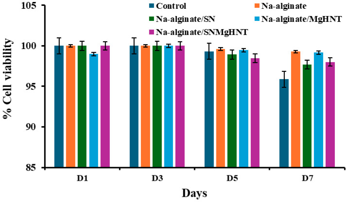
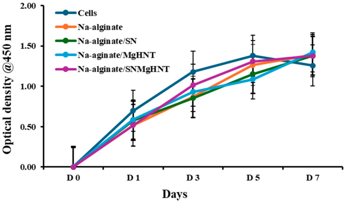
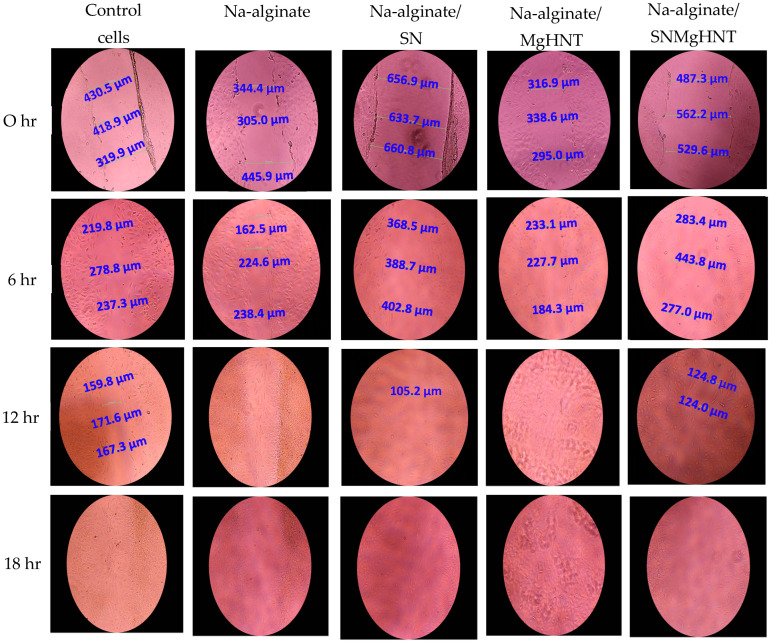
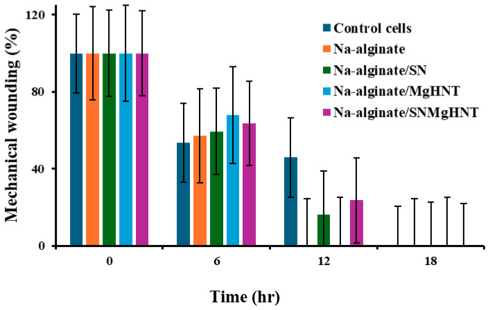
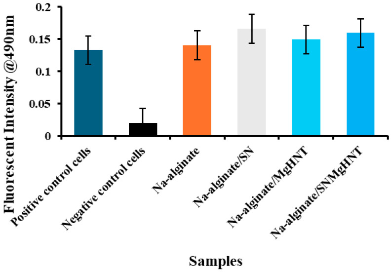
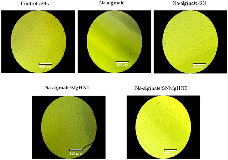
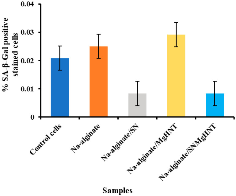
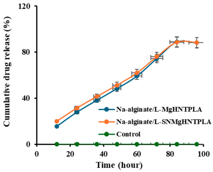
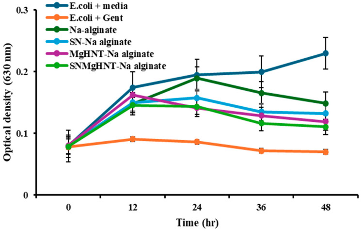
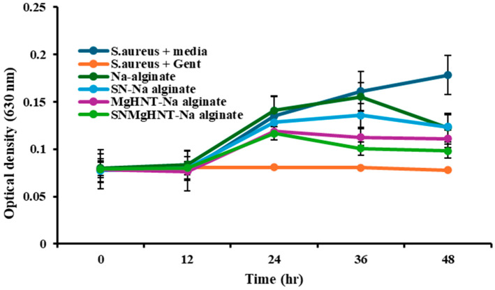
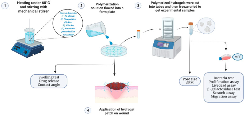
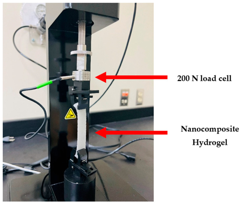
The human body is known as a responsive healing machine, but sometimes, broken bones do not heal, especially if a bacterial infection is present. The present study describes the fabrication and characterization of a nanocomposite hydrogel patch incorporated with silicon nitride and magnesium oxide (MgO) deposited on the halloysite nanotube (HNT) surface using a facile and inexpensive electrodeposition coating process. Scanning electron microscopy (SEM) was used to observe the surface morphology of the MgO/HNT surface coating and the nanocomposite patch. Material characterization, including SEM, contact angle, pore size analysis, and tensile properties, was performed to determine the composite's structure and material properties. E. coli and S. aureus bacterial cultures were used to test the antimicrobial properties. Cellular response to MgO/HNTs was studied using mouse embryonic fibroblasts. The nanocomposite hydrogel patch was discovered to possess inherent properties when tested against bacterial cultures, and it was found to enhance fibroblast cell migration and proliferation. The nanocomposite hydrogel patch also showed sustained drug release. Materials involved in the fabrication helped in the swelling properties by which the nanocomposite hydrogel patch has approximately 400% of its initial weight discovered during the swelling test.
Keywords: antimicrobial properties; halloysite nanotube; hydrogels; nanocomposite; silicon nitride.
Conflict of interest statement
The authors declare that they have no known competing financial interests or personal relationships that could have appeared to influence the work reported in this paper.
Figures
Similar articles
-
A self-healing crosslinked-xanthan gum/soy protein based film containing halloysite nanotube and propolis with antibacterial and antioxidant activity for wound healing.Int J Pharm. 2024 May 10;656:124073. doi: 10.1016/j.ijpharm.2024.124073. Epub 2024 Apr 1. Int J Pharm. 2024. PMID: 38569977
-
Natural halloysite nanotubes /chitosan based bio-nanocomposite for delivering norfloxacin, an anti-microbial agent in sustained release manner.Int J Biol Macromol. 2020 Nov 1;162:1849-1861. doi: 10.1016/j.ijbiomac.2020.08.060. Epub 2020 Aug 8. Int J Biol Macromol. 2020. PMID: 32781129
-
Fluorine-ion-releasing injectable alginate nanocomposite hydrogel for enhanced bioactivity and antibacterial property.Int J Biol Macromol. 2019 Feb 15;123:866-877. doi: 10.1016/j.ijbiomac.2018.11.108. Epub 2018 Nov 14. Int J Biol Macromol. 2019. PMID: 30447366
-
Preclinical functional characterization methods of nanocomposite hydrogels containing silver nanoparticles for biomedical applications.Appl Microbiol Biotechnol. 2020 Jun;104(11):4643-4658. doi: 10.1007/s00253-020-10521-2. Epub 2020 Apr 6. Appl Microbiol Biotechnol. 2020. PMID: 32253473 Review.
-
Nano-interfacial decoration of Halloysite Nanotubes for the development of antimicrobial nanocomposites.Adv Colloid Interface Sci. 2020 Jan;275:102063. doi: 10.1016/j.cis.2019.102063. Epub 2019 Nov 9. Adv Colloid Interface Sci. 2020. PMID: 31739982 Review.
Cited by
-
Hydrogel Microarray for Bioanalytical Applications: Preliminary Study on Material Properties.Materials (Basel). 2025 Jul 1;18(13):3118. doi: 10.3390/ma18133118. Materials (Basel). 2025. PMID: 40649606 Free PMC article.
References
-
- Ishida K., Shimohata T., Kanda Y., Nguyen A.Q., Masuda R., Yamazaki K., Uebanso T., Mawatari K., Kashimoto T., Takahashi A. Characteristic Metabolic Changes in Skeletal Muscle Due to Vibrio vulnificus Infection in a Wound Infection Model. Elias JE. mSystems. 2023;8:e00682-22. doi: 10.1128/msystems.00682-22. - DOI - PMC - PubMed
-
- Ranghar S., Sirohi P., Verma P., Agarwal V. Nanoparticle-based drug delivery systems: Promising approaches against infections. Braz. Arch. Biol. Technol. 2013;57:209–222. doi: 10.1590/S1516-89132013005000011. - DOI
MeSH terms
Substances
Grants and funding
LinkOut - more resources
Full Text Sources
Molecular Biology Databases
