. 2025 Feb 8;18(4):753.
doi: 10.3390/ma18040753.

Evaluation of Custom Microalgae-Based Bioink Formulations for Optimized Green Bioprinting

Affiliations

Evaluation of Custom Microalgae-Based Bioink Formulations for Optimized Green Bioprinting

Olubusuyi Ayowole et al. Materials (Basel). .

Abstract

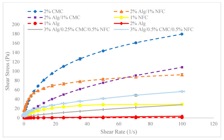
Green bioprinting, from the context of merging 3D bioprinting with microalgae cell organization, holds promise for industrial-scale optimization. This study employs spectrophotometric analysis to explore post-bioprinting cell growth density variation within hybrid hydrogel biomaterial scaffolds. Three hydrogel biomaterials-Alginic acid sodium salt (ALGINATE), Nanofibrillated Cellulose (NFC)-TEMPO, and CarboxyMethyl Cellulose (CMC)-are chosen for their scaffolding capabilities. Bioink development and analysis of their impact on cell proliferation and morphology are conducted. Chlorella microalgae cell growth within hydrogel compositions is probed using absorbance measurements, with additional assessment of shear thinning properties. Notably, NFC exhibits reduced shear thinning compared to CMC. Results reveal that while mono-hydrogel substrates with pronounced adhesion inhibit Chlorella cell proliferation, alginate fosters increased cell concentration alongside a slight viscosity rise.

Keywords: absorbance; bioink; bioprinting; hydrogel; microalgae.


Conflict of interest statement

The authors declare no conflict of interest.

Figures

Figure 1
Figure 1
Techniques of 3D bioprinting.
Figure 2
Figure 2
Alginate sample (a), NFC sample (b), and CMC sample (c).
Figure 3
Figure 3
Centrifuged cell suspension (a), and hydrogel substrate in tube before seeding with cell suspension (b).
Figure 4
Figure 4
Bioinks made up of hydrogel substrates and cell suspensions.
Figure 5
Figure 5
Absorption spectra of freshly isolated Chl a and Chl b in diethyl ether [40].
Figure 6
Figure 6
Schematic of light transmission through a cuvette.
Figure 7
Figure 7
Dispense assembly of custom bioprinter at UMaine Digital and Additive Manufacturing Lab.
Figure 8
Figure 8
3D model for experimental bioprinting construct.
Figure 9
Figure 9
Comparison of rheological characteristics of hydrogel formulations prepared to serve as substrates for custom bioinks.
Figure 10
Figure 10
Relative variation of absorbance for the first set of bioinks at a wavelength of 450 nm and a cell count of 51.5×10⁶ cells/mL.
Figure 11
Figure 11
Replication of absorbance variation in ‘Bioinks S1-A2, S1-Media, and S1-N1’ with ‘Bioinks S2-A2, S2-Media, and S2-N1’, respectively, at a wavelength of 450 nm and a cell count of 55.3×10⁶ cells/mL.
Figure 12
Figure 12
Replication of absorbance variation in ‘Bioinks S1-A2, S1-Media and S1-N1’ with ‘Bioinks S2-A2, S2-Media, and S2-N1’, respectively, at a wavelength of 650 nm and a cell count of 55.3×10⁶ cells/mL.
Figure 13
Figure 13
Representation of the effect of NFC content in bioink substrates on cell density variation.
Figure 14
Figure 14
Cell density variation in microalgae constructs derived from tri-hydrogel substrate, bio-printed at different printing pressures.
Figure 15
Figure 15
Cell Count–Absorbance calibration curve (absorbance recorded at a wavelength of 450 nm).
Figure 16
Figure 16
Cell Count–Absorbance calibration curve (absorbance recorded at a wavelength of 650 nm).
Figure 17
Figure 17
(a,b). Image of the experimental construct from Bioink SE-N1 (bioprinted from experimental bioink derived from 1% NFC w/v).
Figure 18
Figure 18
(a,b). Image of the experimental construct from Bioink SE-C2 (bioprinted from experimental bioink derived from 2% CMC w/v).

References

    1. Mandal S., Nagi G.K., Corcoran A.A., Agrawal R., Dubey M., Hunt R.W. Algal polysaccharides for 3D printing: A review. Carbohydr. Polym. 2023;300:120267. doi: 10.1016/j.carbpol.2022.120267. - DOI - PubMed
    1. Sun H., Gong Q., Fan Y., Wang Y., Wang J., Zhu C., Mou H., Yang S., Liu J. Unlocking 3D printing technology for microalgal production and application. Adv. Biotechnol. 2024;2:36. doi: 10.1007/s44307-024-00044-6. - DOI - PMC - PubMed
    1. Krujatz F., Dani S., Windisch J., Emmermacher J., Hahn F., Mosshammer M., Murthy S., Steingröwer J., Walther T., Kühl M., et al. Think outside the box: 3D bioprinting concepts for biotechnological applications—Recent developments and future perspectives. Biotechnol. Adv. 2022;58:107930. doi: 10.1016/j.biotechadv.2022.107930. - DOI - PubMed
    1. Zhang Y.S., Yue K., Aleman J., Mollazadeh-Moghaddam K., Bakht S.M., Yang J., Jia W., Dell’Erba V., Assawes P., Shin S.R., et al. 3D Bioprinting for Tissue and Organ Fabrication. Ann. Biomed. Eng. 2017;45:148–163. doi: 10.1007/s10439-016-1612-8. - DOI - PMC - PubMed
    1. Yoon S.-W., Kim S.-Y., Jeon J.-S., Oh S., Chung S.-Y., Kim J.-S., Maeng S.-K. 3D-printed Chlorella vulgaris biocarriers: A novel approach to wastewater treatment. J. Water Proc. Eng. 2024;57:104711. doi: 10.1016/j.jwpe.2023.104711. - DOI