Review
. 2025 Feb 13;16(2):220.
doi: 10.3390/genes16020220.

Chemical Evolution of Life on Earth

Affiliations
Review

Chemical Evolution of Life on Earth

Lei Lei et al. Genes (Basel). .

Abstract

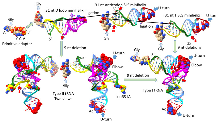
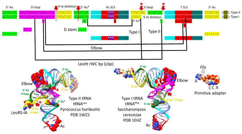
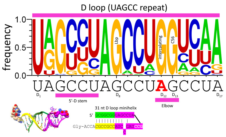
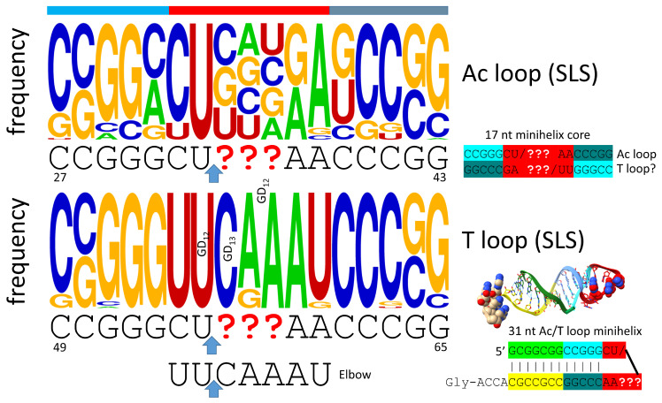
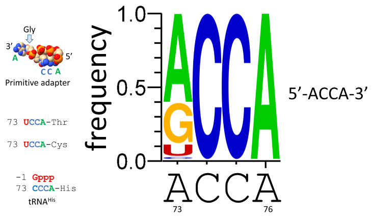
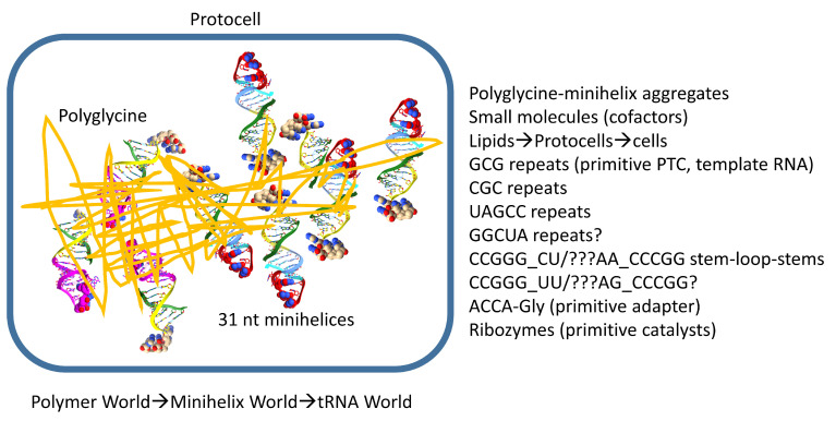
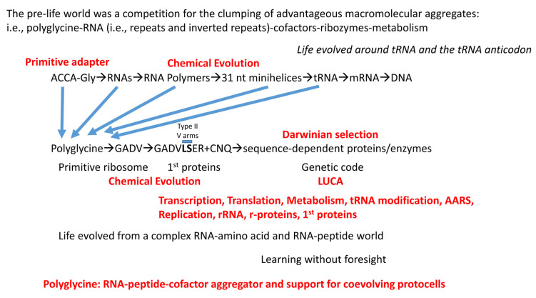
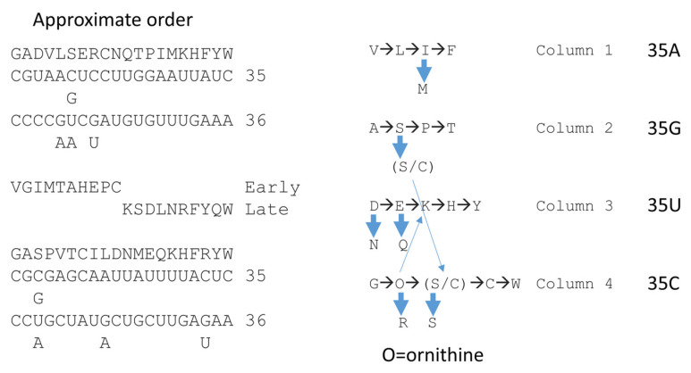
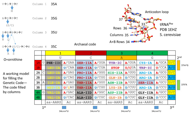
Background/Objectives: The origin of genes and genetics is the story of the coevolution of translation systems and the genetic code. Remarkably, the history of the origin of life on Earth was inscribed and preserved in the sequences of tRNAs. Methods: Sequence logos demonstrate the patterning of pre-life tRNA sequences. Results: The pre-life type I and type II tRNA sequences are known to the last nucleotide with only a few ambiguities. Type I and type II tRNAs evolved from ligation of three 31 nt minihelices of highly patterned and known sequence followed by closely related 9 nt internal deletion(s) within ligated acceptor stems. The D loop 17 nt core was a truncated UAGCC repeat. The anticodon and T 17 nt stem-loop-stems are homologous sequences with 5 nt stems and 7 nt U-turn loops that were selected in pre-life to resist ribozyme nucleases and to present a 3 nt anticodon with a single wobble position. The 7 nt T loop in tRNA was selected to interact with the D loop at the "elbow". The 5'-acceptor stem was based on a 7 nt truncated GCG repeat. The 3'-acceptor stem was based on a complementary 7 nt CGC repeat. In pre-life, ACCA-Gly was a primitive adapter molecule ligated to many RNAs, including tRNAs, to synthesize polyglycine. Conclusions: Analysis of sequence logos of tRNAs from an ancient Archaeon substantiates how the pre-life to life transition occurred on Earth. Polyglycine is posited to have aggregated complex molecular assemblies, including minihelices, tRNAs, cooperating molecules, and protocells, leading to the first life on Earth.

Keywords: anticodon; chemical evolution; genetic code; last universal common (cellular) ancestor; minihelices; origin of life; polyglycine; pre-life; tRNA; type II tRNA.


Conflict of interest statement

The authors declare no conflicts of interest.

Figures

Figure 1
Figure 1
Evolution of type II and type I tRNAs from ligation of three 31 nt minihelices and internal RNA processing. ACCA-Gly was a pre-life adapter molecule that could function alone or ligated to various RNAs, including 31 nt minihelices and tRNAs. Black arrows indicate how ligations of three 31 nt minihelices generated the 93 nt tRNA precursor molecule that was processed into type II and type I tRNAs. In the 93 nt tRNA precursor, type II tRNAs were formed by a single internal 9 nt deletion within ligated 3′ and 5′ acceptor stems. Type I tRNAs were formed by an additional 9 nt deletion in the V arm region. The purpose of the initial molecules was to synthesize polyglycine. Colors: (green) 7 nt 5′-acceptor stems and 5 nt 5′-acceptor stem remnants; (magenta) 17 nt D loop core; (cyan-red-cornflower blue) 17 nt anticodon stem-loop-stem and homologous T stem-loop-stem; (yellow) 7 nt 3′-acceptor stem and 5 nt 3′-acceptor stem remnant (type I V loop). Red arrows indicate the sites of the 9 nt internal deletions. Red arrows with asterisks represent the more 3′-9 nt internal deletion unique to type I tRNA. Blue arrows indicate U-turns. Some bases are emphasized using space-filling representation.
Figure 2
Figure 2
The pre-life 93 nt tRNA precursor was formed from ligation of three 31 nt minihelices (top sequence). Type II tRNA was formed by a single 9 nt internal deletion within ligated 3′- and 5′-acceptor stems. Type I tRNA was formed by an additional 9 nt internal deletion within ligated 3′- and 5′-acceptor stems (second sequence line). Interactions and sequence changes to support the tRNA fold are indicated. Red arrows indicate internal 9 nt deletion sites and endpoints. The 5′-9 nt deletion is common to type II and type I tRNAs. The closely related 3′-9 nt deletion was only for type I tRNAs. Red arrows with asterisks indicate processing of an early type II tRNA to a type I tRNA. Colors are consistent between figures. (As) indicates acceptor stem; (Ac) indicates anticodon; (Lbp) indicates the Levitt reverse Watson–Crick base pair. See the text for details.
Figure 3
Figure 3
The 17 nt D loop core was based on a UAGCC repeat (UAGCCUAGCCUAGCCUA). The sequence and an approximate structure of the D loop minihelix are shown. Colors are meant to be consistent between figures. Magenta bars match segments in the structure and sequence below. GD8 forms the Levitt reverse Watson–Crick base pair to type I CV5 or type II CVn. Red AD12 was substituted by GD12 to form elbow contacts to the T loop. GD13 forms a Watson–Crick pair with tRNA-56C.
Figure 4
Figure 4
The anticodon (Ac) stem-loop-stem (SLS) and the T stem-loop-stem are homologs. The blue arrows indicate the position of the loop U-turn. In tRNA, the T loop (UU/CAAAU) evolved to interact with the D loop at the elbow. The T stem-loop-stem sequence is also very similar to the anticodon stem-loop-stem complement. Colors are meant to be consistent between figures. At the elbow: U55 interacts with GD12; GD13 binds C56 (Watson–Crick pair); GD12 intercalates between A57 or G57 and A58. The bar above the figure indicates the stem-loop-stem structure (cyan–red–cornflower blue).
Figure 5
Figure 5
The anticodon stem-loop stem (two views). Colors and arrows are consistent between figures. WC for Watson–Crick.
Figure 6
Figure 6
Evolution of the type II V arm and alignment to the type I V loop. The tRNASer type II V arm single base insert may not be properly placed. Yellow (3′-acceptor stem) and green (5′-acceptor stem) bars indicate locations of sequences in the structure. tRNALeu and tRNASer type II V arms evolved to form stem-loop-stems with different cognate sequences and trajectories that could be accurately discriminated by LeuRS-IA and SerRS-IIA. Red arrows with asterisks are the same as in other figures and, also, in the structure.
Figure 7
Figure 7
Evolution of the 3′-D stem (5′-As*) and the type I tRNA V loop (3′-As*). The color rectangles indicate the location of sequence within the structure. Arrows are consistent with other figures. Typical sequences of the D stem are indicated.
Figure 8
Figure 8
Alignment of type II V arms to type I V loops. The sequence alignment shows how type II V arms and type I V loops align with one another and their derivation from ligation of 3′- and 5′-acceptor stems. The tRNALeu (CAA) V arm of Pyrococcus horikoshii is shown [23,38]. Sequence logos of 105 14 nt archaeal tRNALeu V arms and 34 15 nt archaeal tRNASer V arms are shown. In Archaea, tRNALeu and tRNASer are discriminated by the following factors: (1) the distinct trajectory set point scores of the type II V arms (cyan asterisks); (2) the tRNALeu V6-UAG-V8 V arm-end loop sequence determinant for LeuRS-IA (red asterisks); and (3) SerRS-IIA binding the type II V arm stems of tRNASer with a trajectory set point score of one unpaired base (cyan asterisk).
Figure 9
Figure 9
ACCA-Gly was the primordial adapter molecule. See the text for details.
Figure 10
Figure 10
A proposed role for polyglycine in the pre-life world. Polyglycine is posited to have been the main aggregator of macromolecules that led to chemical selection, protocell enhancements, and formation of the first true cells. A list of some of the components aggregated by polyglycine that contributed to assembly of the first cells is shown. PTC for peptidyl–transferase center.
Figure 11
Figure 11
A working model for chemical evolution of the first cells. See the text for details.
Figure 12
Figure 12
tRNAomes, tRNA modifications, and tRNA-linked reactions that coevolved with the genetic code.
Figure 13
Figure 13
Protein barrels and sheets coevolved with the genetic code.
Figure 14
Figure 14
AARS, ribosomes, and translation factors coevolved with the genetic code.
Figure 15
Figure 15
Splitting amino acids by genetic code columns helps to explain the order of addition of amino acids into the code. In the approximate order, we provide our determinations and two versions by another group [7]: (1) early and late additions and (2) another determination based on comparisons of amino acid usages at pre-LUCA and LUCA. tRNA-35 and tRNA-36 anticodon bases are indicated. Breaking the genetic code into code columns (tRNA-35) causes the information to make better sense. Blue arrows indicate derivations of one amino acid from another.
Figure 16
Figure 16
Correlation of amino acid additions with the archaeal genetic code. AARS enzymes are colored to emphasize patterns of evolution within code columns. Grey shading indicates an AARS editing active site that is separate from the aminoacylating active site. Blue shading indicates an editing reaction within the AARS aminoacylating active site. Wobble tRNA-34 bases shown in red are not utilized in Archaea. tRNA-34U in blue indicates that the Elp3 acetyltransferase-initiated modification (i.e., tRNA-34cnm5U) was necessary to suppress superwobbling. tRNA-37m1G was necessary to read tRNA-36A. tRNA-37t6A was necessary to read tRNA-36U. To encode isoleucine, CAU was used with C modified to agmatidine. To encode methionine, CAU was used with C unmodified (initiation) or C lightly modified (elongation; Cm). The structure of the code requires several first proteins coevolved with the code.

Similar articles

References

    1. Iyer L.M., Aravind L. Insights from the architecture of the bacterial transcription apparatus. J. Struct. Biol. 2012;179:299–319. doi: 10.1016/j.jsb.2011.12.013. - DOI - PMC - PubMed
    1. Lei L., Burton Z.F. Early Evolution of Transcription Systems and Divergence of Archaea and Bacteria. Front. Mol. Biosci. 2021;8:651134. doi: 10.3389/fmolb.2021.651134. - DOI - PMC - PubMed
    1. Lei L., Burton Z.F. The 3 31 Nucleotide Minihelix tRNA Evolution Theorem and the Origin of Life. Life. 2023;13:2224. doi: 10.3390/life13112224. - DOI - PMC - PubMed
    1. Burton Z.F. The 3-Minihelix tRNA Evolution Theorem. J. Mol. Evol. 2020;88:234–242. doi: 10.1007/s00239-020-09928-2. - DOI - PubMed
    1. Lei L., Burton Z.F. Origin of Type II tRNA Variable Loops, Aminoacyl-tRNA Synthetase Allostery from Distal Determinants, and Diversification of Life. DNA. 2024;4:252–275. doi: 10.3390/dna4030017. - DOI

LinkOut - more resources