Review
. 2025 Feb 15;14(4):1293.
doi: 10.3390/jcm14041293.

Spinal Cord Infarction: Clinical and Neuroradiological Clues of a Rare Stroke Subtype

Affiliations
Review

Spinal Cord Infarction: Clinical and Neuroradiological Clues of a Rare Stroke Subtype

Marialuisa Zedde et al. J Clin Med. .

Abstract

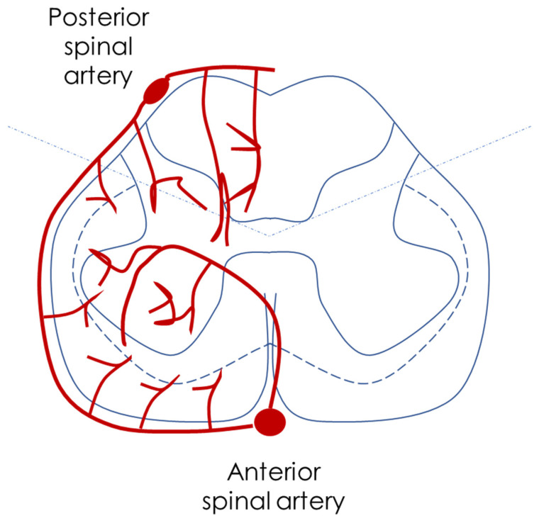
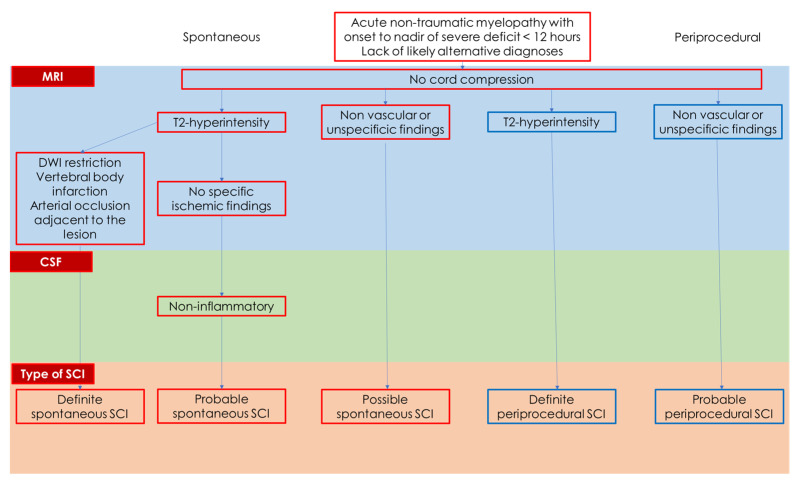
Spinal cord infarction (SCI) of arterial origin is a rare vascular event, and its incidence is probably underestimated. There are no strong epidemiological data, and the diagnostic pathway is complex and sometimes incomplete. Furthermore, many cases may be misdiagnosed as other forms of acute and subacute myelopathies. The focus of this review is the clinical and neuroradiological issues in diagnosing SCI and their respective reliability in a clinical setting. The new proposed diagnostic criteria of SCI, although not covering all aspects, highlight the need for a comprehensive approach, including even atypical cases, as the lack of cord compression on Magnetic Resonance Imaging (MRI) is the only mandatory feature for diagnosis. Some MRI features are supportive of the diagnosis, particularly when the anterior spinal artery territory is involved and diffusion-weighted imaging (DWI) is used. Several etiologies can be considered, considering traditional vascular risk factors and diseases affecting the aorta and its main branches, yet a significant proportion of cases remain without a definite etiology. The strongest predictor of SCI diagnosis is a clinical variable, i.e., a time to nadir of severe deficits < 12 h.

Keywords: DWI; MRI; aorta; dissection; owl’s eyes; periprocedural; spinal cord infarction; time.


Conflict of interest statement

The authors declare no conflicts of interest.

Figures

Figure 1
Figure 1
This is a schematic drawing of the main supply of the spinal cord, from the aorta and its branches to the radicular arteries.
Figure 2
Figure 2
This is a schematic drawing of the arterial sources to the ASA system.
Figure 3
Figure 3
This is a schematic drawing of the arterial pattern of the spinal cord, as described in the text, highlighting the preferential supply of the anterior horns by the ASA and the watershed axial zone.
Figure 4
Figure 4
This is a schematic drawing of the issues considered in the diagnostic criteria proposed by Zalewski et al. [29].
Figure 5
Figure 5
A 78-year-old patient with multiple vascular risk factors underwent vascular surgery for a thoraco-abdominal aortic aneurysm, with complications during awakening due to inability to extubate, which was performed three days later. After extubation and awakening, hyposthenia was noted in all four limbs, asymmetric (but predominantly affecting the lower limbs), with areflexia in the lower limbs and hypo-responsive proprioceptive reflexes in the upper limbs. The patient was catheterized preoperatively, with sphincter function not fully assessable. Sensory levels were unclear. An MRI study was performed 5 days post-surgery under the best possible conditions (hemodynamically stable patient but in fragile balance, on high-flow therapy, GFR < 30 mL/min, etc.). Multifocal intramedullary lesions in the high cervical–thoracic segment (at least 4) were hyperintense on T2 with different lateralization on axial scans (red lined figures) and involvement of the anterior horns (the left axial image corresponds to the highest lesion at C6-C7, and the right one to the lesion at the D3 level). DWI/ADC findings are consistent with timing (5 days post-surgery). Contrast administration was not performed due to renal failure, and the non-contrast study was already diagnostic given the clinical context.
Figure 6
Figure 6
A 38-year-old patient in good health with moderate habitual physical activity. While performing gymnastics at home, the patient experienced the sudden onset of lumbar sensitive deficit, with rapid radiation to both lower limbs, associated with painful symptoms (described as cramp-like) in the same areas. This was followed by hyposthenia in both lower limbs, progressively worsening to the point of being unable to maintain an upright posture. Upon arrival at the emergency department, urinary retention was documented, and a catheter was placed. The patient underwent a CT angiography of the aorta, with a report showing no pathological findings, as well as a spinal MRI that was reported as normal (with a minimal osteophytic protrusion at the D10-D11 level extending posteriorly) (red lined figure). The following day, the patient had a repeat spinal MRI, which showed an intramedullary lesion hyperintense on T2W sequences at the D12-L1 level, with almost full axial extension (red lined figure), without bone alterations suggestive of trauma (DWI not available). The initial diagnosis was post-traumatic injury, followed by a suspicion of myelitis (subsequent diagnostic workup was negative for immune-mediated diseases). Outcome: ASIA D paraplegia from cord contusion at D12-L1 with motor level L4 on the right and L5 on the left, and sensory level L3 bilaterally. Neurogenic bowel and bladder. Most appropriate diagnostic hypothesis based on clinical and neuroradiological data: spinal ischemia. Spinal angiographic study (DSA) was performed after referral for a second opinion 8 months after the event: negative. The images on the right side of the slide are from the MRI performed 24 h after symptom onset.
Figure 7
Figure 7
An 80-year-old patient reported severe acute back pain, followed by immediate onset of paraplegia with sensory level at D3-D4. An emergent CT angiography of the thoracic and abdominal aorta diagnosed an aortic dissection. Even in the pre-contrast sagittal reconstruction scan, an eccentric, crescent-shaped, isodense thickening was clearly visible in the wall of the thoracic aorta, just after the origin of the left subclavian artery, delineated by coarse calcifications on the intimal side of the aortic wall, highly suggestive of a mural hematoma. In the axial scans, a slightly hyperdense component was also outlined, possibly indicating ongoing bleeding. The post-contrast study confirmed the finding, with better delineation of the endoluminal dissection of severe and complicated aortic atheromatosis, where the fracture of the fibrous cap (in the segment without calcifications) could have created the entry point for the formation of the mural hematoma (red arrows).
Figure 8
Figure 8
Same patient as in Figure 6. On the MRI performed three days after the onset, multifocal intramedullary lesions are evident, with extravasation at the level of the dorsal spinal cord. The first lesion spans from D3 to D4 for nearly two vertebral bodies, and the second spans D5-D6-D7 for at least three vertebral bodies, showing hyperintensity in T2 and STIR sequences, consistent with ischemic lesions. Axial scans and DWI are compromised by motion and respiratory artifacts.

Similar articles

Cited by

References

    1. Li X.Y., Kong X.M., Yang C.H., Cheng Z.F., Lv J.J., Guo H., Liu X.H. Global, regional, and national burden of ischemic stroke, 1990–2021: An analysis of data from the global burden of disease study 2021. eClinicalMedicine. 2024;75:1027582024. doi: 10.1016/j.eclinm.2024.102758. - DOI - PMC - PubMed
    1. GBD 2021 Nervous System Disorders Collaborators Global, regional, and national burden of disorders affecting the nervous system, 1990–2021: A systematic analysis for the Global Burden of Disease Study 2021. Lancet Neurol. 2024;23:344–381. doi: 10.1016/S1474-4422(24)00038-3. - DOI - PMC - PubMed
    1. Aho K., Harmsen P., Hatano S., Marquardsen J., Smirnov V.E., Strasser T. Cerebrovascular disease in the community: Results of a WHO collaborative study. Bull. World Health Organ. 1980;58:113–130. - PMC - PubMed
    1. [(accessed on 7 November 2024)]. Available online: https://icd.who.int/en.
    1. Sacco R.L., Kasner S.E., Broderick J.P., Caplan L.R., Connors J.J., Culebras A., Elkind M.S., George M.G., Hamdan A.D., Higashida R.T., et al. An updated definition of stroke for the 21st century: A statement for healthcare professionals from the American Heart Association/American Stroke Association. Stroke. 2013;44:2064–2089. doi: 10.1161/STR.0b013e318296aeca. - DOI - PMC - PubMed

LinkOut - more resources