Review
. 2025 Feb 8;30(4):791.
doi: 10.3390/molecules30040791.

Towards Completion of the "Periodic Table" of Di-2-Pyridyl Ketoxime

Affiliations
Review

Towards Completion of the "Periodic Table" of Di-2-Pyridyl Ketoxime

Christina Stamou et al. Molecules. .

Abstract

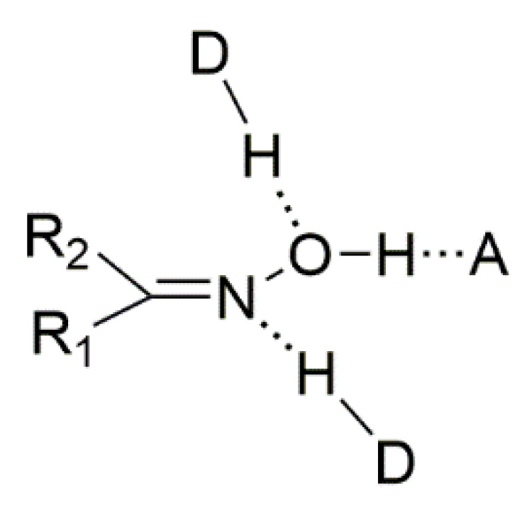
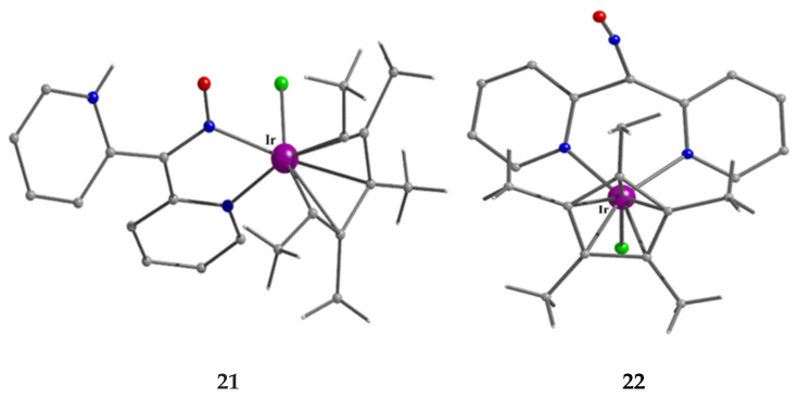
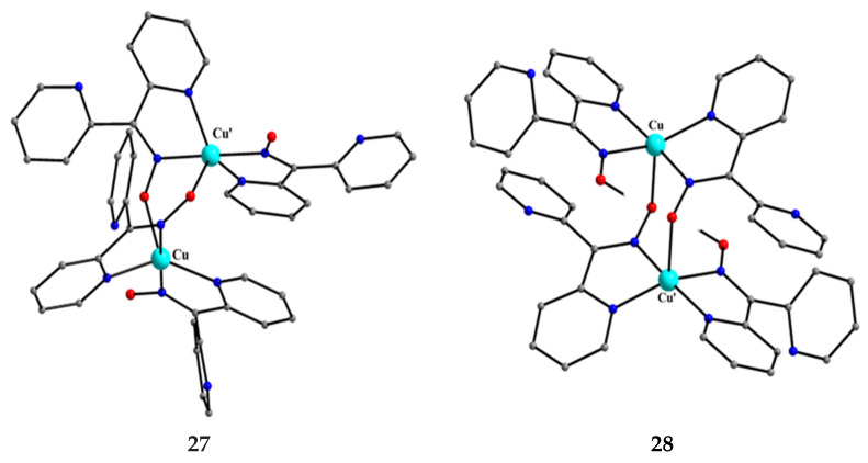
The oxime group is important in organic and inorganic chemistry. In most cases, this group is part of an organic molecule possessing one or more donor sites capable of forming bonds to metal ions. One family of such compounds is the group of 2-pyridyl (aldo)ketoximes. Metal complexes of 2-pyridyl oximes continue to attract the intense interest of many inorganic chemistry groups around the world for a variety of reasons, including their interesting structures, physical and biological properties, and applications. A unique member of 2-pyridyl ketoximes is di-2-pyridyl ketoxime (dpkoxH), which contains two 2-pyridyl groups and an oxime functionality that can be easily deprotonated giving the deprotonated ligand (dpkox-). The extra 2-pyridyl site confers a remarkable flexibility resulting in metal complexes with exciting structural and reactivity features. Our and other research groups have prepared and characterized many metal complexes of dpkoxH and dpkox- over the past 30 years or so. This work is an attempt to build a "periodic table" of dpkoxH, which is near completion. The filled spaces of this "periodic table" contain metal ions whose dpkoxH/dpkox- complexes have been structurally characterized. This work reviews comprehensively the to-date published coordination chemistry of dpkoxH with emphasis on the syntheses, reactivity, relationship to metallacrown chemistry, structures, and properties of the metal complexes; selected unpublished results from our group are also reported. The sixteen coordination modes adopted by dpkoxH and dpkox- have provided access to monomeric and dimeric complexes, trinuclear, tetranuclear, pentanuclear, hexanuclear, heptanuclear, enneanuclear, and decanuclear clusters, as well as to a small number of 1D coordination polymers. With few exceptions ({MIILnIII2} and {NiII2MnIII2}; M = Ni, Cu, Pd, and Ln = lanthanoid), most complexes are homometallic. The metals whose ions have yielded complexes with dpkoxH and dpkox- are Cr, Mn, Fe, Co, Ni, Cu, Zn, Ru, Rh, Pd, Ag, Cd, Re, Os, Ir, Au, Hg, lanthanoids (mainly Pr and Nd), and U. Most metal complexes are homovalent, but some mixed-valence Mn, Fe, and Co compounds have been studied. Metal ion-assisted/promoted transformations of dpkoxH, i.e., reactivity patterns of the coordinated ligand, are also critically discussed. Some perspectives concerning the coordination chemistry of dpkoxH and research work for the future are outlined.

Keywords: coordination chemistry; di-2-pyridyl ketoxime’s metal complexes; magnetic properties; reactivity; structures; synthesis.


Conflict of interest statement

The authors declare no conflicts of interest.

Figures

Figure 7
Figure 7
Two routes for the metal-mediated synthesis of oximes (without C-, N-, and O-functionalization). This drawing was adapted from Ref. [14]. M = metal center.
Figure 8
Figure 8
The three types of classical H bonds formed by the oxime group. H-bond acceptors can be the oxime N and O atoms, while the H-bond donor can be the O atom. A = acceptor, D = donor.
Figure 9
Figure 9
H-bonded dimer (I) and catemers formed by O-H∙∙∙N (II) and O-H∙∙∙O (III) H bonds.
Figure 16
Figure 16
The structures of the molecules [Ni(mef)2(dpkoxH)2], [Ni(tolf)2(dpkoxH)2], [Ni(dilf)2(dpkoxH)2], and the cation [Ni(dicl)(diclH)(dpkoxH)2]+ that are present in the crystals of 2, 3, 4, and 6, respectively.
Figure 17
Figure 17
The structures of the molecules [Zn(mef)2(dpkoxH)2] and [Zn(difl)2(dpkoxH)2] that are present in the crystals of 14 and 16, respectively.
Figure 18
Figure 18
Docking representations of [Zn(difl)2(dpkoxH)2] (16) in orange and free diflH in blue interacting with calf-thymus DNA. Reproduced from Ref. [38]. Copyright 2017 Elsevier.
Figure 21
Figure 21
The molecular structures of [ZnCl2(dpkoxH)2] (11) and [ZnBr2(dpkoxH)2] (13).
Figure 22
Figure 22
The molecular structure of the cation [(Ph)RuIIICl(dpkoxH)]+ that is present in the hexafluorophosphate salt 18.
Figure 23
Figure 23
The molecular structures of the isomeric cations that are present in complexes 21 (major isomer) and 22 (minor isomer); see text for discussion.
Figure 24
Figure 24
The molecular structure of [ReI(CO)3Cl(dpkoxH)] (20).
Figure 27
Figure 27
The structure of the centrosymmetric molecule [MnII,II2(O2CF3)2(hfac)2(dpkoxH)2] that is present in the crystal of 25.
Figure 28
Figure 28
The structure of the molecule [CuII,II2(dpkox)4] and the cation [CuII,II2(dpkox)2(dpkoxH)2]2+ that are present in complexes 27 and 28, respectively.
Figure 29
Figure 29
Schematic illustration of the structure of the cationic complex [CuII,II2Cl4(H2O)2(dpkoxH2)2]Cl2 (29). The symbol with the three nitrogen atoms, the hydroxyl group, and the proton is a short representation of the cationic ligand dpkoxH2+ in which the non-coordinated 2-pyridyl N atom is protonated. The dashed bold lines indicate weak coordination bonds.
Figure 30
Figure 30
The structure of the cation [CuII,II2Br4(dpkoxH2)2]2+ that is present in complex 30.
Figure 31
Figure 31
The molecular structure of [CuII,II2(hfac)2(dpkox)2] (31).
Figure 32
Figure 32
The molecular structure of [CuI,I2Cl2(dpkoxH)2] (32).
Figure 33
Figure 33
The molecular structure of [RuI,I2(CO)4(dpkox)2] (33).
Figure 34
Figure 34
The molecular structure of [AgI,I2(NO3)2(dpkoxH)2] (34).
Figure 35
Figure 35
The molecular structure of [HgI,I2(SCN)4(dpkoxH)2] (35).
Figure 36
Figure 36
The structures of the cation [CrIII,III,III3O(piv)4(H2O)(dpkox)2]+ and the molecule [CrIII,III,III3O Cl(piv)4(dpkox)2] that are present in the crystals of 36 and 37, respectively.
Figure 37
Figure 37
(Left) χMT vs. T plot for a powdered sample of 37 in a 3 KG field (χM is the molar magnetic susceptibility and T is the absolute temperature). The solid line is the fit of the experimental data to the appropriate 2-J model [45]. (Right) X-band EPR spectra of solid 37 recorded at 295 K (dashed line) and 20 K (solid line). Reproduced from Ref. [45]. Copyright 2007 Elsevier.
Figure 38
Figure 38
The molecular structure of [MnII,IV,II3(OMe)2Cl2(dpkox)4] (38) and [MnII,IV,II3(OMe)2(NCO)2(dpkox)4] (40).
Figure 39
Figure 39
The molecular structures of [MnII,IV,II3(ed)Cl2(dpkox)4] (41) and [MnII,IV,II3(perH2)Cl2 (dpkox)4] (43); ed is the dianion of ethanediol and perH2 is the dianion of pentaerythritol (Figure 15).
Figure 40
Figure 40
The structure of the cation [FeII,III,II3(dpkox)6]+ that is present in complex 44.
Figure 41
Figure 41
The molecular structure of [Ni3(shi)2(dpkoxH)2(py)2] (45).
Figure 42
Figure 42
The coordination mode of shi3− in complex [Ni3(shi)2(dpkoxH)2(py)2] (45) and the Harris notation that describes this ligation. The subscript 1 refers to the octahedral metal ion (Ni1 in Figure 41) and the subscript 2 to the square planar metal ion (Ni2 in Figure 41); pl = planar, oct = octahedral.
Figure 43
Figure 43
The molecular structure of complex [Ni3(N3)4(Medpt)2(dpkox)2] (46).
Figure 44
Figure 44
The structures of the molecules [CuII,II,II(OH)(O2CPh)2(dpkox)3] and [CuII,II,II3(OH)Br (tBuPO3H)(dpkox)3] that are present in the crystals of 47 and 49, respectively.
Figure 45
Figure 45
The structures of the molecules [Ru3(CO)8(dpkox)2] and [Os3(H)(CO)9(dpkox)] that are present in the crystals of 50 and 51, respectively.
Figure 46
Figure 46
The metalloligands used for the synthesis of {NiLn2} and {MIILn2} (M = Pd, Cu) complexes based on dpkox.
Figure 47
Figure 47
The molecular structures of [NiDy2(hfac)6(dpkox)2(phen)] (54) and [NiGd2(hfac)6(dpkox)2(py)2] (57).
Figure 48
Figure 48
The molecular structures of [PdIIDy2(hfac)6(dpkox)2] (61) and [CuIIGd2(hfac)6(dpkox)2] (63).
Figure 49
Figure 49
Temperature and frequency dependence of the out-of-phase molar magnetic alternating current (ac) susceptibility, χΙΙac, for complexes [NiDy2(hfac)6(dpkox)2(phen)] (54) (a) and [NiDy2(hfac)6(dpkox)2(py)2] (59) (b). Reproduced from Ref. [64]. Copyright 2005 Elsevier.
Figure 50
Figure 50
(a) Plot of the 3d-4f exchange parameters (J) in the {NiLn2} clusters 5760 and the {CuIILn2} complexes 62, 63, [CuIITb2(hfac)6(dpkox)2] (not listed in Table 4) and [CuIIHo2(hfac)6(dpkox)2] (also not listed in Table 4) as a function of the atomic number, Z. (b) Plot of the cell volume in the above-mentioned complexes as a function of Z. The {CuIITb2} and {CuIIHo2} clusters are isomorphous with 62 and 63. Reproduced from Ref. [65]. Copyright 2013 American Chemical Society.
Figure 51
Figure 51
(a) Magnetization (M) curves and (b) their derivatives for complex [PdDy2(hfac)6(dpkox)2] (61) measured at 0.4 K using a pulse-field magnetometer. Reproduced from Ref. [66]. Copyright 2011 Elsevier.
Figure 52
Figure 52
Frequency and temperature dependence of the ac molar magnetic susceptibility for [CuIIDy2(hfac)6(dpkox)2] (62). (a) χ’ac is in-phase part; (b) χ’’ac is the out-of-phase part. The inset shows the Cole-Cole plot at 8 K. Reproduced from Ref. [67]. Copyright 2006 American Chemical Society.
Figure 53
Figure 53
Selected High-Frequency EPR (HF-EPR) spectra at 4.2 K of complexes [CuIITb2(hfac)6(dpkox)2] (a) and [CuIIHo2(hfac)6(dpkox)2] (b), not listed in Table 4; the two clusters are isomorphous with 62 and 63. The spectra are offset in a linear scale of the frequency. Dotted lines are drawn from linear fitting in the frequency vs. field plot. Reproduced from Ref. [68]. Copyright 2010 The Chemical Society of Japan.
Figure 54
Figure 54
The molecular structure of [MnII,II,II3MnIVO(3,4-D)4(dpkox)4] (64). Only the H atoms of the –CH2- groups are shown. The three chlorine atoms in one of the 3,4-D ligands are a consequence of a crystallographic disorder issue.
Figure 55
Figure 55
The molecular structure of [MnII,II2MnIII,III2Br2(O2CPh)2(dpkox)2{(py)2C(O)2}2] (67), [MnII,II2MnIII,III2Cl2(O2CPh)2(dpkox)2{(py)2C(O)2}2] (69) and [MnII,II2MnIII,III2(NO3)2(O2CMe)2 (dpkox)2{(py)2C(O)2}2] (70).
Figure 56
Figure 56
The coordination mode of (py)2C(O)2 in complexes [MnII,II2MnIII,III2Y2(O2CR)2(dpkox)2{(py)2C(O)2}2] and the detailed Harris notation that describes this mode. The subscript 2 refers to MnIII and 1 to MnII. R = Ph in 67 and 69, and Me in 68 and 70; Y = Br in 67 and 68, Cl in 69, and NO3 in 70.
Figure 57
Figure 57
The molecular structures of the polymorph [FeIII4O2Cl2(O2CMe)2(dpkox)4] (71) [with a 2CH2Cl2∙H2O lattice solvent set] and the azido cluster [FeIII4O2(N3)2(O2CMe)2(dpkox)4] (73).
Figure 58
Figure 58
The structure of the cation [CoII,II2CoIII,III2(OH)2(O2CMe)2(dpkox)4(MeOH)2]2+ that is present in complex 74. The H atoms of the hydroxido, acetato, and methanol ligands have been drawn.
Figure 60
Figure 60
The structure of the cation [Ni4(dpkox)6(MeOH)2]2+ that is present in the crystal of 77.
Figure 65
Figure 65
The molecular structure of [Ni4(SCN)2(shiH)2(dpkox)2(DMF)(MeOH)] (81).
Figure 66
Figure 66
Drawing showing the connectivity pattern and the arrangement around the NiII centers in [Ni4(SCN)2(shiH)2(dpkox)2(DMF)(MeOH)] (81). The coordination bonds are indicated with bold lines.
Figure 69
Figure 69
The molecular structure of [Zn4(OH)2(dpkox)6] (89).
Figure 71
Figure 71
The cation [Ni5(dpkox)5(H2O)7]2+ that is present in the crystal structure of 91.
Figure 72
Figure 72
The Ni5 skeleton of 91 showing the very distorted tetrahedral metal topology (left) and the {NiII53-ONR)42-ONR)}5+ core of 91 (right).
Figure 80
Figure 80
The structure of [CuII6(dpkox)6(MeCN)6]6+ that is present in the cation 99a; the encapsulated (trapped) ClO4 of 99a has not been drawn.
Figure 81
Figure 81
Stereoview of the cation [CuII6(ClO4)3(dpkox)6(MeCN)4]3+ (99b) showing the cavity of the ring and the encapsulated ClO4 ion. Reproduced from Ref. [86]. Copyright 2005 Elsevier.
Figure 82
Figure 82
The molecular structure of [Zn6(OH)2(dpkox)4(fluf)6] (100).
Figure 83
Figure 83
The structure of the cation [MnII4MnIII6MnIV2O6(OH)4(OMe)2(dpkox)12]4+ that is present in complex 101.
Figure 86
Figure 86
The molecular structure of [Ni10(mcpa)2(shi)5(dpkox)3(MeOH)3(H2O)] (104). Atoms Ni4 and Ni8 have square planar geometries.
Figure 87
Figure 87
The five coordination modes of the shi3− ligands in the structure of [Ni10(mcpa)2(shi)5(dpkox)3(MeOH)2(H2O)] (104) and the Harris notation that describes these modes. The coordination bonds are drawn with bold lines. The numbering scheme of the NiII atoms is presented for clarity. The ligation modes 4.2112 and 6.3221 are, to-date, novel in the coordination chemistry of salicylhydroxamic acid.
Figure 90
Figure 90
A small portion of one zigzag chain that is present in the crystal structure of {[CuI(SCN)(dpkoxH)]}n (107).
Figure 98
Figure 98
The molecular structure of one of the solvates of [Pr2(NO3)4(L)2] (121); the structural formula of the anionic ligand L is illustrated in Figure 99.
Figure 99
Figure 99
The structural formula and the abbreviation of the anionic ligand that is present in [Pr2(NO3)4(L)2] (121).
Figure 100
Figure 100
The molecular structure of [AuICl(dpkoxH)] (122).
Figure 101
Figure 101
The present form of the “periodic table” of dpkoxH. Color code: Pale red; metals whose complexes with dpkoxH and/or dpkox have been published, or structurally characterized by our group and remain unpublished. Yellow; metals with unpublished coordination chemistry of dpkoxH/dpkox.
Figure 1
Figure 1
The structural formulae of aldoximes and ketoximes.
Figure 2
Figure 2
The syn-anti isomerism of the simple oxime group assuming that R1 takes precedence over R2 according to the Cahn-Ingold-Prelog system.
Figure 3
Figure 3
The structural formulae of simple 2-pyridyl (aldo)ketoximes (left; R = H, Me, Ph, …) and di-2-pyridyl ketoxime.
Figure 4
Figure 4
Few oxime derivatives which find use in medicine and agriculture (see text for details).
Figure 5
Figure 5
The to-date crystallographically observed coordination modes of the oxime and oximate groups, and the Harris notation [2] that describes these modes. The 1.0011 ligation mode represents one formally oximate group and one formally neutral oxime group; this moiety is present in some complexes, including bis(dimethylglyoximato)nickel(II).
Figure 6
Figure 6
Reactivity modes of the coordinated oxime or oximate groups. M = metal ion, Nu = nucleophile, E = electrophile.
Figure 10
Figure 10
The to-date crystallographically confirmed coordination modes of neutral and anionic 2-pyridyl oximes, and the Harris notation [2] that describes these modes. R (H, Me, Ph, …) is a non-donor group. The 2.111 mode of neutral oximes and the 4.311 mode of the 2-pyridyl oximate ligands have been found only in cases where R = H. M and M’ are metal ions.
Figure 11
Figure 11
An example of the MC-crown ether analogy. The symbol shi represents the trianion of salicylhydroxamic acid (shiH3, Figure 12). The metal ion can be only a first-row transition metal ion. The drawing was adapted from Ref. [3].
Figure 12
Figure 12
The structural formula and abbreviation of salicylhydroxamic acid, one of the most commonly used ligands for the construction of MCs (vide infra).
Figure 13
Figure 13
The molecules dpi, adpm, and adpe that have resulted from the synthetic investigation of the [VIIICl3(THF)3]/dpkoxH reaction system. The molecules are coordinated in the {VIVO}2+ (vanadyl) complexes L, M, N, and O as described in the text.
Figure 14
Figure 14
The to-date crystallographically observed coordination modes of dpkoxH2+, dpkoxH, and dpkox, and the Harris notation [2] that describes these modes. M is a metal ion and Ln is a trivalent lanthanoid.
Figure 15
Figure 15
The structural formulae of most of the ancillary ligands discussed in the text. For the anionic ligands, the negative charge is indicated only in their abbreviations and not at the corresponding atoms in the structural formulae.
Figure 19
Figure 19
The molecular structure of [CuIICl(dpkox)(dpkoxH)] (8).
Figure 20
Figure 20
The N4-tetradentate chelating viewpoint of the moiety (dpkox∙∙∙H∙∙∙dpkox) in complex [CuIICl(dpkox)(dpkoxH)] (8).
Figure 25
Figure 25
The molecular structure of [AuIIICl(dpkox)2] (23).
Figure 26
Figure 26
The structure of the molecule [CrII,II2(dpkox)4] in the crystal of 24.
Figure 59
Figure 59
The structure of the cation [CoIII4(OH)2(O2CMe)4(dpkox)4]2+ that is present in complex 76. The H atoms of the hydroxidο and acetato ligands have been drawn.
Figure 61
Figure 61
The structure of the cation [Ni4(O2CMe)2(dpkox)4]2+ that is present in the crystal of 78.
Figure 62
Figure 62
The molecular structure of [Ni4(SO4)2(dpkox)4(MeOH)4] (79).
Figure 63
Figure 63
The pseudo 12-MC-4 ring of complex [Ni4(SO4)2(dpkox)4(MeOH)4] (79). The coordination bonds are drawn with bold lines.
Figure 64
Figure 64
The structure of the cation [Ni4(ea)2(eaH)2(dpkox)4]2+ that is present in the crystal of its perchlorate salt 80.
Figure 67
Figure 67
The molecular structure of [Ni4(N3)4(dpt)2(dpkox)4] (82).
Figure 68
Figure 68
The molecular structures of [Zn4(OH)2(O2CMe)2(dpkox)4] (83), [Zn4(OH)2(O2CPh)2(dpkox)4] (84), [Zn4(OH)2(N3)2(dpkox)4] (86), and [Zn4(OH)2(acac)2(dpkox)4] (88). The dashed line indicates a weak bonding interaction of one carboxylato O atom to Zn2/Zn2’ (Zn-O = ~2.6 Å).
Figure 70
Figure 70
Drawing shown the connectivity pattern and the arrangement of donor atoms around the MnIII and NiII centers in [Ni2MnIII2(O2CMe)2(shi)2(dpkox)2(DMF)2] (90). The coordination bonds are indicated with bold lines.
Figure 73
Figure 73
The molecular structure of [Ni5(O2CMe)7(dpkox)3(H2O)] (92).
Figure 74
Figure 74
The molecular structure of [Ni5(O2CMe)2(shi)2(dpkox)2(DMF)2] (93).
Figure 75
Figure 75
The pentanuclear cation [CuII5(dpkox)7]3+ that is present in the crystal structure of 94.
Figure 76
Figure 76
The {CuII53-ΝO)32-NO)4}3+ core of the cation of complex 94; NO represents the oximato group.
Figure 77
Figure 77
The pentanuclear cations that are present in the crystal structures of the ionic complexes [Zn5Cl2(dpkox)6][ZnCl(NCS)3] (95) and [Zn5(NCS)2(dpkox)6(MeOH)][Zn(NCS)4] (96).
Figure 78
Figure 78
The structure of the cation [MnII3MnIII3O2(O2CPh)6(dpkox)2{(py)2C(OH)(O)}2]+ that is present in complex 97 (left) and its core (right). The coordination bonds are drawn with bold lines. The quadruply bridging oxygens represent the oxido (O2−) groups, while the triply bridging oxygens come from the O atoms of the 3.3011 (py)2C(OH)(O) ligands.
Figure 79
Figure 79
The structure of the cation [CuII6(OH)2Cl2(dpkox)6]2+ that is present in the crystal of 98.
Figure 84
Figure 84
The molecular structure of [Ni7(N3)2(O2CMe)6(dpkox)6(H2O)2] (102).
Figure 85
Figure 85
The structure of the cation [Ni9(N3)9(dpkox)6(dpt)6]3+ that is present in cluster 103; the H-bonded encapsulated N3 ion (dashed lines) is also shown. An identical numbering scheme (i.e., without primes) is used for the NiII atoms generated by symmetry.
Figure 88
Figure 88
The tetranuclear unit that creates the 1D coordination polymer {[MnII2MnIII2(N3)6(dpkox)2{(py)2C(O)2}(MeOH)2]}n (106). Two terminal azido groups on Mn2 and Mn4 are becoming end-to-end in the 1D chain providing the intercluster linkage.
Figure 89
Figure 89
Chain structure of {[MnII2MnIII2(N3)6(dpkox)2{(py)2C(O)2}(MeOH)2]}n (106). Adjacent tetranuclear units are linked via an end-to-end (2.11) azido group, which connects two MnIII atoms; these groups are terminal in each tetranuclear unit and are becoming end-to-end to generate the polymer. Reproduced from Ref. [71]. Copyright 2008 American Chemical Society.
Figure 91
Figure 91
A portion of one zigzag chain that is present in the crystal structure of {[CdBr2(dpkox)]}n (109).
Figure 92
Figure 92
The 113Cd NMR spectra of {[CdX2(dpkoxH)]}n (X = Cl, 108; X = Br, 109) (left) and {[CdI2(dpkoxH)]}n (110) (right).
Figure 93
Figure 93
The molecular structures of [Cd(1.100-NO3)2(dpkoxH)2] (111) and [Cd(1.110-NO3)2(dpkoxH)2] (112).
Figure 94
Figure 94
Solid-state MAS 113Cd NMR spectra of the samples isolated during the preparations of complexes 111 and 112.
Figure 95
Figure 95
The molecular structure of the centrosymmetric complex [Pr4(OH)2(NO3)4(dpkox)6(MeOH)2] (118).
Figure 96
Figure 96
The molecular structure of the mixed-valence coordination cluster [PrIII8PrIVO4(OH)4(NO3)4(dpkox)12(H2O)4] (120).
Figure 97
Figure 97
The topology (dashed multicolored lines) and the inorganic {PrIII8PrIV3-O)43-OH)4}16+ core in 120. O1X (and symmetry equivalents) and OX2 (and symmetry equivalents) are oxido and hydroxido oxygens.
Figure 102
Figure 102
The 1.1010 (Harris notation) coordination mode confirmed in the just prepared and structurally characterized hexagonal bipyramidal complex [UVIO2(dpkox)2(MeOH)2]. The coordination bonds are drawn with solid lines.

Similar articles

References

    1. March J. Advanced Organic Chemistry. 4th ed. Wiley; New York, NY, USA: 1992. pp. 73, 128, 367, 388, 389, 405, 406, 894, 918, 934, 943, 1038–1040, 1154, 1199.
    1. Coxall R.A., Harris S.G., Henderson D.K., Parsons S., Tasker P.A., Winpenny R.E.P. Inter-Ligand Reactions: In Situ Formation of New Polydentate Ligands. J. Chem. Soc. Dalton Trans. 2000:2349–2356. doi: 10.1039/b001404o. - DOI
    1. Mezei G., Zaleski C.M., Pecoraro V.L. Structural and Functional Evolution of Metallacrowns. Chem. Rev. 2007;107:4933–5003. doi: 10.1021/cr078200h. - DOI - PubMed
    1. Milios C.J., Stamatatos T.C., Perlepes S.P. The coordination chemistry of pyridyl oximes. Polyhedron. 2006;25:134–194. doi: 10.1016/j.poly.2005.07.022. - DOI
    1. Tschugaeff L. Ueber ein neues, empfindliches reagens out nickel. Ber. Dtsch. Chem. Ges. 1885;38:2520–2522. doi: 10.1002/cber.19050380317. - DOI

LinkOut - more resources