. 2025 Feb 11;18(2):238.
doi: 10.3390/ph18020238.

Protective Effects of Oleanolic Acid on Human Keratinocytes: A Defense Against Exogenous Damage

Affiliations

Protective Effects of Oleanolic Acid on Human Keratinocytes: A Defense Against Exogenous Damage

Marzia Vasarri et al. Pharmaceuticals (Basel). .

Abstract

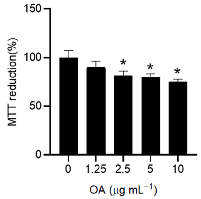
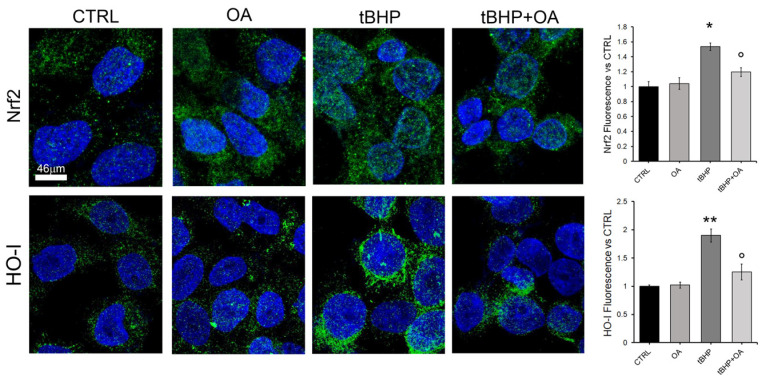
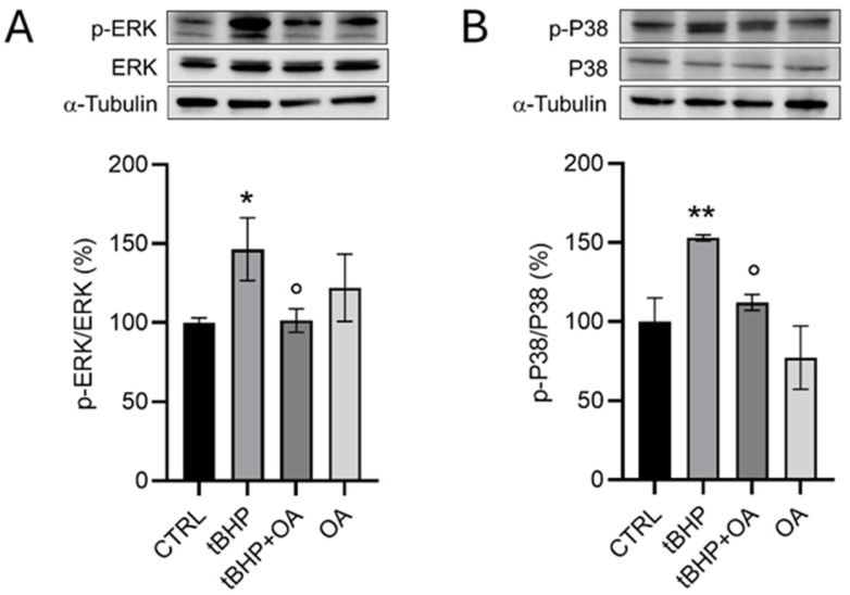
Background/objectives: Aging leads to increased oxidative stress and chronic inflammation in the skin, which contribute to various disorders such as dermatitis and cancer. This study explores the cytoprotective effects of oleanolic acid (OA), a natural triterpenoid compound known for its potential in mitigating oxidative damage, on human keratinocyte (HaCaT) cells exposed to oxidative stress from tert-butyl hydroperoxide (tBHP). Methods: Using in vitro experiments, we assessed cell viability, reactive oxygen species (ROS) levels, nitric oxide (NO) production, and protein expression following OA pre-treatment. Advanced imaging techniques were employed to visualize protein localization. Results: Results demonstrated that OA significantly improved cell viability and reduced intracellular ROS levels compared with those in controls. Additionally, OA inhibited inducible nitric oxide synthase (iNOS) expression and subsequent nitric oxide release, indicating a modulation of inflammatory responses. Notably, while tBHP activated the Nrf2/HO-1 signaling pathway, OA did not enhance this response, suggesting that OA exerts cytoprotective effects through mechanisms independent of Nrf2 activation. Conclusion: OA shows promise in protecting HaCaT cells from tBHP-induced oxidative stress, highlighting its potential role in promoting skin health and addressing aging-related damage. The study proposes that OA operates through pathways distinct from Nrf2 and MAPKs, paving the way for new therapeutic strategies aimed at improving skin health against oxidative stress.

Keywords: HaCaT cells; oleanolic acid; oxidative stress; tert-butyl-hydroperoxide.


Conflict of interest statement

The authors declare no conflicts of interest.

Figures

Figure 1
Figure 1
Effect of OA on HaCaT cell viability. HaCaT cells were exposed to increasing doses of OA (0–10 µg mL−1) in a serum-free medium for 24 h. Viable cells were detected as metabolically active cells using the MTT assay. Results are expressed as the mean ± standard deviation (SD) of three independent experiments. Statistical analysis was performed by one-way ANOVA followed by the post hoc Tukey’s HSD test (n = 3). * p < 0.05 compared with untreated control cells (0).
Figure 2
Figure 2
Effect of OA on the viability (A) and intracellular ROS formation (B) in HaCaT cells exposed to the pro-oxidative agent tBHP. HaCaT cells were pre-treated with OA (1.25 µg mL−1) for 2 h and then exposed to tBHP (200 µM) for 3 h in serum-free medium. Untreated cells or those treated with OA or tBHP alone were used as controls. Data are expressed as the mean ± SD of three separate experiments. Statistical analysis was performed using one-way ANOVA followed by Tukey’s HSD test (n = 3): ** p < 0.01 vs. CTRL; ° p < 0.05, °°° p < 0.001 vs. tBHP-treated cells.
Figure 3
Figure 3
Analysis of iNOS expression by Western blot analysis (A) and NO production and release into the culture medium by the Griess test (B) in HaCaT cells exposed to the pro-oxidant tBHP. HaCaT cells were pre-treated with OA (1.25 µg mL−1) for 2 h and then exposed to tBHP (200 µM) for 3 h in serum-free medium. Untreated cells or cells treated with OA or tBHP alone were used as controls. Data are reported as the mean ± SD of three different experiments. For the Western blot analysis, statistics were performed using the Kruskal–Wallis test followed by Conover’s post hoc test (n = 3): * p < 0.05 vs. CTRL; ° p < 0.05 vs. tBHP-treated cells. For the Griess test, statistics were performed by using one-way ANOVA followed by the post hoc Tukey HSD test (n =3): * p < 0.05 vs. CTRL; ° p < 0.05 vs. tBHP-treated cells.
Figure 4
Figure 4
Analysis of Nrf2 expression by Western blot assay in HaCaT cells exposed to the pro-oxidative agent tBHP. HaCaT cells were pre-treated with OA (1.25 µg mL−1) for 2 h and then exposed to tBHP (200 µM) for 3 h in serum-free medium. Untreated cells or cells treated with OA or tBHP alone were used as controls. Data are reported as the mean ± SD of three different experiments. Statistical analysis was performed using the Kruskal–Wallis test followed by Conover’s post hoc test (n = 3): * p < 0.05 vs. CTRL; ° p < 0.05 vs. tBHP-treated cells.
Figure 5
Figure 5
Nrf2 and HO levels in HaCaT cells. Representative confocal images of HaCaT cells pre-treated with OA (1.25 µg mL−1) for 2 h and then exposed to tBHP (200 µM) for 3 h in serum-free medium. Nuclei cells were labelled by HOECHST 33342 (blue fluorescence), and Nrf2 and HO were stained with rabbit anti-Nrf2 or anti-HO primary antibodies followed by treatment with Alexa 488-conjugated anti-rabbit secondary antibodies (green fluorescence). Quantification of the green mean fluorescence intensity signals is reported on the right and was performed by ImageJ Fiji software (Version 1.54m). One-way ANOVA test: * p < 0.05; ** p < 0.01 vs. CTRL; ° p < 0.01 vs tBHP. Values are the average of 5 independent experiments± SE.
Figure 6
Figure 6
Nrf2 and HO internalization. (A,B) Representative images from super-resolution confocal microscopy (STED). HaCaT cells were pre-treated with OA (1.25 µg mL−1) for 2 h and then exposed to tBHP (200 µM) for 3 h in serum-free medium. Then, HaCaT cells were marked using HOECHST 33342 for nuclei (blue fluorescence), and Nrf2 (A) or HO-1 (B) was visualized with their specific primary antibody and anti-rabbit Alexa 546 secondary antibodies (red fluorescence). Reconstructions of the z-stack analysis of the cells shown in panels were performed by Leica software (Versions 5.1.0).
Figure 7
Figure 7
Analysis of (A) p-ERK and (B) p-p38 as markers of the MAPK signaling pathway in HaCaT cells exposed to the pro-oxidative agent tBHP by Western blot assay. HaCaT cells were pre-treated with OA (1.25 µg mL−1) for 2 h and then exposed to tBHP (200 µM) for 3 h in serum-free medium. Untreated cells or cells treated with OA or tBHP alone were used as controls. Data are reported as the mean ± SD of three different experiments. Statistical analysis was performed using the Kruskal–Wallis test followed by Conover’s post hoc test (n = 3): * p < 0.05; ** p < 0.01 vs. CTRL; ° p < 0.05 vs. tBHP-treated cells.

Similar articles

Cited by

References

    1. Agrawal R., Hu A., Bollag W.B. The Skin and Inflamm-Aging. Biology. 2023;12:1396. doi: 10.3390/biology12111396. - DOI - PMC - PubMed
    1. Fisher G.J., Kang S., Varani J., Bata-Csorgo Z., Wan Y., Datta S., Voorhees J.J. Mechanisms of photoaging and chronological skin aging. Arch. Dermatol. 2002;138:1462–1470. doi: 10.1001/archderm.138.11.1462. - DOI - PubMed
    1. Liu H.-M., Cheng M.-Y., Xun M.-H., Zhao Z.-W., Zhang Y., Tang W., Cheng J., Ni J., Wang W. Possible Mechanisms of Oxidative Stress-Induced Skin Cellular Senescence, Inflammation, and Cancer and the Therapeutic Potential of Plant Polyphenols. Int. J. Mol. Sci. 2023;24:3755. doi: 10.3390/ijms24043755. - DOI - PMC - PubMed
    1. Leyane T.S., Jere S.W., Houreld N.N. Oxidative Stress in Ageing and Chronic Degenerative Pathologies: Molecular Mechanisms Involved in Counteracting Oxidative Stress and Chronic Inflammation. Int. J. Mol. Sci. 2022;23:7273. doi: 10.3390/ijms23137273. - DOI - PMC - PubMed
    1. Aranda-Rivera A.K., Cruz-Gregorio A., Arancibia-Hernández Y.L., Hernández-Cruz E.Y., Pedraza-Chaverri J. RONS and Oxidative Stress: An Overview of Basic Concepts. Oxygen. 2022;2:437–478. doi: 10.3390/oxygen2040030. - DOI

LinkOut - more resources