

Nanofibrous Scaffolds' Ability to Induce Mesenchymal Stem Cell Differentiation for Soft Tissue Regenerative Applications
- PMID: 40006052
- PMCID: PMC11859969
- DOI: 10.3390/ph18020239
Nanofibrous Scaffolds' Ability to Induce Mesenchymal Stem Cell Differentiation for Soft Tissue Regenerative Applications
Abstract
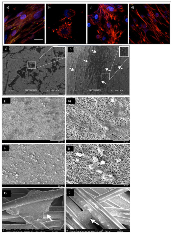
Mesenchymal stem cells (MSCs) have gained recognition as a highly versatile and promising cell source for repopulating bioengineered scaffolds due to their inherent capacity to differentiate into multiple cell types. However, MSC implantation techniques have often yielded inconsistent clinical results, underscoring the need for advanced approaches to enhance their therapeutic efficacy. Recent developments in three-dimensional (3D) bioengineered scaffolds have provided a significant breakthrough by closely mimicking the in vivo environment, addressing the limitations of traditional two-dimensional (2D) cell cultures. Among these, nanofibrous scaffolds have proven particularly effective, offering an optimal 3D framework, growth-permissive substrates, and the delivery of trophic factors crucial for MSC survival and regeneration. Furthermore, the selection of appropriate biomaterials can amplify the paracrine effects of MSCs, promoting both proliferation and targeted differentiation. The synergistic combination of MSCs with nanofibrous scaffolds has demonstrated remarkable potential in achieving repair, regeneration, and tissue-specific differentiation with enhanced safety and efficacy, paving the way for routine clinical applications. In this review, we examine the most recent studies (2013-2023) that explore the combined use of MSCs and nanofibrous scaffolds for differentiation into cardiogenic, epithelial, myogenic, tendon, and vascular cell lineages. Using PubMed, we identified and analyzed 275 relevant articles based on the search terms "Nanofibers", "Electrospinning", "Mesenchymal stem cells", and "Differentiation". This review highlights the critical advancements in the use of nanofibrous scaffolds as a platform for MSC differentiation and tissue regeneration. By summarizing key findings from the last decade, it provides valuable insights for researchers and clinicians aiming to optimize scaffold design, MSC integration, and translational applications. These insights could significantly influence future research directions and the development of more effective regenerative therapies.
Keywords: differentiation; electrospinning; mesenchymal stem cells; nanofibrous scaffolds.
Conflict of interest statement
The authors declare no conflicts of interest.
Figures


Similar articles
-
Polycaprolactone-co-polylactic acid nanofiber scaffold in combination with 5-azacytidine and transforming growth factor-β to induce cardiomyocyte differentiation of adipose-derived mesenchymal stem cells.Cell Biochem Funct. 2022 Oct;40(7):668-682. doi: 10.1002/cbf.3728. Epub 2022 Aug 4. Cell Biochem Funct. 2022. PMID: 35924670
-
Stem cell differentiation to epidermal lineages on electrospun nanofibrous substrates for skin tissue engineering.Acta Biomater. 2011 Aug;7(8):3113-22. doi: 10.1016/j.actbio.2011.04.017. Epub 2011 Apr 23. Acta Biomater. 2011. PMID: 21550425
-
Osteogenic differentiation and bone regeneration of iPSC-MSCs supported by a biomimetic nanofibrous scaffold.Acta Biomater. 2016 Jan;29:365-379. doi: 10.1016/j.actbio.2015.10.007. Epub 2015 Oct 9. Acta Biomater. 2016. PMID: 26441129
-
Recent Advances in Endocrine, Metabolic and Immune Disorders: Mesenchymal Stem Cells (MSCs) and Engineered Scaffolds.Endocr Metab Immune Disord Drug Targets. 2018;18(5):466-469. doi: 10.2174/1871530318666180423102905. Endocr Metab Immune Disord Drug Targets. 2018. PMID: 29692270 Review.
-
Mesenchymal Stem Cell Spheroids: A Promising Tool for Vascularized Tissue Regeneration.Tissue Eng Regen Med. 2024 Jul;21(5):673-693. doi: 10.1007/s13770-024-00636-2. Epub 2024 Apr 5. Tissue Eng Regen Med. 2024. PMID: 38578424 Free PMC article. Review.
References
-
- Dominici M., Le Blanc K., Mueller I., Slaper-Cortenbach I., Marini F., Krause D., Deans R., Keating A., Prockop D., Horwitz E. Minimal criteria for defining multipotent mesenchymal stromal cells. The International Society for Cellular Therapy position statement. Cytotherapy. 2006;8:315–317. doi: 10.1080/14653240600855905. - DOI - PubMed
Publication types
Grants and funding
LinkOut - more resources
Full Text Sources
