. 2025 Jan 22;13(2):109.
doi: 10.3390/vaccines13020109.

Development of a Recombinase-Mediated Cassette Exchange System for Gene Knockout and Expression of Non-Native Gene Sequences in Rickettsia

Affiliations

Development of a Recombinase-Mediated Cassette Exchange System for Gene Knockout and Expression of Non-Native Gene Sequences in Rickettsia

Benjamin Cull et al. Vaccines (Basel). .

Abstract

Background/objectives: Incidence of vector-borne diseases, including rickettsioses and anaplasmosis, has been increasing in many parts of the world. The obligate intracellular nature of rickettsial pathogens has hindered the development of robust genetic tools for the study of gene function and the identification of therapeutic targets. Transposon mutagenesis has contributed to recent progress in the identification of virulence factors in this important group of pathogens.

Methods: Combining the efficiency of the himar1 transposon method with a recombinase-mediated system, we aimed to develop a genetic tool enabling the exchange of the transposon with a cassette encoding non-native sequences.

Results: This approach was used in Rickettsia parkeri to insert a himar1 transposon encoding fluorescent protein and antibiotic resistance genes for visualization and selection, flanked by mismatched loxP sites to enable subsequent recombinase-mediated cassette exchange (RMCE). RMCE mediated by a plasmid-encoded Cre recombinase was then employed to replace the transposon with a different cassette containing alternate fluorescent and selection markers and epitopes of Anaplasma phagocytophilum antigens. The resulting genetically modified R. parkeri was trialed as a live-attenuated vaccine against spotted fever rickettsiosis and anaplasmosis in mice.

Conclusions: The use of this system provides a well-established and relatively efficient way of inserting non-native sequences into the rickettsial genome, with applications for the study of gene function and vaccine development.

Keywords: Anaplasma; Rickettsia; genetic tools; live-attenuated vaccine; transposon mutagenesis.


Conflict of interest statement

The authors declare no conflicts of interest. The funders had no role in the design of this study; in the collection, analyses, or interpretation of data; in the writing of the manuscript; or in the decision to publish the results.

Figures
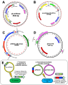
Figure 1
Figure 1
Transposon mutagenesis and recombinase-mediated cassette exchange in Rickettsia parkeri. (A,B) pLoxHimar plasmids designed for transposon mutagenesis of rickettsiae with (A) mCherry and spectinomycin/streptomycin resistance or (B) GFPuv and rifampicin resistance. (C,D) Plasmids for (C) recombinase-mediated cassette exchange (RMCE) and (D) expression of the Cre recombinase. (E) Schematic showing the process of transposon mutagenesis followed by insertion replacement by RMCE.
Figure 2
Figure 2
Comparison of murine infection with Rickettsia parkeri transposon mutants and wild type. (A) Details of disrupted genes in R. parkeri mutants and weight change in C3H/HeJ mice 7 days post infection (n = 2 mice/infection). Insertion site refers to position in R. parkeri Tate’s Hell genome. (B) Tissue load of R. parkeri mutants and wild type at day 7 as determined by qPCR quantification of gltA.
Figure 3
Figure 3
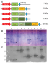
Expression of epitope arrays in Escherichia coli. (A) Diagram showing arrangement and expected molecular mass of different epitope arrays. (B,C) Expression of epitope arrays in E. coli BL21(DE3). Extracts from induced and uninduced cultures were separated by SDS-PAGE and stained with Coomassie (B) or probed with anti-6HisTag-HRP conjugate (C). 1. YchF uninduced; 2. YchF induced; 3. YchF-Asp55-Asp62-virB9 uninduced; 4. YchF-Asp55-Asp62-virB9 induced; 5. YchF-virB9 uninduced; 6. YchFvirB9 induced; 7. YchF-Asp55 uninduced; 8. YchF-Asp55 induced; 9. YchF-Asp62 uninduced; 10. YchF-Asp62 induced; 11. Untransformed BL21(DE3) control. Asterisks mark the expected band size for each array.
Figure 4
Figure 4
Challenge of epitope-immunized C3H/HeJ mice with A. phagocytophilum or R. parkeri. Mice (2 per group) were immunized with purified epitope protein produced in E. coli. Two injections of 50 μg were given 4 weeks apart, and pathogen challenge was performed 19 days after the booster dose. (A) Average weights of each mouse group following challenge with A. phagocytophilum (groups 1–5) or R. parkeri (group 6). (B,C) qPCR quantification of A. phagocytophilum (B) and R. parkeri (C) in tissues of challenged mice on day 8 post infection. Means were compared using a two-way ANOVA with Dunnett’s multiple comparisons test, * indicates p < 0.05.
Figure 5
Figure 5
Detection of mKate-epitope fusion proteins expressed from pRAM18dSGK shuttle vectors in transformed WT R. parkeri. (AC) Western blots of protein extracted from R. parkeri transformed with various iterations of epitope cassettes in pRAM18 shuttle vectors. Promoter OmpA or OmpB is shown above the constructs in A and B, whilst all transformants in C used the OmpA promoter. Western blotting against GFPuv confirmed successful transformation with shuttle vectors (A). mKate expression was detected in R. parkeri transformed with plasmids containing [virB9-mKate] and [Asp62-mKate], but not [Asp55-Asp62-virB9-mKate] or [Asp62-virB9-mKate] (B,C). Asterisks mark the expected size of mKate-epitope fusion proteins. Numbers on left of blots indicate protein size in kDa; expected size of GFPuv is 27 kDa, virB9-mKate and Asp62-mKate 33 kDa, Asp62-virB9-mKate 37 kDa, and Asp55-Asp62-virB9-mKate 46 kDa. In (C) P = pellet and S = supernatant, and the positive control is R. parkeri expressing mKate only from pRAM18dSFA. (D,E) Fluorescent microscopy of R. parkeri-infected ISE6 cells gave similar results with mKate visible in pRAM18dSGK[OmpA-virB9-mKate]-transformed rickettsiae (D) but not in those transformed with pRAM18dSGK[OmpA-Asp55-Asp62-virB9-mKate] (E).
Figure 6
Figure 6
Detection of mKate-epitope fusion proteins in the R. parkeri G8::lox mutant. (A) Schematic showing recombinase-mediated cassette exchange in the G8::lox mutant. RMCE results in the replacement of the GFPuv/rif intragenic transposon insertion in RPATATE_1142 with a cassette containing the epitope, mKate, and spectinomycin/streptomycin (“Spec”) resistance sequences. Unlabeled red arrows indicate location of promoters (see Figure 1). (B) Western blot detection of GFPuv and mKate in G8::lox mutants transformed with pRAM18dSGK shuttle vectors containing mKate and epitope sequences and in G8::lox mutants that have undergone RMCE to replace GFPuv/rif transposon with mKate-epitope sequence. Abbreviations used for epitopes: A55 = Asp55; A62 = Asp62; vB9 = virB9. Asterisks mark the expected size of mKate-epitope fusion proteins. (C) RT-PCR of DNA/RNA extracts from R. parkeri G8::lox[virB9-mKate] with primers to virB9 and virB9-mKate. NTC = no template control; noRT = control without reverse transcriptase added; RNA = RNA with reverse transcriptase; DNA = positive control using DNA. (DF) Fluorescent microscopy of ISE6 cells infected with R. parkeri G8::lox expressing GFPuv and mKate from pRAM18dSGK plasmid. mKate-epitope fusion protein was expressed in rickettsiae transformed with plasmids containing virB9-mKate (D) and Asp62-mKate (E) sequences but not in those containing Asp55-mKate sequences (F). (G) Fluorescent microscopy of R. parkeri G8::lox mutant after RMCE, showing replacement of GFPuv-containing insertion with virB9-mKate cassette.
Figure 7
Figure 7
Outline of mouse vaccination-challenge experiment.
Figure 8
Figure 8
Mouse responses to pathogen challenge following vaccination with R. parkeri G8::lox[virB9-mKate]. (A,B) Weights of C3H/HeJ mice challenged with (A) R. parkeri or (B) A. phagocytophilum. (C) Spleen weights of challenged mice. Means were compared using a one-way ANOVA with Tukey’s multiple comparisons test, * indicates p < 0.05. Vaccination status is denoted by color (black = unvaccinated control; green = vaccinated; blue = boosted) and pathogen challenge is shown by shape (circle = unchallenged control; triangle = R. parkeri; square = A. phagocytophilum).
Figure 9
Figure 9
Tissue load of pathogens in challenged mice vaccinated with R. parkeri G8::lox[virB9-mKate]. Copy numbers of (A) R. parkeri and (B) A. phagocytophilum in tissues of challenged mice. Means were compared using a one-way ANOVA with Tukey’s multiple comparisons test: ns = not significant; * p < 0.05; ** p < 0.01; **** p <0.0001.
Figure 10
Figure 10
Analysis of response to infection with A. phagocytophilum and R. parkeri in mice vaccinated with R. parkeri G8::lox[virB9-mKate]. (A) Giemsa results from blood cultures of A. phagocytophilum-challenged mice. Data were analyzed by two-way ANOVA: No significant difference by vaccination status, p < 0.0001 by time. (B) PER1/2 PCR results from day 11 blood cultures of A. phagocytophilum-challenged mice. Expected band size is 451 bp. A, B, and C are individual mice from each group. (C) IFA on R. parkeri-infected ISE6 cells using sera from unvaccinated, unchallenged mice (negative control), sera taken from mice 3 dpi with WT R. parkeri (Rp-exposed), and sera from vaccinated and boosted mice (taken on day 35). 40× magnification. (D) IFA on A. phagocytophilum-infected HL60 cells using sera from unvaccinated, unchallenged mice (negative control), sera taken from mice 7 dpi with WT A. phagocytophilum (Ap-exposed), and sera from vaccinated and boosted mice (taken on day 35). 40× magnification. (E) ELISA against wells coated with A. phagocytophilum, WT R. parkeri, or R. parkeri G8::lox[virB9-mKate] and probed with serum from five groups of mice (listed on x-axis). Well contents are indicated by color of bars. Dotted line represents the positive cut-off value calculated as mean + 3 standard deviations of the negative control. Absorbance was adjusted to a blank well containing no bacteria to which no sera were added.

Similar articles

References

    1. Blanton L.S. The Rickettsioses: A Practical Update. Infect. Dis. Clin. N. Am. 2019;33:213–229. doi: 10.1016/j.idc.2018.10.010. - DOI - PMC - PubMed
    1. Binder A.M., Nichols Heitman K., Drexler N.A. Diagnostic Methods Used to Classify Confirmed and Probable Cases of Spotted Fever Rickettsioses—United States, 2010–2015. MMWR Morb. Mortal. Wkly. Rep. 2019;68:243–246. doi: 10.15585/mmwr.mm6810a3. - DOI - PMC - PubMed
    1. Osterloh A. The Neglected Challenge: Vaccination against Rickettsiae. PLoS Negl. Trop. Dis. 2020;14:e0008704. doi: 10.1371/journal.pntd.0008704. - DOI - PMC - PubMed
    1. Qin A., Tucker A.M., Hines A., Wood D.O. Transposon Mutagenesis of the Obligate Intracellular Pathogen Rickettsia prowazekii. Appl. Environ. Microbiol. 2004;70:2816–2822. doi: 10.1128/AEM.70.5.2816-2822.2004. - DOI - PMC - PubMed
    1. Noriea N.F., Clark T.R., Hackstadt T. Targeted Knockout of the Rickettsia rickettsii OmpA Surface Antigen Does Not Diminish Virulence in a Mammalian Model System. mBio. 2015;6:e00323-15. doi: 10.1128/mBio.00323-15. - DOI - PMC - PubMed

LinkOut - more resources