. 2025 Jan 22;17(2):143.
doi: 10.3390/v17020143.

SARS-CoV-2 Impairs Osteoblast Differentiation Through Spike Glycoprotein and Cytokine Dysregulation

Affiliations

SARS-CoV-2 Impairs Osteoblast Differentiation Through Spike Glycoprotein and Cytokine Dysregulation

Rosa Nicole Freiberger et al. Viruses. .

Abstract

Pulmonary and extrapulmonary manifestations have been reported following infection with SARS-CoV-2, the causative agent of COVID-19. The virus persists in multiple organs due to its tropism for various tissues, including the skeletal system. This study investigates the effects of SARS-CoV-2 infection, including both ancestral and Omicron viral strains, on differentiating mesenchymal stem cells (MSCs), the precursor cells, into osteoblasts. Although both viral strains can productively infect osteoblasts, precursor cell infection remained abortive. Viral exposure during osteoblast differentiation demonstrates that both variants inhibit mineral and organic matrix deposition. This is accompanied by reduced expression of runt-related transcription factor 2 (RUNX2) and increased levels of interleukin-6 (IL-6), a cytokine that negatively regulates osteoblast differentiation. Furthermore, the upregulation of receptor activator of nuclear factor kappa B ligand (RANKL) strongly suggests that the ancestral and Omicron variants may disrupt bone homeostasis by promoting osteoclast differentiation, ultimately leading to the formation of bone-resorbing cells. This process is dependent of spike glycoprotein since its neutralization significantly reduced the effect of infective SARS-CoV-2 and UV-C inactivated virus. This study underscores the capacity of ancestral and Omicron SARS-CoV-2 variants to disrupt osteoblast differentiation, a process essential for preserving the homeostasis and functionality of bone tissue.

Keywords: COVID-19; SARS-CoV-2; bone; osteoblasts.


Conflict of interest statement

The authors declare no conflicts of interest. The funders had no role in the design of this study; in the collection, analyses, or interpretation of data; in the writing of the manuscript; or in the decision to publish the results.

Figures
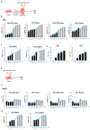
Figure 1
Figure 1
SARS-CoV-2 replication kinetics. (A) Experimental timeline schedule for differentiated osteoblasts (OBs). (B) Kinetics of SARS-CoV-2 replication in differentiated osteoblasts infected with the ancestral strain (Wh; left) and Omicron strain (BA.5; right) at a multiplicity of infection (MOI) of 0.1 (black bars) and 1 (shadowed bars), measured by mRNA levels of ORF1ab and nucleocapsid (N) in culture supernatants via RT-qPCR. (C) Kinetics of transcription of SARS-CoV-2 subgenomics (sg) RNA in differentiated osteoblasts infected with the ancestral strain (Wh; left) and Omicron strain (BA.5; right) at a multiplicity of infection (MOI) of 0.1 and 1, measured by mRNA levels of nucleocapsid 2 (N2). (D) SARS-CoV-2 titration (Wh and BA.5 strains) in culture supernatants from differentiated osteoblasts, reported as the number of plaque-forming units per mL (PFU/mL). (E) Experimental timeline schedule for mesenchymal stem cells (MSCs). (F) Kinetics of SARS-CoV-2 replication in mesenchymal stem cells (MSCs), measured as described in panel B. (G) Kinetics of transcription of SARS-CoV-2 subgenomics (sg) RNA in mesenchymal stem cells (MSCs) as described for panel C. dpi: days postinfection. Data are expressed as mean ± SD from four independent experiments.
Figure 2
Figure 2
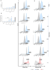
ACE2 expression in mesenchymal stem cells (MSCs) and osteoblasts (OBs). (A) Experimental timeline. (B) Analysis of ACE2 surface expression in mesenchymal stem cells (MSCs) and differentiated osteoblasts (OBs) using flow cytometry. (C) Representative flow cytometry histograms illustrating ACE2 surface expression, as described in panel B. Data are expressed as mean ± SD from three independent experiments.
Figure 3
Figure 3
Effect of Wh and BA.5 SARS-CoV-2 variants on osteoblast differentiation. Mesenchymal stem cells (MSCs) were infected with the ancestral strain (Wh) or the Omicron strain (BA.5) at a multiplicity of infection (MOI) of 0.1 or 1, and then cultured in osteoblast differentiation medium. (A) Representative microscopy images showing alkaline phosphatase (ALP) activity detected by BCIP-NBT substrate deposition, calcium (Ca) deposition visualized by Alizarin Red S staining, and collagen (Col) deposition identified by Sirius Red staining at 14 days post-infection (dpi) (upper set of images) and 21 dpi (lower set of images). (BD) Spectrophotometric quantification of ALP activity (B), calcium deposition (C), and collagen deposition (D). NI: noninfected (fully differentiated positive control); dpi: days post-infection. Ten microscopic fields per condition were quantified for each experiment. Scale bar: 100 µm. Data are expressed as mean ± SD from three independent experiments. * p < 0.01, ** p < 0.005, *** p < 0.0005, **** p < 0.0001 vs. NI.
Figure 4
Figure 4
Effects of SARS-CoV-2 on cell viability and mROS production. (A) Assessment of cell viability, shown as the percentage of cells stained with Ghost Dye Violet450, following infection with the ancestral SARS-CoV-2 variant (Wh) at a MOI of 1, in MSCs and at four distinct time points: 1, 7, 14, and 21 days post-infection (dpi) during osteoblast differentiation. (B) Quantification of mitochondrial reactive oxygen species (mROS) production, expressed as the percentage of cells stained with MitoSOX Red, measured by flow cytometry in MSCs and at the same time points during osteoblast differentiation. (C) Representative flow cytometry histograms of Ghost Dye staining at the time points indicated in panel A. (D) Representative flow cytometry histograms of MitoSOX staining, illustrating mROS levels at the time points indicated in panel B. NI: noninfected cells. dpi: days post-infection. mROS: mitochondrial reactive oxygen species. SARS-CoV-2: severe acute respiratory syndrome coronavirus 2. MSCs: mesenchymal stem cells. NI: non-infected. C+: positive control. Data are expressed as mean ± SD from three independent experiments. ** p < 0.005 vs. NI, **** p < 0.0001.
Figure 5
Figure 5
SARS-CoV-2 modulates RUNX2, PPARγ, IFNβ1, IL-6, and RANKL expression during osteoblast differentiation. Mesenchymal stem cells (MSCs) were infected with the ancestral strain (Wh) or the Omicron strain (BA.5) at a multiplicity of infection (MOI) of 0.1 or 1, and then cultured in osteoblast differentiation medium. The transcription levels of RUNX2, PPARγ, IFNβ1, and RANKL were assessed by RT-qPCR at 1 and 21 dpi. IL-6 expression was measured in culture supernatants by ELISA at 1, 7, and 21 dpi. Panels show the following: RUNX2 (A), PPARγ (B), PPARγ/RUNX2 ratio (C), IFNβ1 (D), IL-6 (E), and RANKL (F). NI: noninfected; dpi: days post-infection. Data are expressed as mean ± SD from three independent experiments. * p < 0.01, ** p < 0.005, *** p < 0.0005, **** p < 0.0001 vs. NI.
Figure 6
Figure 6
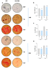
UV-inactivated SARS-CoV-2 inhibits osteoblast differentiation. Effect of UV-inactivated SARS-CoV-2 Wh strain on osteoblast differentiation. (A) Representative microscopy images showing alkaline phosphatase (ALP) activity (visualized by BCIP-NTB substrate deposition), calcium deposition (Ca, stained with Alizarin Red S), and collagen deposition (Col, stained with Sirius Red) at 14 and 21 days post-infection (dpi). (BD) Spectrophotometric quantification of ALP activity (B), calcium deposition (C), and collagen deposition (D). NI: noninfected; d: days post-stimulation. Ten microscopic fields per condition were quantified for each experiment. Scale bar: 100 µm. Data are expressed as mean ± SD from three independent experiments. * p < 0.01, ** p < 0.005, *** p < 0.0005 vs. NI.
Figure 7
Figure 7
The Spike (S) glycoprotein inhibits osteoblast differentiation. Mesenchymal stem cells (MSCs) were infected with the ancestral variant (Wh) or UV-C-inactivated virus at a multiplicity of infection (MOI) of 0.1. Infectious virus or UV-C-inactivated virus was pre-incubated for 1 h at 37 °C with an anti-spike glycoprotein antibody (anti-S) or an anti-snakebite antibody as a control (anti-SB). The cells were then cultured in osteoblast differentiation medium. (A) Representative microscopy images showing calcium deposition (stained with Alizarin Red S) and collagen deposition (stained with Sirius Red) at 21 days post-infection (dpi). (B,C) Spectrophotometric quantification of calcium deposition (B) and collagen deposition (C). NI: non-infected; NT: non-treated. Ten microscopic fields per condition were quantified for each experiment. Scale bar: 100 µm. Data are expressed as mean ± SD from three independent experiments. * p < 0.01, ** p < 0.005, *** p < 0.0005, vs. NI.
Figure 8
Figure 8
Effect of Wh and BA.5 SARS-CoV-2 strains on adipocyte differentiation. Mesenchymal stem cells (MSCs) were infected with the ancestral strain (Wh) or the Omicron strain (BA.5) at a multiplicity of infection (MOI) of 0.1 or 1, and then cultured in adipocyte differentiation medium. (A) Representative images of lipid droplets stained with Bodipy 493/503 at 7 days post-differentiation. (B,C) Quantification of the experiment shown in panel A: number of cells per field (B) and percentage of adipocytes per field (C). (D) PPARγ expression was measured by RT-qPCR at 1 and 7 days post-infection (dpi). NI: non-infected; dpi: days post-infection. Ten microscopic fields per condition were quantified for each experiment. Scale bar: 50 µm. Data are expressed as mean ± SD from three independent experiments.

References

    1. Guan W.J., Ni Z.Y., Hu Y., Liang W.H., Ou C.Q., He J.X., Liu L., Shan H., Lei C.L., Hui D.S.C., et al. Clinical Characteristics of Coronavirus Disease 2019 in China. N. Engl. J. Med. 2020;382:1708–1720. doi: 10.1056/NEJMoa2002032. - DOI - PMC - PubMed
    1. Hill P.A. Bone remodelling. Br. J. Orthod. 1998;25:101–107. doi: 10.1093/ortho/25.2.101. - DOI - PubMed
    1. Buckwalter J.A., Cooper R.R. Bone structure and function. Instr. Course Lect. 1987;36:27–48. - PubMed
    1. Vimalraj S. Alkaline phosphatase: Structure, expression and its function in bone mineralization. Gene. 2020;754:144855. doi: 10.1016/j.gene.2020.144855. - DOI - PubMed
    1. Mohamed A.M. An overview of bone cells and their regulating factors of differentiation. Malays. J. Med. Sci. 2008;15:4–12. - PMC - PubMed

Publication types

LinkOut - more resources