Review
. 2025 Feb 21:12:rbae137.
doi: 10.1093/rb/rbae137. eCollection 2025.

Biomaterials for neuroengineering: applications and challenges

Affiliations
Review

Biomaterials for neuroengineering: applications and challenges

Huanghui Wu et al. Regen Biomater. .

Abstract

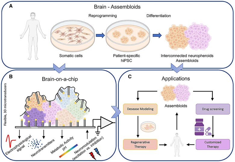
Neurological injuries and diseases are a leading cause of disability worldwide, underscoring the urgent need for effective therapies. Neural regaining and enhancement therapies are seen as the most promising strategies for restoring neural function, offering hope for individuals affected by these conditions. Despite their promise, the path from animal research to clinical application is fraught with challenges. Neuroengineering, particularly through the use of biomaterials, has emerged as a key field that is paving the way for innovative solutions to these challenges. It seeks to understand and treat neurological disorders, unravel the nature of consciousness, and explore the mechanisms of memory and the brain's relationship with behavior, offering solutions for neural tissue engineering, neural interfaces and targeted drug delivery systems. These biomaterials, including both natural and synthetic types, are designed to replicate the cellular environment of the brain, thereby facilitating neural repair. This review aims to provide a comprehensive overview for biomaterials in neuroengineering, highlighting their application in neural functional regaining and enhancement across both basic research and clinical practice. It covers recent developments in biomaterial-based products, including 2D to 3D bioprinted scaffolds for cell and organoid culture, brain-on-a-chip systems, biomimetic electrodes and brain-computer interfaces. It also explores artificial synapses and neural networks, discussing their applications in modeling neural microenvironments for repair and regeneration, neural modulation and manipulation and the integration of traditional Chinese medicine. This review serves as a comprehensive guide to the role of biomaterials in advancing neuroengineering solutions, providing insights into the ongoing efforts to bridge the gap between innovation and clinical application.

Keywords: 3D printing; biomaterials; brain-on-a-chip; nanopattern; neuroengineering; organoid.


Figures

None
Graphical abstract
Figure 1.
Figure 1.
The application of biomaterials in neuroengineering. (A) Brain organoids: 3D printing has been applied to make human brain tissue. Matrigel helps grow these tissues. Specially treated mouse stem cells in a gel bead turn into nerve cells more effectively. (B) Organ-on-a-chip: the ‘brain on a chip’ combines brain-like tissue with tiny chips to mimic the human brain. It helps scientists’ study how the brain develops and reacts to diseases and drugs outside the body. (C) Neuromorphic devices: These devices copy how neurons and synapses work, creating artificial networks that can advance a new type of computing. (D) Biomimetic simulated inductive materials: Patterns on glass with gold are made by using a special process. Adding tiny particles and molecules to this pattern improves how well electrodes pick up nerve signals. These materials also mimic the body’s environment to help nerve cells grow. (E) BBB and sustained release drugs: advanced materials like nanomedicines and special gels are applied to help drugs get past the brain’s protective barrier and release slowly. (F) Human–computer interaction: This area focuses on using safe and smart materials to create new ways to diagnose and treat brain-related diseases. (Created with BioRender.com.)
Figure 2.
Figure 2.
The close relationship between biomaterials and neuroengineering. The close relationship between biomaterials and neuroengineering is a pivotal area of research, as it involves the development and application of biomaterials designed to interact with the nervous system. This interdisciplinary field is crucial for creating innovative solutions for neurological disorders and injuries, including (i) neural regeneration, stimulation, modulation and manipulation; (ii) brain-on-a-chip systems; (iii) brain–computer interface.
Figure 3.
Figure 3.
Brain-on-a-chip overview. (A) The reprogramming of human somatic cells into induced hiPSCs facilitates the differentiation into various neuronal types while preserving essential topological features of the brain, including its 3D architecture, adequate heterogeneity and modular connectivity. Consequently, this approach enables the in vitro cultivation of interconnected neurospheroids and assembloids. (B) Coupled to microtransducers, these systems are capable of recording the electrophysiological activity of the biological structures and monitoring other pertinent parameters, including neurotransmitter concentrations and fluctuations in metabolic activity. Furthermore, such devices should possess bidirectional functionality, enabling them to modulate electrophysiological activity through the application of either excitatory or inhibitory stimuli. (C) An accurate biological model of the brain, such as this, can be utilized not only for fundamental scientific research but also for drug screening, enabling the development of personalized and patient-specific therapies. Additionally, it facilitates the study of the pathogenesis of brain disorders in vitro, thereby aiding in the identification of potential therapeutic solutions. Reproduced from Ref. [108] with permission of Frontiers, © 2022.
Figure 4.
Figure 4.
Schematic diagram of 3D bioprinting and ‘BBB-on-a-chip’ systems. (A) 3D bioprinting technologies for neural tissue bioprinting. Reproduced from Ref. [105] with permission of AIP Publishing, © 2022. (B) The BBB model development and ‘BBB-on-chip’ design. Reproduced from Ref. [176] with permission of MDPI, © 2021. (C) Tree dimensional sketch of a porous PDMS membrane specifying the adopted terminology: pore size (PS), pore to pore distance (P-P), thickness (T) and area (W1 × W2). Reproduced from Ref. [181] with permission of Nature Publishing Group, © 2018. (D) Top view and 60°tilted representative SEM images of a UPP membrane with 3 µm pore size and 300 nm thickness. White arrows demonstrate the ultrathin thickness of the membrane (≈300 nm) (scale bar = 10 and 4 µm, respectively). Reproduced from Ref. [196] with permission of John Wiley and Sons, © 2020. (E) Brief procedure to micro-contact printing for fabricating microdot arrays. Reproduced from Ref. [212] with permission of Frontiers, © 2016.
Figure 5.
Figure 5.
Schematic diagram of micropatterning. (A) Micropatterning provides the opportunity to independently control various aspects of the cellular environment, including substrate composition, mechanical properties, geometry and topography. Certain micropatterning techniques are also capable of modulating these variables in a dynamic manner. Reproduced from Ref. [224] with permission of the Company of Biologists, © 2021. (B) Fabrication procedure of the RGD nanopattern using block copolymer micelle nanolithography. Reproduced from Ref. [241] with permission of Elsevier, © 2020. (C) Scheme of protocol for transferring carboxyl-rich micro-patterns onto glass coverslips. Reproduced from Ref. [229] with permission of Public Library of Science (PLoS), © 2019.
Figure 6.
Figure 6.
Schematic diagram and application of surface-modified materials. (A) The fabrication process for the preparation of the G-an and the detection of DA by the G-an. Reproduced from Ref. [245] with permission of Springer Nature, © 2015. (B) (i) Schematic diagram of the FGPC/AuNPs/acupuncture needle. (ii) Schematic diagram of real-time NO measurement in acupoint ST 36 stimulated by L-arginine. Reproduced from Ref. [249] with permission of Springer Nature, © 2017. (C) Schematic diagram of real time and in vivo monitoring of 5-HT by means of the PEDOT/CNTmodified acupuncture needle. Reproduced from Ref. [250] with permission of Springer Nature, © 2016. (D) Schematic representation of construction of nanocomposite hydrogel. Reproduced from Ref. [257] with permission of Springer Nature, © 2021.
Figure 7.
Figure 7.
The classification and their properties of hydrogels. Natural hydrogels, such as collagen, chitosan, cellulose, gelatine, hyaluronic acid and alginate, are derived from biological sources and are known for their biocompatibility and biodegradability. They exhibit porosity, which is beneficial for cell infiltration and nutrient exchange, and have swelling properties that can be controlled by their chemical structure. Synthetic hydrogels, including PEG, PIC, PAA and PVA-based materials, offer customizability, controlled degradation rates and a wide range of mechanical properties. They can be engineered to be responsive to various stimuli, such as temperature, pH and light, and provide reproducibility and scalability in production. The choice between natural and synthetic hydrogels depends on the specific requirements of the application, with natural hydrogels often preferred for their biocompatibility and biodegradability, while synthetic hydrogels offer more control over their properties and can be tailored to specific needs.
Figure 8.
Figure 8.
Schematic diagram of artificial synapses. (A) Types of 2D materials-based synaptic devices. Reproduced from Ref. [390] with permission of John Wiley and Sons, © 2021. (B) (i) Schematic of the CsBi3I10-based organic synaptic transistor. (ii) EPSC triggered by an optical pulse at different VDS varied from −0.01 to −1 V. Reproduced from Ref. [431] with permission of American Chemical Society, © 2021. (C) (i) Schematic diagram of dual-organic-transistor-based tactile-perception element (DOT-TPE). (ii) The relative changes in current for pressure-sensitive transistors under varying pressure conditions, as well as the corresponding postsynaptic current responses of the synaptic transistors, were investigated. Reproduced from Ref. [432] with permission of Whiley-VCH, © 2017.
Figure 9.
Figure 9.
The application and their characteristics of brain–computer interfaces. Brain–computer interfaces (BCIs) can be divided into three main categories: (1) Noninvasive BCI: This type can decode scalp EEG signals. However, it suffers from fast signal attenuation and difficulty in signal extraction. (2) Minimally Invasive BCI: This type decodes cortical surface EEG signals, which have a higher signal-to-noise ratio. (3) Invasive BCI: This type decodes intracortical EEG signals, offering advantages such as fast conduction and strong signals, but it carries a risk of infection.
Figure 10.
Figure 10.
Schematic diagram of nerve electrodes. (A) Schematic detailing the Utah MEA configuration and schematic detailing the Michigan MEA configuration. Reproduced from Ref. [468] with permission of Wiley-VCH, © 2019. (B) (i) Schematic illustration for fabrication of silver nanowire (AgNW)-based microelectrodes using a photolithographic process. (ii) SEM images of silver nanowire-based microelectrodes. Reproduced from Ref. [479] with permission of American Chemical Society, © 2014. (C) The next-generation neuro-nano interfaces. Reproduced from Ref. [471] with permission of John Wiley and Sons, © 2017.
Figure 11.
Figure 11.
The clinical application of nano-TCM. The clinical application of nano-TCM involves the integration of nanotechnology with TCM to enhance the therapeutic potential of TCM components. This includes using nanoparticles for targeted and controlled drug delivery, which can improve the bioavailability, solubility and stability of TCM extracts. Nanomedicine can also increase the absorption of medicinal components by altering their size and surface properties, leading to enhanced efficacy and reduced toxicity. Nano-acupoint applies extremely fine needles made from nanomaterials that can be used for acupoint stimulation. These nanoneedles can potentially penetrate the skin with less pain and with higher precision than conventional needles. Moreover, equipped with nanosensors, these needles can detect subtle changes in the skin’s electrical properties, temperature or the release of biochemical markers. This capability enables real-time monitoring of the body’s response to acupoint stimulation, providing valuable feedback that can guide the treatment process.

Similar articles

Cited by

References

    1. Braun J, Hurtak F, Wang-Chen S, Ramdya P. Descending networks transform command signals into population motor control. Nature 2024;630:686–94. - PMC - PubMed
    1. Fu X, Hu Z, Li W, Ma L, Chen J, Liu M, Liu J, Hu S, Wang H, Huang Y, Tang G, Zhang B, Cai X, Wang Y, Li L, Ma J, Shi SH, Yin L, Zhang H, Li X, Sheng X. A silicon diode-based optoelectronic interface for bidirectional neural modulation. Proc Natl Acad Sci USA 2024;121:e2404164121. - PMC - PubMed
    1. Micera S, Menciassi A, Cianferotti L, Gruppioni E, Lionetti V. Organ neuroprosthetics: connecting transplanted and artificial organs with the nervous system. Adv Healthc Mater 2024;13:e2302896. - PubMed
    1. Gao Y, Yang Z, Li X. Regeneration strategies after the adult mammalian central nervous system injury-biomaterials. Regen Biomater 2016;3:115–22. - PMC - PubMed
    1. Vassiliadis P, Beanato E, Popa T, Windel F, Morishita T, Neufeld E, Duque J, Derosiere G, Wessel MJ, Hummel FC. Non-invasive stimulation of the human striatum disrupts reinforcement learning of motor skills. Nat Hum Behav 2024;8:1581–98. - PMC - PubMed

LinkOut - more resources