

Meibomian Gland Alterations in Keratoconus Patients After Corneal Cross-Linking
- PMID: 40007675
- PMCID: PMC11849734
- DOI: 10.14744/bej.2024.93695
Meibomian Gland Alterations in Keratoconus Patients After Corneal Cross-Linking
Abstract
Objectives: The objective of the study was to evaluate the changes in the meibomian glands (MGs) and ocular surface parameters after corneal cross-linking (CXL) in keratoconus patients.
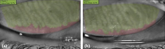
Methods: Forty-eight eyes of 48 keratoconus patients that underwent epi-off CXL were included in this prospective study. Upper and lower lid MGs were assessed with non-contact meibography at preoperatively, 1st, 3rd, 6th, and 12th month after CXL. Uncorrected distance visual acuity (UCVA), corrected distance visual acuity (BCVA), spherical equivalent (SE), and corneal tomography findings (K1, K2, Kmean, and Kmax) were recorded at each visit. Ocular surface staining score (Oxford grade), ocular surface disease index (OSDI) questionnaire, and non-invasive tear break-up time (NI-TBUT) were evaluated at preoperatively and 12 months after CXL.
Results: K1, K2, Kmean, and Kmax were decreased at post-operative 12th month compared to baseline (p=0.004, p<0.001, p<0.001, and p<0.001, respectively). UCVA, BCVA, and SE did not change between preoperatively and post-operative 12 months (p=0.142, p=0.306, and p=1.000, respectively). NI-TBUT showed similarity between pre-operative and 12 months values (p=0.180), while OSDI scores significantly decreased (p<0.001). MG loss in the upper and lower lids did not show significant difference compared to pre-operative values at any of the follow-up visits (p=0.121 and p=0.117, respectively).
Conclusion: CXL treatment did not significantly affect the NI-TBUT and MGs morphology, while improving ocular symptoms.
Keywords: Corneal cross-linking; Keratoconus; Meibography.
Conflict of interest statement
Conflict of Interest: None declared.
Figures
Similar articles
-
Evaluation of Corneal Parameters and Meibomian Gland Alterations After Corneal Cross-Linking in Patients With Progressive Keratoconus.Eye Contact Lens. 2023 Mar 1;49(3):110-115. doi: 10.1097/ICL.0000000000000964. Epub 2022 Dec 20. Eye Contact Lens. 2023. PMID: 36729083
-
Evaluation of tear parameters and meibomian gland morphology in keratoconus patients after epithelial-on corneal cross-linking.Eur J Ophthalmol. 2022 Aug 5:11206721221118740. doi: 10.1177/11206721221118740. Online ahead of print. Eur J Ophthalmol. 2022. PMID: 35929885
-
[Topography-guided transepithelial corneal collagen cross-linking by sequential ultraviolet A irradiation in different diameters for progressive keratoconus in adults].Zhonghua Yan Ke Za Zhi. 2023 Oct 11;59(10):791-804. doi: 10.3760/cma.j.cn112142-20221216-00642. Zhonghua Yan Ke Za Zhi. 2023. PMID: 37805413 Chinese.
-
Corneal collagen cross-linking: a review of 1-year outcomes.Eye Contact Lens. 2014 Nov;40(6):345-52. doi: 10.1097/ICL.0000000000000094. Eye Contact Lens. 2014. PMID: 25343263 Review.
-
Efficacy comparison of combining cross-linking and refractive laser ablation in progressive keratoconus: systematic review and meta-analysis.Can J Ophthalmol. 2024 Dec;59(6):e661-e672. doi: 10.1016/j.jcjo.2024.02.017. Epub 2024 Mar 18. Can J Ophthalmol. 2024. PMID: 38513713
References
-
- Wollensak G, Spoerl E, Seiler T. Riboflavin/ultraviolet-a-induced collagen crosslinking for the treatment of keratoconus. Am J Ophthalmol. 2003;135:620–7. - PubMed
-
- Mazzotta C, Hafezi F, Kymionis G, Caragiuli S, Jacob S, Traversi C, et al. In vivo confocal microscopy after corneal collagen crosslinking. Ocul Surf. 2015;13:298–314. - PubMed
-
- Jordan C, Patel DV, Abeysekera N, McGhee CN. In vivo confocal microscopy analyses of corneal microstructural changes in a prospective study of collagen cross-linking in keratoconus. Ophthalmology. 2014;121:469–74. - PubMed
-
- Kymionis GD, Diakonis VF, Kalyvianaki M, Portaliou D, Siganos C, Kozobolis VP, et al. One-year follow-up of corneal confocal microscopy after corneal cross-linking in patients with post laser in situ keratosmileusis ectasia and keratoconus. Am J Ophthalmol. 2009;147:774–8. 778.e1. - PubMed
-
- Recalde JI, Acera A, Rodríguez-Agirretxe I, Sánchez-Tena MA, San-Cristóbal J, Durán JA. Ocular surface disease parameters after collagen cross-linking for keratoconus. Cornea. 2017;36:148–52. - PubMed
LinkOut - more resources
Full Text Sources
Miscellaneous