. 2025 Feb 21:20:2305-2322.
doi: 10.2147/IJN.S502850. eCollection 2025.

Dual-Responsive Methotrexate-Human Serum Albumin Complex-Encapsulated Liposomes for Targeted and Enhanced Atherosclerosis Therapy

Affiliations

Dual-Responsive Methotrexate-Human Serum Albumin Complex-Encapsulated Liposomes for Targeted and Enhanced Atherosclerosis Therapy

Xueqin Wang et al. Int J Nanomedicine. .

Abstract

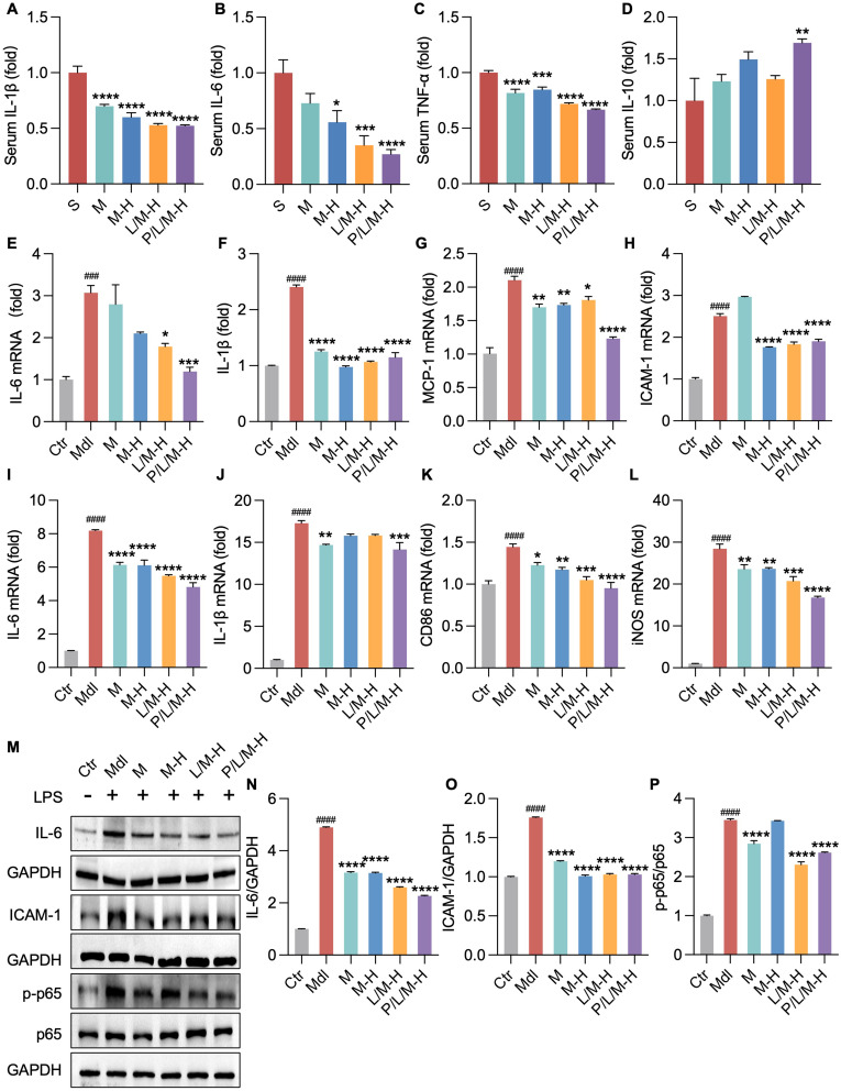
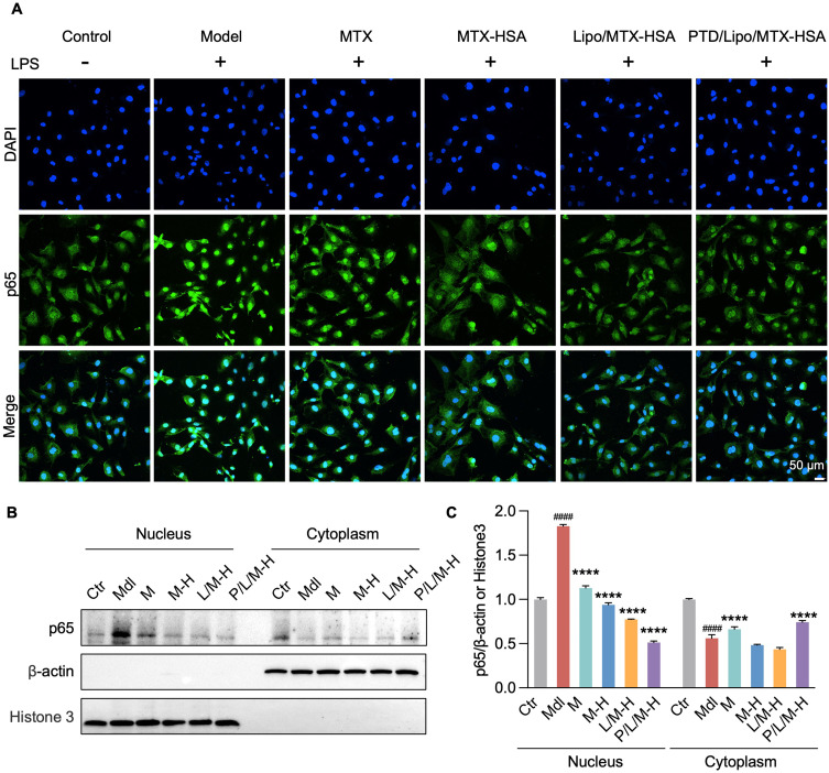
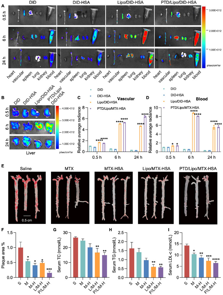
Introduction: In plaque sites of atherosclerosis (AS), the physiological barrier caused by the thick fiber cap due to the overmigration of vascular smooth muscle cells (VSMCs) prevents efficient drug delivery to damaged macrophages. How to ensure precise targeted delivery of drugs to plaque sites and their on-demand release to dysfunctional cells under the thick fibrous cap are feasible solutions to enhance AS treatment.

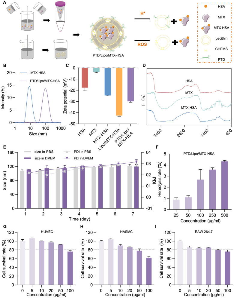
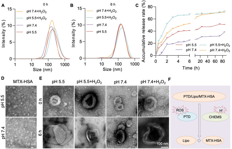
Methods: A small complex of methotrexate (MTX)-human serum albumin (HSA) with strong, thick fibrous cap penetration ability was encapsulated in a cholesterol hemisuccinate (CHEM) prepared pH-sensitive liposome, modifying with ROS-responsive PEG2000-TK-DSPE (PTD), termed PTD/Lipo/MTX-HSA.

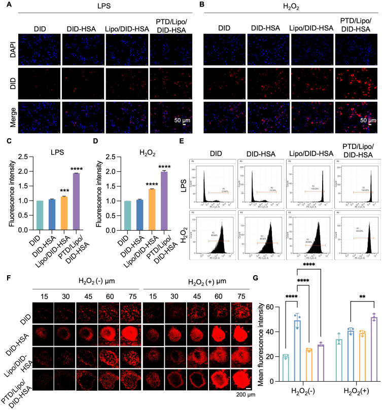
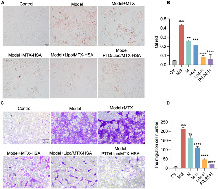
Results: PTD/Lipo/MTX-HSA can achieve precise targeting and on-demand release in response to plaques environments of AS. The designed formulation accelerated the release of the small-sized MTX-HSA complex in response to excess ROS and acidic pH conditions, and it better penetrated the macrophage spheroids. Furthermore, it has precise targeting ability in the AS mouse model and can produce good anti-inflammatory efficacy by inhibiting p65 entry into the nucleus turn out inflammatory factor.

Conclusion: Our formulations work with safety in mind, and it also highlights the potential of precisely targeted and on-demand-released dual-responsive smart nanoplatforms as promising therapeutic options to penetrate deeper plaques for the effective treatment of AS.

Keywords: dual-responsive smart liposomes; methotrexate-human serum albumin complex; penetration of deeper plaques.


Conflict of interest statement

The authors have declared no conflicts of interest in this work.

Figures

Figure 1
Figure 1
Characterization of the nanomaterials. (A). PTD/Lipo/MTX-HSA fabrication and pattern plots; (B) MTX-HSA and PTD/Lipo/MTX-HSA particle size plots; (C) Zeta potential of PTD/Lipo/MTX-HSA; (D) FT-IR spectra of HSA, MTX and MTX-HSA; (E) PTD/Lipo/MTX-HSA sequential PDI and size distributions in PBS and DMEM containing 10% FBS for 7 days; (F) Hemolysis plots of PTD/Lipo/MTX-HSA. (G–I) Results of the CCK-8 assay of PTD/Lipo/MTX-HSA-treated HUVECs, HASMCs and Raw264.7 cells.
Figure 2
Figure 2
Dual responsiveness of nanoparticles to pH and ROS. (A) Particle size of PTD/Lipo/MTX-HSA under different conditions at 0 h; (B) Particle size of PTD/Lipo/MTX-HSA under different conditions at 6 h; (C) Drug release rate of PTD/Lipo/MTX-HSA under different conditions; (D) TEM of MTX-HSA at pH 5.5 and pH 7.4 (scale 10 nm); (E) TEM of PTD/Lipo/MTX-HSA at pH 5.5 and pH 7.4 with/without 1 mm H2O2 at 0 and 6 h (scale 100 nm); (F) Schematic diagrams of different environments.
Figure 3
Figure 3
In vitro targeting of nanomaterials. (A and B) are the confocal microscopy results of DID, DID-HSA, Lipo/DID-HSA and PTD/Lipo/DID-HSA in the presence of LPS or H2O2, respectively (scale 50 μm); (C) Fluorescence statistics for A; (D) Fluorescence statistics for B; (E) Flow cytometry results of DID, DID-HSA, Lipo/DID-HSA and PTD/Lipo/DID-HSA in the presence of LPS or H2O2, respectively; (F) Fluorescence penetration efficiency of different formulations in Raw264.7 cell spheroids at 8 h (scale 200 μm); (G) The fluorescence statistics at 75 μm of F. (***<0.001 vs DID; ****<0.0001 vs DID).
Figure 4
Figure 4
Antilipid uptake effects of the nanomaterials in vitro. (A) The control, model (ox-LDL), ox-LDL+MTX, ox-LDL+MTX-HSA, ox-LDL+Lipo/MTX-HSA and ox-LDL+PTD/Lipo/MTX-HSA preparations were treated with HASMCs, and white light imaging of oil red O staining was performed (scale 20 μm); (B) is a statistical graph of A; (C) The control, model (LPS), LPS+MTX, LPS+MTX, LPS+MTX-HSA, LPS+Lipo/MTX-HSA and LPS+PTD/Lipo/MTX-HSA groups were treated with HASMCs and stained with crystal violet. Images were taken under an optical microscope (scale 50 μm). (D) is a statistical graph of C. (Ctr, control; Mdl, model (LPS/ox-LDL); M, model+MTX; M-H, model+MTX-HSA; L/M-H, model+Lipo/MTX-HSA; P/L/M-H, model+PTD/Lipo/MTX-HSA; ####<0.0001 vs control; *<0.05; **<0.01; ***<0.001; ****<0.0001 vs Model).
Figure 5
Figure 5
Anti-inflammatory effects of the nanomaterials in vitro. (A), (B), (C) and (D) are the ELISA results for serum IL-1β, IL-6, TNF-α and IL-10, respectively. (S, saline; M, MTX; M-H, MTX-HSA; L/M-H, Lipo/MTX-HSA; P/L/M-H, PTD/Lipo/MTX-HSA; *<0.05; **<0.01; ***<0.001; ****<0.0001 vs saline); E, F, G, H are the PCR results for the control, model (LPS), LPS+MTX, LPS+MTX-HSA, LPS+Lipo/MTX-HSA and LPS+PTD/Lipo/MTX-HSA-treated HUVECs ((E), IL-6; (F), IL-1β; (G), MCP-1; (H), ICAM-1); (IL) are the PCR results for the control, LPS, LPS+MTX, LPS+MTX-HSA, LPS+Lipo/MTX-HSA and LPS+PTD/Lipo/MTX-HSA-treated Raw264.7 cells ((I), IL-6 (J), IL-1β; (K), CD86; (L), iNOS); (M) shows the WB results; (N) (IL-6), (O) (ICAM-1) and (P) (p-p65/p65) are the statistical plots of I. (Ctr, control; Mdl, model; M, model+MTX; M-H, model+MTX-HSA; L/M-H, model+Lipo/MTX-HSA; P/L/M-H, model+PTD/Lipo/MTX-HSA; ###<0.001; ####<0.0001 vs control; *<0.05; **<0.01; ***<0.001; ****<0.0001 vs Model).
Figure 6
Figure 6
Anti-inflammatory pathway of nanomaterials in vitro. (A) Control, model (LPS), LPS+MTX, LPS+MTX-HSA, LPS+Lipo/MTX-HSA, and LPS+PTD/Lipo/MTX-HSA groups for immunofluorescence imaging; DAPI is shown in blue; p65 is shown in green; merge is the combination of the two (scale 50 μm); (B) shows the WB results; (C) is the statistical plot of B. (Ctr, control; Mdl, model; M, model+MTX; M-H, model+MTX-HSA; L/M-H, model+Lipo/MTX-HSA; P/L/M-H, model+PTD/Lipo/MTX-HSA; ####<0.0001 vs the control; ****<0.0001 vs model).
Figure 7
Figure 7
In vivo targeting and antilipidemic effects of the nanomaterials. (A). In vivo heart, vascular, spleen, lung, kidney and blood imaging thermograms of DID, DID-HSA, Lipo/DID-HSA and PTD/Lipo/DID-HSA at 0.5, 6 and 24 h. (B) In vivo liver imaging thermograms of DID, DID-HSA, Lipo/DID-HSA and PTD/Lipo/DID-HSA at 0.5, 6 and 24 h. (C and D). Statistical graphs of vascular and blood images. (*<0.05 vs DID; ****<0.0001 vs DID); (E) Oil red O staining of whole sections after treatment with saline, MTX, MTX-HSA, Lipo/MTX-HSA and PTD/Lipo/MTX-HSA (scale 0.5 cm); (F) Statistical diagram of vascular plaques; (G), (H) and (I) are the serum TC, TG and LDL-c results of in vivo experiments with saline, MTX, MTX-HSA, Lipo/MTX-HSA and PTD/Lipo/MTX-HSA preparations, respectively (S, saline; M, MTX; M-H, MTX-HSA; L/M-H, Lipo/MTX-HSA; P/L/M-H, PTD/Lipo/MTX-HSA; *<0.05; **<0.01; ***<0.001; ****<0.0001 vs saline).

Similar articles

References

    1. Yuan J, Li L, Yang Q, et al. Targeted treatment of ischemic stroke by bioactive nanoparticle-derived reactive oxygen species responsive and inflammation-resolving nanotherapies. ACS Nano. 2021;15:16076. - PubMed
    1. Hu R, Dai C, Dong C, et al. Living Macrophage-Delivered Tetrapod PdH Nanoenzyme for Targeted Atherosclerosis Management by ROS Scavenging, Hydrogen Anti-inflammation, and Autophagy Activation. ACS Nano. 2022;16(10):15959. doi:10.1021/acsnano.2c03422 - DOI - PubMed
    1. Jain T, Nikolopoulou EA, Xu Q, Qu A. Hypoxia inducible factor as a therapeutic target for atherosclerosis. Pharmacol Ther. 2018;183:22. doi:10.1016/j.pharmthera.2017.09.003 - DOI - PubMed
    1. Pan H, Ho SE, Xue C, et al. Atherosclerosis is a smooth muscle cell–driven tumor-like disease. Circulation. 2024;2024:1. - PMC - PubMed
    1. Bennett MR, Sinha S, Owens GK. Vascular Smooth Muscle Cells in Atherosclerosis. Circ Res. 2016;118(4):692. doi:10.1161/CIRCRESAHA.115.306361 - DOI - PMC - PubMed

MeSH terms