

Primary leiomyosarcoma of the scalp: a case report and review of the literature
- PMID: 40007995
- PMCID: PMC11850309
- DOI: 10.3389/fonc.2025.1533114
Primary leiomyosarcoma of the scalp: a case report and review of the literature
Abstract
Background and importance: Leiomyosarcoma is a rare and aggressive malignant tumor with a high potential for relapse and metastasis. Correct and timely diagnosis is critical for effective treatment, yet it is often challenging due to the diverse clinical presentations. This case report highlights the significance of early identification and the consequences of delayed diagnosis in scalp leiomyosarcoma.
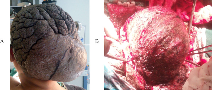
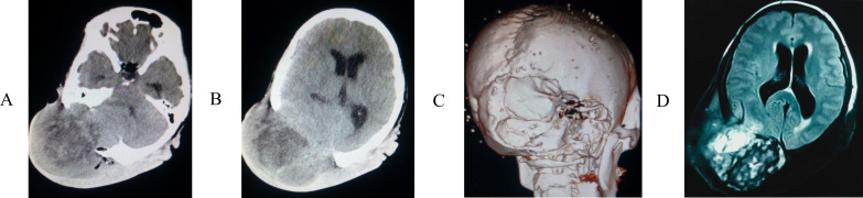
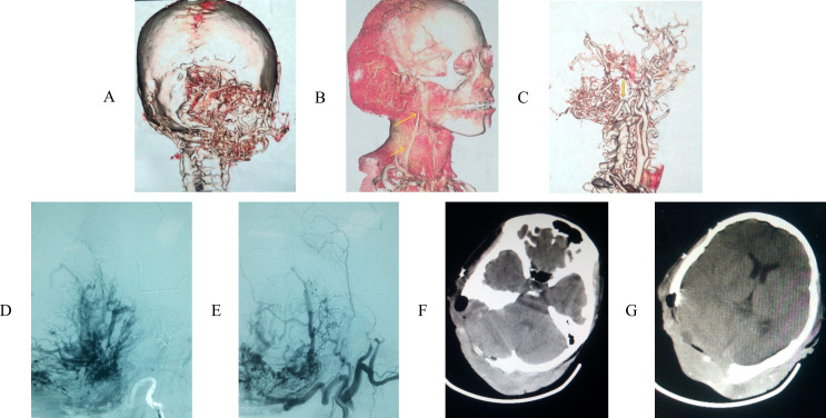
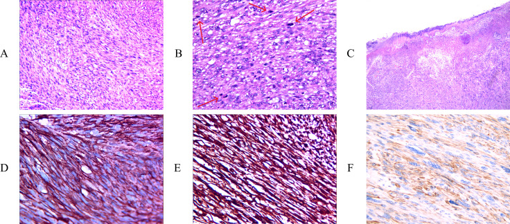
Clinical presentation: We present the case of a 39-year-old woman with a scalp neoplasm. Initially, the diagnosis was missed, leading to a delay in surgical intervention. The tumor demonstrated a locally aggressive course, infiltrating the skull and dura mater. Upon admission, the scalp tumor was promptly excised. This case provides valuable insights into the varied symptoms and presentations of scalp leiomyosarcoma, which can aid in the recognition of this condition.
Conclusion: This report underscores the importance of considering leiomyosarcoma in the differential diagnosis of scalp masses, particularly when the etiology is unclear. Early recognition and intervention are essential to prevent locally invasive growth and potential metastasis, emphasizing the need for a high index of suspicion among healthcare professionals.
Keywords: case report; leiomyosarcoma; sarcoma; scalp; surgery.
Copyright © 2025 Gao, Liu, Liu, Yang and Yang.
Conflict of interest statement
The authors declare that the research was conducted in the absence of any commercial or financial relationships that could be construed as a potential conflict of interest.
Figures
Similar articles
-
Skull metastasis from uterine leiomyosarcoma: a case report.Acta Neurol Taiwan. 2006 Jun;15(2):109-13. Acta Neurol Taiwan. 2006. PMID: 16871898
-
Cutaneous scalp metastases from retroperitoneal leiomyosarcoma: a case report.J Cutan Pathol. 2014 Aug;41(8):680-5. doi: 10.1111/cup.12329. Epub 2014 Apr 29. J Cutan Pathol. 2014. PMID: 24628578
-
Paraovarian leiomyosarcoma with scalp metastasis: a case report.Eur J Gynaecol Oncol. 2009;30(5):566-7. Eur J Gynaecol Oncol. 2009. PMID: 19899418
-
Leiomyosarcoma of the spermatic cord with scalp metastasis: case report and literature review.Coll Antropol. 2014 Jun;38(2):763-6. Coll Antropol. 2014. PMID: 25145020 Review.
-
Intractable Repeated Intracerebral Hemorrhage Due to Primary Dural Leiomyosarcoma: Case Report and Literature Review.World Neurosurg. 2019 Feb;122:116-122. doi: 10.1016/j.wneu.2018.10.132. Epub 2018 Nov 1. World Neurosurg. 2019. PMID: 30391601 Review.
References
-
- Workman AD, Farquhar DR, Brody RM, Parasher AK, Carey RM, Purkey MT, et al. . Leiomyosarcoma of the head and neck: A 17&8208;year single institution experience and review of the National Cancer Data Base. Head Neck. (2018) 40:756–62. - PubMed
Publication types
LinkOut - more resources
Full Text Sources