. 2025 Apr;25(4):e2400412.
doi: 10.1002/mabi.202400412. Epub 2025 Feb 26.

Evaluation of Electrospun Poly-4-Hydroxybutyrate as Biofunctional and Degradable Scaffold for Pelvic Organ Prolapse in a Vaginal Sheep Model

Affiliations

Evaluation of Electrospun Poly-4-Hydroxybutyrate as Biofunctional and Degradable Scaffold for Pelvic Organ Prolapse in a Vaginal Sheep Model

Krista L C van Rest et al. Macromol Biosci. 2025 Apr.

Abstract

Pelvic organ prolapse (POP) affects many women, especially after menopause. POP occurs due to the descent of weakened supportive tissue. Current prolapse surgeries have high failure rates, due to disturbed wound healing caused by lower tissue regeneration and estrogen depletion. Absorbable poly-4-hydroxybutyrate (P4HB) knit implants exhibited improved cell and tissue response leading to less complications from prolapse surgery. This study aims to enhance wound healing and improve surgical outcomes by using an electrospun (ES) P4HB scaffold (ES P4HB) that emulates natural tissue structure. Further 17β-estradiol (E2)-a prominent wound healing factor-is incorporated into the scaffold (ES P4HB-E2). Parous Dohne Merino sheep underwent posterior vaginal wall implantation of either P4HB (n = 6) or 17β-estradiol relasing P4HB-E2 (n = 6) scaffolds, or underwent native tissue repair (NTR) (n = 4). Vaginal explants were compared for short-term host response in terms of gross necropsy, histomorphology, biomechanics, tissue-integration, and degradation of P4HB at 3-months post-implantation. Both scaffolds show promising results with enhanced mechanical properties and increased macrophage infiltration compared to NTR, but without differences between scaffolds. Thus, it seems electrospun P4HB scaffolds already improve tissue integration and healing. Further long-term studies are needed before these scaffolds can be used in clinical practice.

Keywords: absorbable scaffold; electrospinning; estradiol; poly‐4‐hydroxybutyrate (P4HB); vaginal sheep model.


Conflict of interest statement

The authors declare no conflict of interest.

Figures

Figure 1
Figure 1
Vaginal implantation of ES P4HB or ES P4HB‐E2 scaffolds in the posterior compartment and explantation images of the vaginal tissue showing the healed incision and cross‐sectional view of scaffold‐tissue integration.
Figure 2
Figure 2
Scanning electron microscopy (SEM) images A), fiber size distribution B), and water contact angle C) of ES P4HB and ES P4HB‐E2 scaffolds. The scale bars indicate 50 µm.
Figure 3
Figure 3
Stiffness A) and ultimate tensile strength (UTS) B) of ES P4HB and ES P4HB‐E2 scaffolds before implantation. Error bars represent means ± standard deviations (SD). Two‐way ANOVA and multiple comparisons between individual groups using Tukey's test were used to test for differences between groups and time points. Values differing significantly from the control are indicated by asterisks: *p < 0.05.
Figure 4
Figure 4
Explant properties; gross necropsy showing vaginal explants A) and exposures B), biomechanics C), and in vivo degradation D). (A) shows representative images of vaginal explants and (B) shows exposures through the incision site. (C) shows stiffness (A) and ultimate tensile strength (UTS) (B) of ES P4HB and ES P4HB‐E2 explants at 3‐months postimplantation compared with NTR and vaginal control tissue (tissue harvested from posterior middle vagina). (C) shows molecular weight (Mw) of ES P4HB and ES P4HB‐E2 scaffolds before and 3‐months (3 m) after implantation. Error bars represent means ± standard deviations (SD). Two‐way ANOVA and multiple comparisons between individual groups using Tukey's test were used to test for differences between groups and time points. Values differing significantly from the control are indicated by asterisks: *p < 0.05.
Figure 5
Figure 5
Scanning electron microscopy images of ES P4HB and ES P4HB‐E2 explants in low (the scale bar is 100 µm) (left) and high magnification (the scale bar is 20 µm) (right) showing the scaffold‐tissue integration at 3‐months postimplantation A) and nanofibrous structure of the scaffold that mimics the tissue ECM structure in high magnification (the scale bars are 2 µm and 1 µm) B). Arrows indicate the placement of the scaffold within the vaginal tissue and stars indicate the fibrous structure of the scaffolds.
Figure 6
Figure 6
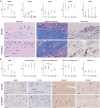
Mean and median scores for presence of A) PMNCs, FBGCs, vessels, collagen, and elastin, and B) smooth muscle cells (αSMA), neovascularization (CD‐34), macrophages types I (M1) and type II (M2) and M1/M2 ratio. Error bars represent means ± standard deviations (SD) or median ± interquartile range. Two‐way ANOVA and Tukey's multiple comparisons test between individual groups were used to test for differences between groups. ** indicates p < 0.01, *** indicates p < 0.001. All statistical results with p < 0.05 are written out in text in the manuscript. Images show representative histomorphology slides, with indication as follows; * = scaffold location, blue arrow = FBGC, black arrow = vessel, red arrow = PNMC.

References

    1. Brito L. G. O., Pereira G. M. V., Moalli P., Shynlova O., Manonai J., Weintraub A. Y., Deprest J., Bortolini M. A. T., Int. Urogynecol. J. 2022, 33, 15. - PubMed
    1. Deprest J. A., Cartwright R., Dietz H. P., Brito L. G. O., Koch M., Allen‐Brady K., Manonai J., Weintraub A. Y., Chua J. W. F., Cuffolo R., Sorrentino F., Cattani L., Decoene J., Page A.‐S., Weeg N., Varella Pereira G. M., Mori da Cunha de Carvalho M. G. M. C., Mackova K., Hympanova L. H., Moalli P., Shynlova O., Alperin M., Bortolini M. A. T., Int. Urogynecol. J. 2022, 33, 1699. - PubMed
    1. Wu J. M., Kawasaki A., Hundley A. F., Dieter A. A., Myers E. R., Sung V. W., Am. J. Obstet. Gynecol. 2011, 205, 230. - PMC - PubMed
    1. Smith F. J., Holman C. D., Moorin R. E., Tsokos N., Obstet. Gynecol. 2010, 116, 1096. - PubMed
    1. Guler Z., Roovers J. P., Biomolecules 2022, 12, 94. - PMC - PubMed

LinkOut - more resources