. 2025 Mar 19;91(3):e0133624.
doi: 10.1128/aem.01336-24. Epub 2025 Feb 26.

Inhibition of pneumococcal growth and biofilm formation by human isolates of Streptococcus mitis and Streptococcus oralis

Affiliations

Inhibition of pneumococcal growth and biofilm formation by human isolates of Streptococcus mitis and Streptococcus oralis

João Borralho et al. Appl Environ Microbiol. .

Abstract

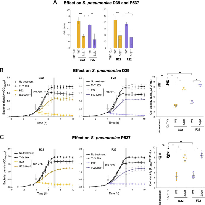
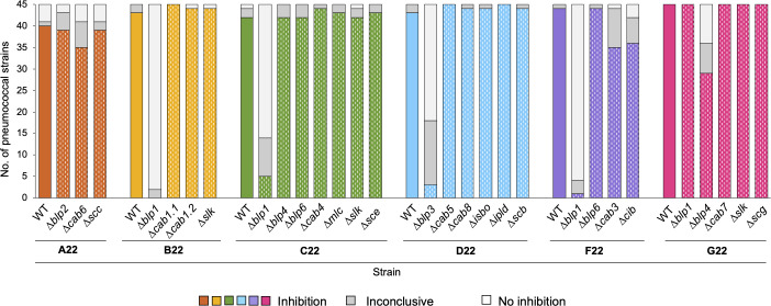
In a world facing the unprecedented threat of antibiotic-resistant bacteria, targeted approaches to control colonization and prevent disease caused by common pathobionts offer a promising solution. Streptococcus pneumoniae (pneumococcus) is a leading cause of infections worldwide, affecting both children and adults despite available antimicrobials and vaccines. Colonization, which occurs in the form of a biofilm in the upper respiratory tract, is frequent and a prerequisite for disease and transmission. The use of live bacterial strains as biotherapeutics for infectious diseases is actively being explored. Here, we investigated the potential of commensal streptococci to control S. pneumoniae. Screening of over 300 human isolates led to the identification of seven strains (one Streptococcus oralis and six Streptococcus mitis, designated A22 to G22) with inhibitory activity against S. pneumoniae of multiple serotypes and genotypes. Characterization of A22 to G22 cell-free supernatants indicated the involvement of secreted proteins or peptides in the inhibitory effect of all S. mitis isolates. Genome analyses revealed the presence of 64 bacteriocin loci, encoding 70 putative bacteriocins, several of which are novel and absent or rare in over 7,000 publicly available pneumococcal genomes. Deletion mutants indicated that bacteriocins partially or completely explained the anti-pneumococcal activity of the commensal strains. Importantly, strains A22 to G22 were further able to prevent and disrupt pneumococcal biofilms, a proxy for nasopharyngeal colonization. These results highlight the intricacy of the interactions among nasopharyngeal colonizers and support the potential of strains A22 to G22 to be used as live biotherapeutics, alone or in combination, to control S. pneumoniae colonization.

Importance: Streptococcus pneumoniae (pneumococcus) infections remain a major public health issue despite the use of vaccines and antibiotics. Pneumococci asymptomatically colonize the human upper respiratory tract, a niche shared with several commensal Streptococcus species. Competition for space and nutrients among species sharing the same niche is well documented and tends to be more intense among closely related species. Based on this rationale, a screening of several commensal streptococci isolated from the human upper respiratory tract led to the identification of strains of Streptococcus mitis and Streptococcus oralis capable of inhibiting most pneumococcal strains, across diverse serotypes and genotypes. This inhibition was partially or wholly linked to the expression of novel bacteriocins. The selected S. mitis and S. oralis strains significantly disrupted pneumococcal biofilms, indicating a potential for using commensals as biotherapeutics to control pneumococcal colonization, a key step in preventing disease and transmission.

Keywords: Streptococcus mitis; Streptococcus oralis; Streptococcus pneumoniae; bacteriocin; biofilm; biotherapeutic; colonization.


Conflict of interest statement

The authors declare no conflict of interest.

Figures

Fig 1
Fig 1
Identification of S. oralis and S. mitis isolates with broad anti-pneumococcal activity. (A) A collection of 313 non-pneumococcal streptococcal isolates was screened for anti-pneumococcal activity against the blp-bacteriocin susceptible S. pneumoniae strain P537 (34) through overlay assays. Isolates inhibiting P537 were further screened for inhibitory activity against 153 pneumococci representing epidemiologically relevant serotypes. Seven isolates, named strains A22 to G22, inhibited at least 90% of that S. pneumoniae collection and were next tested against additional S. pneumoniae strains (n = 77) with diverse BIRs in the blp locus. Combining the inhibition results of the two pneumococcal collections, strains A22 to G22 inhibited 92.5%–99.5% of the 230 pneumococci. (B) Species assignment of strains A22 to G22 was done by constructing a neighbor-joining phylogenetic tree using concatenated sequences of seven housekeeping genes described for the viridans multilocus sequence analysis scheme (36). The Jukes-Cantor model was used for nucleotide distance measure, and bootstrap analysis was performed based on 500 replicates. Scale bar reflects the number of nucleotide substitutions per site.
Fig 2
Fig 2
Supernatants of strains A22 to G22 inhibit S. pneumoniae. (A) Cell-free supernatants of strains A22 to G22 were obtained from the late exponential phase (OD600nm of 1.0) THY-grown cultures and 10-fold concentrated. CFSs were heat treated (by incubation at 37°C, 45°C, 60°C, and 75°C for 1.5 h, at 95°C for 1 h, and at 121°C for 20 min) and protease treated (incubation with 1 mg/mL proteinase K [PK] at 37°C for 3 h) and tested for inhibitory activity against the indicator S. pneumoniae strain P537 through well-diffusion assays. Inhibition halos above 11 mm (red outline) indicate inhibitory activity; inhibition halos of 9–10 mm (yellow outline) indicate residual inhibitory activity; and inhibition halos of 8 mm (well width, green outline) indicate no inhibitory activity. (B) Early exponential phase cultures (OD600nm of 0.3) of S. pneumoniae P537 were treated with 10× CFS, 10× THY, or remained untreated (as indicated by the gray arrow), and OD600nm was monitored every 30 min. Gray bar indicates the time point at which an aliquot was taken for cell viability counts. Mean and standard deviation of at least four biological replicates are shown for each time point. (C) Cell viability of P537 was assessed 3 h post-treatment with 10× CFS. Geometric mean and geometric standard deviation of at least four biological replicates are represented for each condition. **P-value < 0.01, Mann-Whitney U test with Benjamini and Hochberg correction for FDR; statistical comparison was done with treatment with 10× THY; ns, not significant.
Fig 3
Fig 3
The genomes of strains A22 to G22 encode multiple bacteriocin loci. A schematic representation of gene clusters is shown. Genes are colored by function, and flanking genes are represented in gray. Names of putative bacteriocins and immunity proteins are indicated in blue and red, respectively. Other relevant proteins are indicated in black. Numbers after gene names indicate allelic variants. Locus size (excluding the flanking genes) is indicated next to the locus name. Putative DNA-binding sites (SigX, RpoD, BlpR, ComE, and AC-, ABC-box elements) and terminator regions are also represented. Deletion-mutant strains were constructed for bacteriocin-containing regions, indicated by light purple boxes. This was done for all strains with the exception of strain E22 and locus scf from strain F22. Locus tags of the represented regions (genes from left to right) are the following: blp1—SMIB22_19330–19210, SMIC22_19450–19330, SMIE22_04050–04170, SMIF22_19410–19290, SMIG22_20130–2010; blp2—SORA22_03770–04130; blp3—SMID22_17960–17760; blp4—SMIC22_15990–15830, SMIE22_15420-15250, SMIG22_16660–16500; blp6—SMIC22_12570–12540, SMIF22_12710–12680; cab1—SMIB22_00640–00940; cab2—SMIE22_00500-00750; cab3—SMIF22_00400–00560; cab4—SMIC22_00520–00670; cab5—SMID22_00350–00420; cab6—SORA22_00820–00900; cab7—SMIG22_00540–00590; cab8—SMID22_18840–18800; cib—SMIE22_18370-18400, SMIF22_18970–19000; scb—SMID22_02310–02360; scc—SORA22_14540–14500; sce—SMIC22_05670–05730; scf—SMIF22_03120–03070; scg—SMIG22_15150–15110; mld— SMIE22_18640-18490; ipld—SMID22_05170–05260; mlc—SMIC22_19190–19110; slk—SMIB22_16070–16020, SMIC22_16240–16190, SMIE22_15680-15620, SMIG22_16890–16840; isbo—SMID22_12430–12340.
Fig 4
Fig 4
The bacteriocin-like peptide loci of strains A22 to G22 are expressed and controlled by the BlpC pheromone. Gene expression was measured by qRT-PCR after exposure of strains for 5 min to synthetic cognate BlpC. Loci iblp5.1, iblp5.2, and blp6 lack a cognate BlpC; hence, their gene stimulation was done with the addition of blp1 BlpC of the same strain. The Y axis shows fold change differences after BlpC treatment compared to the untreated culture. Error bars represent the standard deviation of the mean of three biological replicates.
Fig 5
Fig 5
Locus blp1 from S. mitis strains B22 and F22 negatively affects the growth and cell viability of S. pneumoniae D39 and P537 strains. The CFS of S. mitis strains B22 and F22 and the corresponding deletion mutants lacking the bacteriocin immunity regions of locus blp1 were tested against S. pneumoniae D39 and P537. (A) Well-diffusion assay using 10× CFS or 10× THY (control) against S. pneumoniae D39 (left) and P537 (right). After overnight incubation with CFS, plates were inspected for inhibition halos. The diameter of the inhibition halos was measured independently by two researchers in four independent experiments. Mean and standard deviation are represented for each condition. The inhibition halo diameter includes the well width of 8 mm. *P-value < 0.05, **P-value < 0.01, and ***P-value < 0.001, Student’s t-test. (B and C) The effect of 10× CFS on planktonic growth of S. pneumoniae D39 (B) and P537 (C) was assessed. Early exponential phase cultures (OD600nm of 0.3) of D39 and P537 were treated with 10× CFS, 10× THY, or remained untreated (as indicated by the gray arrow) and OD600nm was monitored every 30 min (left and middle panels). Gray Bar indicates the time point in which an aliquot was taken for cell viability counts. Mean and standard deviation of at least four biological replicates are represented for each condition. Cell viability (CFU/mL) of D39 and P537 was assessed 3 h post-treatment (right panel). Geometric mean and geometric standard deviation of at least four biological replicates are represented for each condition. *P-value < 0.05 and **P-value < 0.01, Mann-Whitney U test with Benjamini and Hochberg correction for FDR; ns, not significant.
Fig 6
Fig 6
Bacteriocin-like peptide loci from S. mitis strains contribute to their anti-pneumococcal activity in agar overlay assays. Strains A22, B22, C22, D22, F22, and G22 and their deletion mutants (marked with dashes within the colored bars) were tested for inhibitory activity against 45 S. pneumoniae strains (representatives of 23 serotypes and 42 multilocus sequence types) in agar overlay assays. Plates were inspected for growth inhibition halos around the stabbed strains (A22, B22, C22, D22, F22, and G22 and their mutants). Results were inspected independently by two researchers, in three independent experiments.
Fig 7
Fig 7
The anti-pneumococcal inhibitory activity of S. mitis strain G22 is driven by an additive activity of bacteriocin loci. (A) Strains G22 and its deletion mutants (marked with dashes within the colored bars) were tested for inhibitory activity against 45 S. pneumoniae strains (representatives of 23 serotypes and 42 multilocus sequence types) in agar overlay assays. Plates were inspected for growth inhibition halos around the stabbed strains (G22 and its mutants). Results were inspected independently by two researchers, in three independent experiments. (B) Well-diffusion assay using CFS 10× or THY 10× (control) against S. pneumoniae D39 (left) and P537 (right). After overnight incubation with CFS, plates were inspected for inhibition halos. The diameter of the inhibition halos was measured independently by two researchers in four independent experiments. Mean and standard deviation are represented for each condition. The inhibition halo diameter includes the well width of 8 mm. *P-value < 0.05 and **P-value < 0.01, Student’s t-test with Benjamini and Hochberg correction for FDR. (C and D) The effect of 10× CFS on planktonic growth of S. pneumoniae D39 (C) and P537 (D) was assessed. Early exponential phase cultures (OD600nm of 0.3) of D39 and P537 were treated with 10% (vol/vol) of CFS 10×, THY 10×, or remained untreated (as indicated by the gray arrow) and OD600nm was monitored every 30 min (left and middle panels). Gray bar indicates the time point in which an aliquot was taken for cell viability counts. Mean and standard deviation of at least four biological replicates are represented for each condition. Cell viability (CFU/mL) of D39 and P537 was assessed 3 h post-treatment (right panel). *P-value < 0.05 and **P-value < 0.01, Mann-Whitney U test with Benjamini and Hochberg correction for FDR; astatistical comparison with treatment with THY 10×; ns, not significant.
Fig 8
Fig 8
S. oralis strains A22 and S. mitis strains B22 to G22 inhibit S. pneumoniae in multi-species biofilms. The ability of strains A22 to G22, individually or in a seven-strain cocktail, to prevent or disrupt biofilms of S. pneumoniae strains D39-RCm and 7632-RCm was evaluated with both species inoculated in a 1:1 ratio. (A) Strains A22 to G22 were inoculated simultaneously with S. pneumoniae strains. Growth was evaluated after 24 h. (B) Strains A22 to G22 were grown for 24 h in biofilm prior to inoculation of S. pneumoniae. S. pneumoniae growth was evaluated after 24 h. (C and D) Strains A22 to G22 were added to pre-grown (24 h) S. pneumoniae biofilms as a single treatment or as a 3 day-treatment. S. pneumoniae growth was evaluated 24 h after treatment. In all experiments, single-strain biofilms of the corresponding strains were performed in parallel as control. Y axis represents the ratio between the bacterial density (CFU/mL) of S. pneumoniae biofilm after treatment with strains A22 to G22 and the bacterial density (CFU/mL) of S. pneumoniae biofilm without treatment. Four independent experiments were performed. *P < 0.05; **P < 0.01; ***P < 0.001; and ****P < 0.0001, ratio paired t-test; ns, not significant.

Similar articles

Cited by

References

    1. Wahl B, O’Brien KL, Greenbaum A, Majumder A, Liu L, Chu Y, Lukšić I, Nair H, McAllister DA, Campbell H, Rudan I, Black R, Knoll MD. 2018. Burden of Streptococcus pneumoniae and Haemophilus influenzae type b disease in children in the era of conjugate vaccines: global, regional, and national estimates for 2000-15. Lancet Glob Health 6:e744–e757. doi:10.1016/S2214-109X(18)30247-X - DOI - PMC - PubMed
    1. Drijkoningen JJC, Rohde GGU. 2014. Pneumococcal infection in adults: burden of disease. Clin Microbiol Infect 20 Suppl 5:45–51. doi:10.1111/1469-0691.12461 - DOI - PubMed
    1. Ganaie F, Saad JS, McGee L, van Tonder AJ, Bentley SD, Lo SW, Gladstone RA, Turner P, Keenan JD, Breiman RF, Nahm MH. 2020. A new pneumococcal capsule type, 10D, is the 100th Serotype and has a large cps fragment from an oral streptococcus. MBio 11:e00937-20. doi:10.1128/mBio.00937-20 - DOI - PMC - PubMed
    1. Ganaie F, Maruhn K, Li C, Porambo RJ, Elverdal PL, Abeygunwardana C, van der Linden M, Duus JØ, Sheppard CL, Nahm MH. 2021. Structural, genetic, and serological elucidation of Streptococcus pneumoniae serogroup 24 serotypes: discovery of a new serotype, 24C, with a variable capsule structure. J Clin Microbiol 59:e0054021. doi:10.1128/JCM.00540-21 - DOI - PMC - PubMed
    1. Essink B, Sabharwal C, Cannon K, Frenck R, Lal H, Xu X, Sundaraiyer V, Peng Y, Moyer L, Pride MW, Scully IL, Jansen KU, Gruber WC, Scott DA, Watson W. 2022. Pivotal phase 3 randomized clinical trial of the safety, tolerability, and immunogenicity of 20-valent pneumococcal conjugate vaccine in adults aged ≥18 Years. Clin Infect Dis 75:390–398. doi:10.1093/cid/ciab990 - DOI - PMC - PubMed

MeSH terms

LinkOut - more resources