. 2025 Mar 25;10(3):e0091724.
doi: 10.1128/msphere.00917-24. Epub 2025 Feb 26.

Cell differentiation controls iron assimilation in the choanoflagellate Salpingoeca rosetta

Affiliations

Cell differentiation controls iron assimilation in the choanoflagellate Salpingoeca rosetta

Fredrick Leon et al. mSphere. .

Abstract

Marine microeukaryotes have evolved diverse cellular features that link their life histories to surrounding environments. How those dynamic life histories intersect with the ecological functions of microeukaryotes remains a frontier to understanding their roles in critical biogeochemical cycles. Choanoflagellates, phagotrophs that cycle nutrients through filter feeding, provide models to explore this intersection, for many choanoflagellate species transition between life history stages by differentiating into distinct cell types. Here, we report that cell differentiation in the marine choanoflagellate Salpingoeca rosetta endows one of its cell types with the ability to utilize insoluble ferric colloids. These colloids are a predominant form of iron in marine environments and are largely inaccessible to cell-walled microbes. Therefore, choanoflagellates and other phagotrophic eukaryotes may serve critical ecological roles by cycling this essential nutrient through iron utilization pathways. We found that S. rosetta can utilize these ferric colloids via the expression of a cytochrome b561 iron reductase (cytb561a). This gene and its mammalian ortholog, the duodenal cytochrome b561 (DCYTB) that reduces ferric cations for uptake in gut epithelia, belong to a subgroup of cytochrome b561 proteins with distinct biochemical features that contribute to iron reduction activity. Overall, our findings provide insight into the ecological roles choanoflagellates perform and inform reconstructions of early animal evolution where functionally distinct cell types became an integrated whole at the origin of animal multicellularity.

Importance: This study examines how cell differentiation in a choanoflagellate enables the uptake of iron, an essential nutrient. Choanoflagellates are widespread, aquatic microeukaryotes that are the closest living relatives of animals. Similar to their animal relatives, we found that the model choanoflagellate, S. rosetta, divides metabolic functions between distinct cell types. One cell type uses an iron reductase to acquire ferric colloids, a key source of iron in the ocean. We also observed that S. rosetta has three variants of this reductase, each with distinct biochemical properties that likely lead to differences in how they reduce iron. These reductases are variably distributed across ocean regions, suggesting a role for choanoflagellates in cycling iron in marine environments.

Keywords: cell-type evolution; choanoflagellate; cytochrome b561; iron colloid; marine microeukaryote.


Conflict of interest statement

The authors declare no conflict of interest.

Figures

Fig 1
Fig 1
Thecates from the choanoflagellate S. rosetta are a distinct cell type that upregulates a cytochrome b561 reductase. (A) S. rosetta differentiates into morphologically distinct cell types. This schematic shows four of the cell types from S. rosetta that were stably cultured to produce transcriptome profiles. All of these cell types display the common choanoflagellate cell architecture in which an apical flagellum is encircled by a collar of actin-filled microvilli (indicated on the slow swimmer) that enables choanoflagellates to phagocytose bacteria and particulate matter. When nutrients are abundant, slow swimmers (green) respond to bacterial and algal cues to develop into multicellular rosettes (blue) that form by serial cell division. Cultures of slow swimmers also form chains of cells through serial cell divisions, but those chains are easily disrupted by mechanical force. Under starvation, slow swimmers and rosettes become fast swimmers (gray), which have a reduced cell body and collar with a longer flagellum. Under sustained nutrient deprivation, fast swimmers differentiate into thecates (red), a type of cell that adheres to substrates by constructing an extracellular apparatus called a theca. Thecates still proliferate in low nutrient conditions by dividing into swimmers that build their own theca. By taking the supernatant of thecate cultures in high nutrient conditions, thecates can then differentiate into slow swimmers. (B) The transcriptome profile of thecates stands apart from all other cell types. A principal component analysis of triplicate RNA-seq profiles from each cell type (panel A) shows that 86% of the variance between samples is attributed to the thecate transcriptome profile. All other cell types cluster closely together. (C) Cell-type expression of iron acquisition genes identified S. rosetta cell types. Genes are denoted by unique identifiers for S. rosetta genes and gene names that were given in this work based on their homology with animals. Expression values are averaged triplicate values of transcripts per million (TPM) from cell type transcriptomes (B). Of all genes and paralogs, cytb561a exhibits the most striking differential regulation. (D) Diagram of iron transport pathway based on identified iron acquisition homologs. We hypothesize one or more Cytb561 reductases (R) reduce ferric iron to ferrous iron to be trafficked across the cell membrane by ion transporter Dmt1 (T). Inside the cell, ferrous iron can be metabolized and utilized or stored in vacuoles by vacuolar transporters Vit1/2 (VT). Upon leaving the cell, ferrous iron is exported across the membrane by Fpn as an efflux pump (E] )and finally oxidized back to ferric iron by the oxidase Heph (O).
Fig 2
Fig 2
Thecates relieve iron limitation through the ingestion of ferric colloids. (A) A method to swap iron sources that support choanoflagellate growth. In preparation for iron limitation assays, S. rosetta (strain PRA-390) was passaged in low-nutrient media and depleted of iron. Cells were then washed multiple times and inoculated with iron-limited feeder bacteria. At the same time, ferric EDTA (Fe3+•EDTA) or ferric colloids (Fe3+) were provided. Cultures grew for 48 h at 27°C before assaying cytb561a growth (B) (Fig. 3B; Fig. S3) and expression (Fig. 3C; Fig. S7). (B) Thecates exhibit improved growth with low concentrations of ferric colloids compared with slow swimmers. The cell density of slow swimmer and thecate cultures when grown with titrations of ferric EDTA or ferric colloids. Ferric colloids at 50–100 μM ferric colloids supported higher cell densities for thecates. Meanwhile, higher concentrations of ferric EDTA (≥ 200 µM) supported higher growth in both cell types compared with ferric colloids. However, the cell density of cultures grown with 800 µM ferric EDTA did not exceed that of thecates grown with 100 µM ferric colloids. (C) E. pacifica grows similarly with ferric EDTA or ferric colloids. Because ferric colloids would influence OD600 measurements in a standard growth curve experiment, E. pacifica cultures were grown for 48 h with 100 µM ferric EDTA or ferric colloids and then lightly centrifuged at 500 × g for 1 min at room temperature to settle iron particulates. Afterward, the supernatant OD600 was measured. Gray scale represents matching replicates, the black bar denotes the global mean, and P-values were calculated using Tukey’s multiple comparisons. (D) Thecates ingest ferric colloids through phagocytosis. Time courses of wild-type thecates incubated with fluorescently labeled ferric colloids (see methods on ferric colloid labeling). Fluorescent ferric colloid particles were tracked (magenta outline) and observed being ingested and internalized through phagocytosis. The cell outline and feeding collar are marked by dotted lines. The zero-time point indicates initial contact with the ferric colloid particle. (E) Ingested ferric colloids reside within food vacuoles. Confocal microscopy of wild-type thecates incubated with and without fluorescently labeled ferric colloids. Split channels show membranes (gray), ferric colloids (magenta), and vacuoles (blue). The cell outline and feeding collar are marked by dotted lines. (F) Populations of thecates exhibit widespread ferric colloid ingestion. Fluorescence intensity of thecate cultures incubated with (+) and without (–) fluorescently labeled ferric colloids. Individual cell intensities from a replicate experiment (N = 3) are shaded in the same gray hue, and larger circles indicate the mean calculated by fitting a gamma distribution to a replicate population. P-values were calculated from the mean values with a one-tailed t-test.
Fig 3
Fig 3
cytb561a expression is necessary for the utilization of ferric colloids. (A) The endogenous locus of cytb561a was edited to produce hypomorphic and epitope-tagged alleles. Diagram of endogenous genomic locus of cytb561a shows the position of premature terminal sequence (PTS) and epitope tag (ALFA-tag) insertions. The position of qPCR primers is shown as well, spanning exon-exon junctions. The predicted protein products of each insertion are shown as well, with the cytochrome b561 domain labeled as cytb561. (B) Thecates require cytb561a for increased proliferation with ferric colloids. To account for differences in growth between slow swimmers (green) and thecates (red), the cell density of cultures grown with ferric colloids was normalized to cultures grown with ferric EDTA. With this metric, a ratio greater than one indicates that the cell type displays increased growth with ferric colloids, whereas a ratio less than one indicates the converse. A premature termination sequence introduced at position 151 in cytb561a with CRISPR/Cas9 genome editing produced a mutant allele (cytb561aPTS) with a 3.6-fold reduction in cytb561a mRNA levels and stop codons that would truncate the protein translated from this transcript (Fig. S2; https://doi.org/10.6084/m9.figshare.28225106). Unlike the wild-type strain (circles), thecate cells with cytb561aPTS displayed no improved growth with ferric colloids (triangles). P-values were calculated from a two-way ANOVA. (C) cytb561a expression is part of the thecate regulon and not determined by external iron conditions. The expression of cytb561a was monitored by RT-qPCR in cultures of slow swimmers (green) or thecates (red) that were grown with either ferric EDTA or ferric colloids. The expression of cytb561a was normalized to cofilin (PTSG_01554), a eukaryotic gene that displays high, consistent expression across all S. rosetta cell types. Independent triplicates were performed, and P-values were calculated from a two-way ANOVA. (D) Slow swimmers do not respond to iron availability at the protein level of Cytb561a. Cytb561a protein levels were detected using an endogenously edited epitope tag and normalized to total protein levels via western blot (raw images, Fig. S8). Proteins levels were measured from the cultures of slow swimmers (green) or thecates (red) that were grown with either low iron media (4% PG, 0.27 ± 0.03 µM Fe) or nutrient-replete media (25% RA, 11.7 µM added Fe). P-values were calculated from a two-tailed t-test.
Fig 4
Fig 4
Cytb561 paralogs possess distinct biochemical properties. (A) Phylogeny of iron reductases reveals that Cytb561a is an ortholog of animal DCYTB. Each paralog of S. rosetta falls into distinct clades of cytochrome b561. Circles on each branch are proportional to bootstrap values greater than sixty. Scale bar indicates the average number of substitutions per site. Branch colors correspond to clades as shown in the legend. (B) Predicted structures of S. rosetta Cytb561 paralogs show differences in dimerization and substrate-binding interfaces. Alphafold (version 3) predictions show Cytb561a as the only paralog-forming homodimers. Predicted electrostatic surfaces Cytb561 paralogs show different charge distributions around the substrate binding pocket (arrows). Models are angled 45° to view the lumenal surface where iron binds.
Fig 5
Fig 5
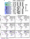
Phylogenetic and geographic distribution of Cytb561 homologs from choanoflagellates. (A) The presence of Cytb561 homologs across choanoflagellate species. Choanoflagellates are divided into two major groups: Loricates, including Tectiform and Nudiform species, are choanoflagellates that produce siliceous coverings, and Craspedids, in which S. rosetta belongs, have organic coverings. The grayscale corresponds to the number of homologs found in the phylogenetic tree in Fig. 4A. Morphology is determined by the observation of cell types in culture (46, 83): Sw for swimming cells, Th for theca formation, Col for colony formation, Lor for lorica or siliceous covering formation. Environment corresponds to the original isolation location and culturable media salinity (Mar for marine and Fre for freshwater) (46, 83). Tree order and species characteristics adapted from (83) and (46). (B) The geographic distribution of Cytb561 homologs. Homologs and congeners of S. rosetta were identified from a repository of metagenomes and metatranscriptomes from marine environments. In these data sets, surface waters are defined as the top 5 m of the ocean, and Deep Chlorophyll maxima (DCM) are found 20–200 m from the surface, depending on the highest intensity from chlorophyll. Homolog abundance was measured as the percentage of total reads from each sample site. Red vertical lines correspond to genomic abundance. Blue horizontal lines correspond to transcript abundance. Black crosses denoted sites where ASVs congeneric to S. rosetta were detected. Overlapping hits at matching locations increase the color saturation. Longitude and latitude degrees are marked on the bottom right map.

Update of

References

    1. Wilken S, Yung CCM, Poirier C, Massana R, Jimenez V, Worden AZ. 2023. Choanoflagellates alongside diverse uncultured predatory protists consume the abundant open-ocean cyanobacterium Prochlorococcus. Proc Natl Acad Sci USA 120:e2302388120. doi:10.1073/pnas.2302388120 - DOI - PMC - PubMed
    1. Brown JM, Labonté JM, Brown J, Record NR, Poulton NJ, Sieracki ME, Logares R, Stepanauskas R. 2020. Single cell genomics reveals viruses consumed by marine protists. Front Microbiol 11:524828. doi:10.3389/fmicb.2020.524828 - DOI - PMC - PubMed
    1. González JM, Suttle CA. 1993. Grazing by marine nanoflagellates on viruses and virus-sized particles: ingestion and digestion. Mar Ecol Prog Ser 94:1–10. doi:10.3354/meps094001 - DOI
    1. Barbeau K, Moffett JW, Caron DA, Croot PL, Erdner DL. 1996. Role of protozoan grazing in relieving iron limitation of phytoplankton. Nature 380:61–64. doi:10.1038/380061a0 - DOI
    1. Güde H. 1985. Influence of phagotrophic processes on the regeneration of nutrients in two-stage continuous culture systems. Microb Ecol 11:193–204. doi:10.1007/BF02010599 - DOI - PubMed

MeSH terms