Review
. 2025 Mar;30(2):181-207.
doi: 10.1007/s00775-025-02102-3. Epub 2025 Feb 26.

Cytochromes P460 and c'-β: exploiting a novel fold for multiple functions

Affiliations
Review

Cytochromes P460 and c'-β: exploiting a novel fold for multiple functions

Hannah R Adams et al. J Biol Inorg Chem. 2025 Mar.

Abstract

Two related classes of ligand-binding heme c-containing proteins with a high degree of structural homology have been identified and characterized over recent decades: cytochromes P460 (cyts P460), defined by an unusual heme-lysine cross-link, and cytochromes c'-β (cyts c'-β), containing a canonical c-heme without the lysine cross-link. The shared protein fold of the cyt P460-cyt c'-β superfamily can accommodate a variety of heme environments with entirely different reactivities. On the one hand, cyts P460 with polar distal pockets have been shown to oxidize NH2OH to NO and/or N2O via proton-coupled electron transfer. On the other hand, cyts c'-β with hydrophobic distal pockets have a proposed gas binding function similar to the unrelated, but more extensively characterized, alpha helical cytochromes c'. Recent studies have also identified 'halfway house' proteins (cyts P460 with non-polar heme pockets and cyts c'-β with polar distal heme pockets) with functions yet to be resolved. Here, we review the structural, spectroscopic and enzymatic properties of the cyt P460-cyt c'-β superfamily with a view to understanding the structural determinants of their different functional properties.

Keywords: Cross-link; Gas binding; Haem; Heme; Nitrification; P460.


Conflict of interest statement

Declarations. Conflict of interest: The authors declare that they have no competing interests. Ethics approval and consent to participate: Not applicable. Consent for publication: Not applicable.

Figures

Fig. 1
Fig. 1
Overall fold (top) and distal heme pocket (bottom) structures of McCP-β (A), TtCP-β (B), NeCP-β (C), McP460 (D), NeP460 (E), and NsP460 (F). The overall protein fold and dimeric structure is well maintained within these different proteins, while the active site and surrounding region is highly variable
Fig. 2
Fig. 2
Overview of the reactions within the Nitrogen Cycle highlighting the role of Cyt P460. Blue lines represent denitrification, red lines represent nitrogen fixation, orange lines represent dissimilatory nitrate reduction to ammonium, black lines represent nitrification, and purple lines represent anammox
Fig. 3
Fig. 3
Proposed mechanism of oxidation of hydroxylamine by cyt P460 showing key compounds in the catalytic cycle using Enemark–Feltham notation. Steps in black are those which have been observed only in cyt P460, while those in green have been observed in both HAO and P460. Gray represents an off-pathway 5-coordinate (5c) {FeNO}7 species. NH2OH binds to the heme of the ferric protein and is oxidized to form an {FeNO}6 species via a 6-coordinate (6c) {FeNO}7 intermediate. This {FeNO}6 species then undergoes nucleophilic attack by a second NH2OH to produce N2O and H2O. The heme is then free to start the cycle over again [18, 42, 43, 45]
Fig. 4
Fig. 4
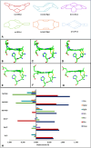
Heme distortions in cyts P460 and cyts c′-β. The distortion of the hemes of NsP460 (6amg) (B), NeP460 (2je3) (C), McP460 (6hiu) (D), McCP-β (6hih) (E), NeCP-β (7s5o) (F), and TtCP-β (7ead) (G) away from planarity. The different types of heme distortions are shown in Panel A. Graphical representation of the displacement of the heme from planarity for each published wild-type structure (H)
Fig. 5
Fig. 5
Room-temperature UV–Vis absorbance spectra of McCP-β (A) and McP460 (B) in their FeIII (red trace) and FeII (blue trace) redox states, together with corresponding resonance Raman spectra (C and D, respectively) using 407 nm laser excitation (or 442 nm for FeII McP460)
Fig. 6
Fig. 6
Ligand bound structures (gold) of cyts P460 and c′-β in comparison to their ligand free states (green – NsP460/blue—McCP). NO (6e17) (A) and NH2OH (6eoy) (B) bound Ala131Glu NsP460 demonstrates movement of Phe 76 to allow ligands to bind to the distal face of the heme. NO bound McCP (7zps) (C), Phe32Val McCP (7zsw) (E), and Phe61Val McCP (7zqz) (G) and CO bound McCP (6zsk) (D), Phe32Val McCP (7zsx) (F), and Phe61Val McCP (7zti) (H) also demonstrate the interaction of the capping Phe residues with movement of Phe 32 upon ligand binding in wt McCP (C, D).and the Phe61Val mutant (G, H). Phe/Val 61 does not show any movement or ligand interaction in either the wt or mutant McCP
Fig. 7
Fig. 7
Crystal structures for cyt P460 and c′-β mutants (gold) and changes in residue positioning in the distal pocket compared to wt protein (pink – NeP460/green – NsP460/blue—McCP-β). The NeP460 Arg44Ala mutation (8gar) causes the lysine crosslink to not form in the crystal structure, suggesting that Arg 44 has a role in the formation of the cross-link (A). Removal of the cross-link and introduction of a Glu over the distal face of the heme in the NsP460 Lys106Leu/Ala131Glu mutant (6w6n) causes a shift in the positioning of Phe 76 in the distal pocket (B). The NsP460 Ala131Glu mutant (6eox) retains its lysine crosslink and causes little movement in Phe 76 with the Glu residue positioned away from the distal heme face (C). The NsP460 Ala131Gln mutant (6eoz) retains its lysine crosslink, but the presence of the Gln residue over the distal face of the heme causes Phe 76 to rotate away from the heme face (D). Introduction of the McCP-β Phe32Val mutation (7zs4) causes little change to the other residues in the distal heme pocket (E), while the Phe61Val mutation (7zrw) gives rise to two alternative conformations of Phe 32 (F)

References

    1. Paoli M, Marles-Wright J, Smith A (2002) Structure-function relationships in heme-proteins. DNA Cell Biol 21:271–280. 10.1089/104454902753759690 - PubMed
    1. Mense SM, Zhang L (2006) Heme: a versatile signaling molecule controlling the activities of diverse regulators ranging from transcription factors to MAP kinases. Cell Res 16:681–692. 10.1038/sj.cr.7310086 - PubMed
    1. Zahn JA, Duncan C, DiSpirito AA (1994) Oxidation of hydroxylamine by cytochrome P-460 of the obligate Methylotroph Methylococcus capsulatus Bath. J Bacteriol 176:5879–5887. 10.1128/jb.176.19.5879-5887.1994 - PMC - PubMed
    1. Zahn JA, Arciero DM, Hooper AB, Dispirito AA (1996) Cytochrome c′ of Methylococcus capsulatus Bath. Eur J Biochem 240:684–691. 10.1111/j.1432-1033.1996.0684h.x - PubMed
    1. Bergmann DJ, Zahn JA, Hooper AB, DiSpirito AA (1998) Cytochrome P460 genes from the Methanotroph Methylococcus capsulatus Bath. J Bacteriol 180:6440–6445. 10.1128/JB.180.24.6440-6445.1998 - PMC - PubMed

Publication types

LinkOut - more resources