. 2025 Feb 25;166(7):1662-1679.
doi: 10.1097/j.pain.0000000000003512.

Modelling inflammation-induced peripheral sensitization in a dish-more complex than expected?

Affiliations

Modelling inflammation-induced peripheral sensitization in a dish-more complex than expected?

Yuening Li et al. Pain. .

Abstract

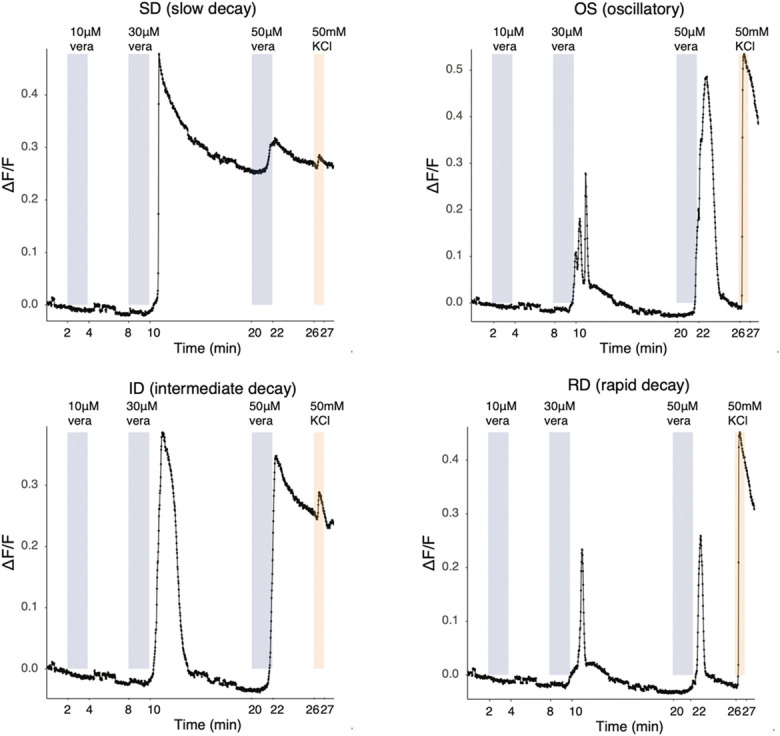
Peripheral sensitization of nociceptors is believed to be a key driver of chronic pain states. Here, we sought to study the effects of a modified version of inflammatory soup on the excitability of human stem cell-derived sensory neurons. For this, we used a preexisting and a novel stem cell line, modified to stably express the calcium sensor GCamP6f. Upon treatment with inflammatory soup, we observed no changes in neuronal transcription or functional responses upon calcium imaging and only a very minor increase in resting membrane potential (RMP) via whole cell patch clamping: control RMP (-71.31 ± 1.1 mV) vs inflammatory soup RMP (-67.74 ± 1.29 mV), uncorrected 2-tailed independent samples t test, P = 0.0383. Similarly, small changes were observed when treating mouse primary sensory neurons with inflammatory soup. A semi-systematic reexamination of past literature further indicated that observed effects of inflammatory mediators on dissociated sensory neuron cultures are generally small. We conclude that modelling inflammation-induced peripheral sensitization in vitro is nontrivial and will require careful selection of mediators and/or more complex, longitudinal multicellular setups. Especially in the latter, our novel GCamP6f-induced pluripotent stem cell line may be of value.

Keywords: Calcium imaging; Gcamp; IPSC; Inflammation; Pain; Patch clamping; Stem cell–derived sensory neurons.


Conflict of interest statement

O.B. is a cofounder, CEO, and shareholder of LIFE & BRAIN GmbH. None of the other authors have any conflicts of interest to declare in relation to this work.

Sponsorships or competing interests that may be relevant to content are disclosed at the end of this article.

Figures

Figure 1.
Figure 1.
Immunostaining analysis demonstrates that pure sensory neurons could be differentiated from both our stem cell lines. (A) Representative images of UKB (day 54) and Kute4 (day 48) iPSC-derived sensory neurons. Scale bars: 100 µm. (B) Percentage of pure iSNs was determined for neurons from each independent differentiation used in subsequent experiments. Neurons aged days 46 to 81 were fixed and stained. Purity of iSNs was measured by taking the ratio of cells double positive for BRN3A and PGP9.5 or NeuN over DAPI+ nuclei. Each dot represents an individual coverslip. IDs T7-T35 refer to individual differentiations starting with UKB or Kute4 iPSC lines, respectively. iPSC, induced pluripotent stem cell; iSNs, induced pluripotent stem cell–derived sensory neurons.
Figure 2.
Figure 2.
A novel iPSC line was generated, which constitutively expresses GCaMP6f. (A) Schematic overview of the genomic insertion of GCaMP6f in the AAVS1 safe harbour locus of the UKB line. The insert contained GCamP6f driven by the CAG promoter, as well as 3′ and 5′ homology arms, a 3′ splice acceptor site (SA), a P2A self-cleaving peptide sequence (P2A), and a puromycin resistance gene (puro) terminated by a poly-adenylation signal (pA). (B) Screenshots of UKB-derived iSNs during calcium imaging, illustrating GCaMP6f fluorescent signal before and after application of 50 mM KCl. For comparison, we also show screenshots of the UKB-line before it was modified, this time capturing calcium signal with the fluorescent dye Fluo-4 before and after application of 40 mM KCl. CAG, cytomegalovirus immediate-early enhancer/ chicken beta-actin; iPSC, induced pluripotent stem cell; iSNs, induced pluripotent stem cell–derived sensory neurons.
Figure 3.
Figure 3.
Representative traces of UKB-derived iSNs responding to veratridine demonstrating that the line's endogenous GCamP6f can be used to detect fast, dynamic responses. UKB iSNs respond to veratridine with distinct patterns, similar to mouse DRG neurons, as characterised by Reference 43. Neurons (82 days old) were subject to 3 different concentrations of veratridine. 50 mM KCl was used as a positive control. DRG, dorsal root ganglion; iSNs, induced pluripotent stem cell–derived sensory neurons.
Figure 4.
Figure 4.
iSNs express common sensory neuronal markers, with low expression of nonneuronal markers. Heatmap showing TPM expression values for selected neuronal (in black) and nonneuronal markers (in grey). T8, T9, T34, and T35 are independent differentiations of 2 iPSC lines: UKB and Kute4, respectively. For the control and modified inflammatory soup conditions, day 60 neurons were treated for 24 hours before RNA was extracted. iSNs, induced pluripotent stem cell–derived sensory neurons; iPSC, induced pluripotent stem cell; TPM, transcripts per million.
Figure 5.
Figure 5.
iSNs express receptors for several ligands within the modified-IS. TPM expression values for our iSN RNA-seq data are shown on the left. Each square is a sample, arranged in the same order as in Figure 4. All genes above the black horizontal line are expressed at TPM > 1 in at least 7 samples or more. Genes below the line fall within the noise range of our particular dataset. On the right, we plotted pseudobulk counts derived from human postmortem DRG, specifically single-nucleus RNA-seq data published by 2 independent groups (Nguyen et al. and Jung et al.). Each square displays data on DRG neurons obtained from a different individual (n = 22 postmortem samples). We estimate that counts less than 50 are within the noise range. HTR1A and HTR6 were not annotated within Jung et al. and are, therefore, labelled with an #. DRG, dorsal root ganglion; IS, inflammatory soup; iSNs, induced pluripotent stem cell–derived sensory neurons; TPM, transcripts per million.
Figure 6.
Figure 6.
iSNs reach a consistent level of maturity beyond differentiation day 50. (A) Heatmap displaying differentially regulated genes across 3 iSN differentiation timepoints (D30, D50, and D70). Colours represent z-scored TPM values (by gene). Columns represent individual samples and were ordered using unsupervised clustering with the hclust function (method = ward.D2). Clustering was performed on all genes, which were expressed according to our cutoff (see Methods), differentially regulated at adj. P < 0.05 and regulated at log2FC > 1.5 or log2FC < −1.5. (B) Volcano plots displaying differentially expressed genes for each time point comparison. Adjusted P cutoff: <0.05, Log2FC cutoff: > 0.5 or < −0.5. Plots created using Enhanced Volcano R package. T = trial indicating independent differentiations of Kute4 and UKB lines. iSNs, induced pluripotent stem cell–derived sensory neurons; TPM, transcripts per million.
Figure 7.
Figure 7.
Modified-IS does not sensitise Kute4-derived iSNs in response to veratridine or pregnenolone sulphate (PS). Inflammatory soup does not change the response percentage (A and B) to 50 μM veratridine (n = 6 coverslips) or 100 μM pregnenolone sulphate (PS, n = 5 coverslips). Maximum response amplitudes were unchanged with modified-IS (C) or, if anything, reduced in response to PS (D). Each big dot in (A and B) represents a coverslip. In (C and D), the small, transparent dots are the maximal values of each cell in each coverslip. The superimposed large-sized dots are the mean values of each experiment. The dotted line is at 0.1, the cutoff for responders. Mann–Whitney nonparametric t-tests were used to compare between N2 and IS, with each coverslip taken as an independent unit (n = 6 for A and C, n = 5 for B and D). (E and F) Plot average traces in response to modified-IS and N2 control medium over the course of the experiment; the shades surrounding the average line are SD at each time point. For each condition, the trace is the average of 3 experiments. See Supplementary Figure 2, http://links.lww.com/PAIN/C199 for individual representative traces. For all experiments, neurons were incubated in N2 (medium control) or modified-IS for 24 hours before the experiment. Recordings took place at room temperature. Neurons were differentiated from Kute4 iPSC for at least 50 days. Different colours indicate independent differentiations (T23-29); for veratridine: grey = T28 72 days old; orange: T29 57 days old; blue: T29 62 days old; for PS experiments: grey = T25 63 days old; orange = T23 74 days old. iPSC, induced pluripotent stem cell; IS, inflammatory soup; iSNs, induced pluripotent stem cell–derived sensory neurons.
Figure 8.
Figure 8.
Modified-IS induces only a very minor change in resting membrane potential in iSNs, while all other electrophysiological properties of the neurons appear unaffected. Representative traces of evoked action potentials in control (A) and inflammatory (B) conditions and of spontaneous action potentials (C). (D) The resting membrane was more depolarised in modified-IS treated compared to control neurons (2 tailed unpaired t test, P = 0.0383). Modified-IS did not elicit any differences on capacitance (E), input resistance (F), spontaneous activity (G), rheobase (H), firing characteristics (I), firing frequency upon current injection (J), number of maximal action potentials (K), peak amplitude (L), time to peak (M), action potential threshold (N), half-width (O), fast afterhyperpolarisation (fAHP) (P), and medium afterhyperpolarisation (mAHP) (Q). Data were tested for normality and 2-tailed independent sample or Mann–Whitney t-tests were used accordingly for: (D, E, F, H, L, M, N, O). Fisher exact test was used to assess any potential changes in spontaneous activity and firing characteristics. Repeated measures mixed effects analyses were used to assess any changes in firing frequency between the 2 groups (J) with Bonferroni posthoc test: current injection resulted in significant changes in firing frequency (P < 0.0001); however, neither treatment (Ctrl vs IS) nor current injection × treatment was significant. All data represent mean ± SEM pooled from 5 different differentiations per line, n = 45 per treatment group. Darker colour represents Kute4 line, lighter colour UKB line. Ctrl: Kute4 n = 22, UKB n = 23; modified-IS: Kute4 n = 21, UKB n = 24. * P < 0.05. IS, inflammatory soup; iSNs, induced pluripotent stem cell–derived sensory neurons.
Figure 9.
Figure 9.
Differentiation batches that are less pure are not more likely to show signs of peripheral sensitization. Comparison of the effect of modified inflammatory soup on electrophysiological parameters reported from the literature between “pure” and “impure” iSNs. (A) Resting membrane potential, (B) input resistance, (C) spontaneous activity, (D) rheobase, (E) action potential threshold, (F) firing characteristics. Pure trials (T8, T9, T10, T25, and T35), Ctrl n = 23, mod. IS n = 22; impure trials (T7, T23 and T34), Ctrl n = 9, mod. IS n = 6. A 2-way ANOVA was used to compare groups displayed in (A, B, D and E), and a Fisher exact test was used for (C and F). IS, inflammatory soup; iSNs, induced pluripotent stem cell–derived sensory neurons.
Figure 10.
Figure 10.
Incubation with modified-IS does not alter the transcriptome of iSNs. (A) Volcano plot displaying differentially expressed genes. Adjusted P cutoff: <0.05, Log2FC cutoff: > 0.5 or < −0.5. (B) PCA plots displaying points coloured by condition (control in blue and modified-IS in red) and shaped to indicate different iPSC lines (square for Kute4, circle for UKB). iPSC, induced pluripotent stem cell; IS, inflammatory soup; iSNs, induced pluripotent stem cell–derived sensory neurons; PCA, principal component analysis.
Figure 11.
Figure 11.
Modified-IS induces only very few, modest changes in the electrophysiological properties of mouse DRG neurons. Representative traces of evoked action potentials in control (A) and inflammatory (B) conditions and of spontaneous action potentials (C). mod-IS resulted in significant changes in input resistance (F) (Mann–Whitney test, P= 0.0153), firing characteristics (I) (Fisher exact test, P = 0.0025) and action potential threshold (N) (unpaired t test P = 0.0151). IS did not elicit any differences on resting membrane potential (D), capacitance (E), spontaneous activity (G), rheobase (H), firing frequency upon current injection (J), number of maximal action potentials (K), peak amplitude (L), time to peak (M), half-width (O), fast afterhyperpolarisation (fAHP) (P), and medium afterhyperpolarisation (mAHP) (Q). Data were tested for normality and two-tailed independent sample or Mann–Whitney t-tests were used accordingly (D, E, F, H, L, M, N, O). Fisher exact test was used to assess any potential changes in spontaneous activity and firing characteristics. Repeated measures mixed effects analyses were used to assess any changes in firing frequency between the 2 groups (J) with Bonferroni posthoc test: current injection resulted in significant changes in firing frequency (P < 0.0001) and current injection x Treatment (P = 0.0002); however, treatment alone (Ctrl vs IS) did not elicit any significant differences. All data represent mean ± SEM. DRG neurons were patched after 5 to 7 days in culture (Ctrl n = 26; IS n = 20) and after 3 days in culture (Ctrl: n = 4, IS n = 6), total Ctrl: n = 29, IS: n = 26. Darker colour represents cells isolated from male mice, lighter colour from female mice. *P < 0.05; **P < 0.01. DRG, dorsal root ganglion; IS, inflammatory soup.
Figure 12.
Figure 12.
Review of previous literature indicates that mixed results are being reported in studies examining peripheral sensitization in primary dissociated cultures. Full data were extracted from a total of 39 studies; 10 measured the resting membrane potential (RMP) with the mean values reported in control wells and those treated with a substance designed to induce peripheral sensitization plotted in (A). Colours indicate effect sizes: 10 mV+ difference between group averages (orange), 2 to 6 mV difference between group averages (blue), 0 to 1 mV difference between group averages (black). When the 2 groups were analysed across all experiments with a nonparametric Mann–Whitney t test, no significant difference was detected (P = 0.27), when they were analysed with a paired parametric t test, the RMP was significantly increased in inflammatory conditions (*P = 0.0116). Nineteen studies conducted patch clamping, reporting changes in a variety of parameters plotted in (B). The blue up arrow indicates a study that reported an increase in a particular parameter, the light-blue down arrow indicates a decrease, and the grey circles indicate that no change was reported. Row labels represent the mediators used in each study, as well as their digital object identifiers. (C) Twenty-two studies conducted calcium imaging, reporting that peripheral sensitization induced changes in the number of responding neurons or in their response amplitude or both or neither. The substances used to induce peripheral sensitization in the 39 studies we extracted data from are summarised in (D). AP, action potential; AHP, afterhyperpolarization.

References

    1. Baskozos G, Dawes JM, Austin JS, Antunes-Martins A, McDermott L, Clark AJ, Trendafilova T, Lees JG, McMahon SB, Mogil JS, Orengo C, Bennett DL. Comprehensive analysis of long noncoding RNA expression in dorsal root ganglion reveals cell-type specificity and dysregulation after nerve injury. PAIN 2019;160:463–85. - PMC - PubMed
    1. Behrendt M, Solinski HJ, Schmelz M, Carr R. Bradykinin-induced sensitization of transient receptor potential channel melastatin 3 calcium responses in mouse nociceptive neurons. Front Cell Neurosci 2022;16:843225. - PMC - PubMed
    1. Bennett DL, Clark AJ, Huang J, Waxman SG, Dib-Hajj SD. The role of voltage-gated sodium channels in pain signaling. Physiol Rev 2019;99:1079–151. - PubMed
    1. Blighe K, Rana S, Lewis M. EnhancedVolcano: publication-ready volcano plots with enhanced colouring and labeling. R Package. 2023. https://github.com/kevinblighe/EnhancedVolcano.
    1. Bray NL, Pimentel H, Melsted P, Pachter L. Near-optimal probabilistic RNA-seq quantification. Nat Biotechnol 2016;34:525–7. - PubMed

LinkOut - more resources