. 2025 Feb 26;16(1):1995.
doi: 10.1038/s41467-025-57182-1.

Broadly neutralizing antibodies isolated from HEV convalescents confer protective effects in human liver-chimeric mice

Affiliations

Broadly neutralizing antibodies isolated from HEV convalescents confer protective effects in human liver-chimeric mice

George Ssebyatika et al. Nat Commun. .

Abstract

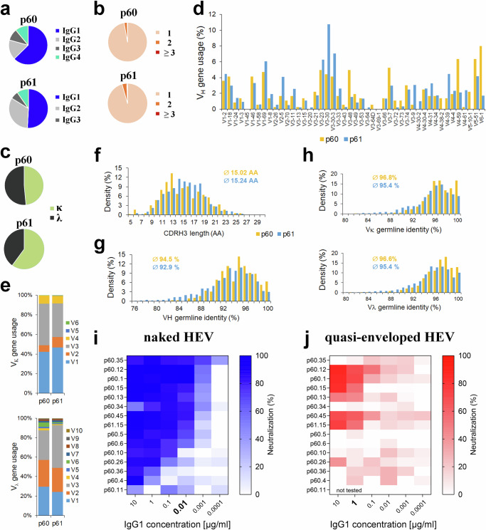
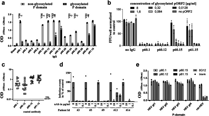
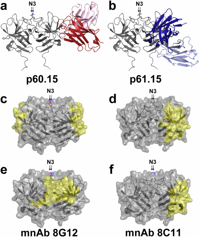
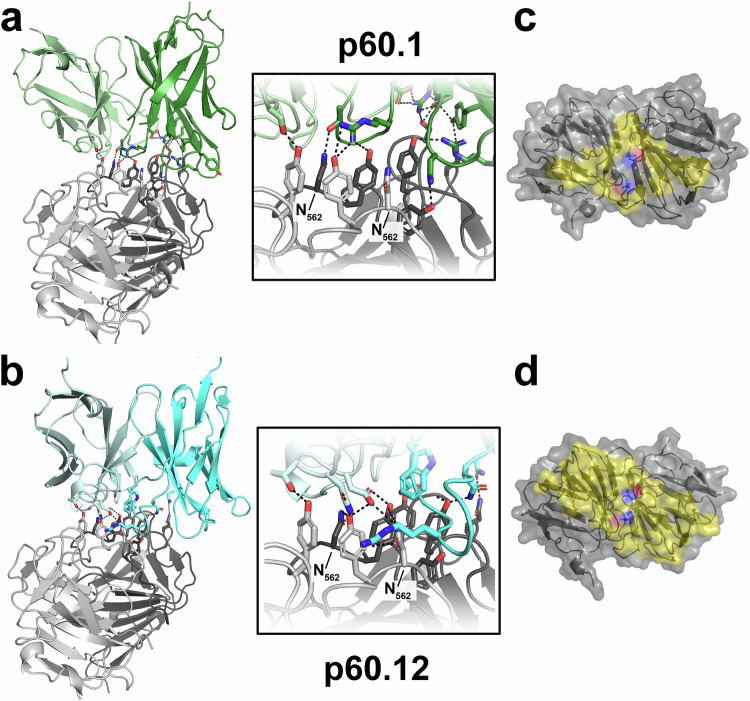
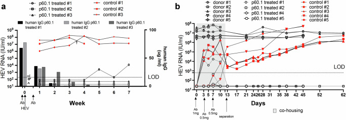
Hepatitis E virus (HEV) causes 3.3 million symptomatic cases and 44,000 deaths per year. Chronic infections can arise in immunocompromised individuals, and pregnant women may suffer from fulminant disease as a consequence of HEV infection. Despite these important implications for public health, no specific antiviral treatment has been approved to date. Here, we report combined functional, biochemical, and X-ray crystallographic studies that characterize the human antibody response in convalescent HEV patients. We identified a class of potent and broadly neutralizing human antibodies (bnAbs), targeting a quaternary epitope located at the tip of the HEV capsid protein pORF2 that contains an N-glycosylation motif and is conserved across members of the Hepeviridae. These glycan-sensitive bnAbs specifically recognize the non-glycosylated pORF2 present in infectious particles but not the secreted glycosylated form acting as antibody decoy. Our most potent bnAb protects human liver-chimeric mice from intraperitoneal HEV challenge and co-housing exposure. These results provide insights into the bnAb response to this important emerging pathogen and support the development of glycan-sensitive antibodies to combat HEV infection.


Conflict of interest statement

Competing interests: G.S., K.D., T.K., and P.B. are listed as inventors on a patent “BROADLY NEUTRALIZING ANTIBODIES AGAINST HEPATITIS E VIRUS” describing the use of glycan-sensitive antibodies p60.1 and p60.12 targeting HEV pORF2 for diagnostics, prevention and treatment of HEV infection (EP 22 162 453.9). These authors declare no restrictions on the publication of data and the remaining authors declare no competing interests.

Figures

Fig. 1
Fig. 1. B cells of convalescent HEV patients encode potent nAbs.
HEV-specific B cell repertoire analysis from two donors showing the distribution of productive IgG subclasses (a), clonality within individual patients (b), and type of light chains (c) as well as heavy (d) and light chain (e) variable (V) gene usage. Frequencies of heavy chain complementarity determining region 3 (CDRH3) length (f) and % germline identity of heavy chain variable segment (VH) (g) and light chain variable segment Vκ/Vλ (h) are shown. Mean values for CDRH3 length and mean germline identity are given. (i + j) The heat maps show neutralization in % as measured by focus forming units per well against the naked (i) and quasi-enveloped (j) particles of the HEV GT 3 Kernow-C1 p6 G1634R strain. (n = 3 biological replicates). Source data are provided within the Source Data file.
Fig. 2
Fig. 2. Characterization of pORF2 nAbs.
a IgG antibody binding to a non-glycosylated HEV GT 3 P domain purified from the cytoplasm (light gray) and an identical P domain glycosylated in the secretory pathway (dark gray; n = 3 biological replicates). Variable heavy chain germline segments (VH) of glycan-sensitive antibodies are indicated above the bar plot. b Neutralization of naked viral particles of HEV Kernow-C1 p6 G1634R using 0.1 µg/ml of the respective nAb after pre-incubation with increasing concentrations of glycosylated P domain. The graph depicts the number of infected cells normalised to the control. (n = 3 biological replicates) c ELISA analysis on plasma of 14 viraemic patients reveals that all tested nAbs bind to patient isolate antigens. (n = 3 biological replicates). Overall, glycan sensitive nAbs show approximately 10x lower optical density (OD) values. d Clinical HEV isolates are efficiently neutralized by bnAb p60.1. A total of five stool filtrates from HEV patients were used to infect stem-cell derived hepatocyte-like cells with or without p60.1 at indicated concentrations (n = 2 biological replicates). 7 days post-infection, HEV infection was analyzed by quantifying pORF2-positive cells using Zen software. e IgG antibody binding to a non-glycosylated HEV P domain derived from the indicated HEV genotype demonstrates broad reactivity for all four tested nAbs (n = 3 biological replicates). Only glycan-sensitive nAbs also recognize the P domain derived from rat HEV. All ELISA experiments shown in panel a, b and e were performed at least in technical triplicates. Source data are provided within the Source Data file. Error bars represent the standard deviation.
Fig. 3
Fig. 3. Crystal structure of P domain in complex with glycan-insensitive bnAbs.
Cartoon view of complex structures of the GT3 P domain (gray) with p60.15 (red; a) and p61.15 scFv (blue; b), which recognize partially overlapping epitopes on the P domain dimer (gray); only the bnAb fragment binding to the right P domain protomer is shown for clarity. The position of the N-linked glycosylation site N3 is indicated. cf Side-view of the footprints (yellow) of human “glycan-insensitive“ bnAbs on the P domain dimer (gray) in comparison to described murine nAbs 8G12 (PDB 4PLJ;; e) and 8C11 (PDB 3RKD;; f). 8G12 is the only antibody that binds at the P domain dimer interface. Sidechain oxygen and nitrogen atoms of N562 are colored in red and blue, respectively, to indicate the position of N3.
Fig. 4
Fig. 4. Crystal structure of P domain in complex with glycan-sensitive bnAbs.
Cartoon view of P domain structures in complex with p60.1 (green; a) and p60.12 (cyan; b) that recognize partially overlapping epitopes at the tip of the P domain dimer (individual protomers colored in light and dark gray). Same view as in Fig. 3a, b. The insets show zoomed views on the interface with a particular focus on the two sidechains of N562. Sidechain oxygen and nitrogen atoms are colored in red and blue, respectively, dashed lines indicate the complex hydrogen binding network stabilizing the high affinity interaction. c + d Top-view of the footprints of human glycan-sensitive bnAbs on the P domain dimer (gray). Sidechain oxygen and nitrogen atoms of N562 are colored in red and blue, respectively, to indicate the position of N3.
Fig. 5
Fig. 5. Human bnAb p60.1 protects against HEV (a) intraperitoneal injection and (b) co-housing transmission.
a Naïve human liver chimeric mice were pre-treated with antibody p60.1 or a PBS control followed by HEV GT 3 challenge 24 hrs later and a second treatment on day 4. The quantity of human antibody in serum was determined by ELISA. (q.l. = upper quantification limit). b Naïve human liver chimeric mice were pre-treated with antibody p60.1 or an irrelevant control antibody prior to 12 days of co-housing with HEV GT 1-infected human liver chimeric mice. In both cases, HEV infection of individual mice was monitored by RT-PCR to detect HEV genomes in feces. Source data are provided within the Source Data file. LOD lower limit of detection.

References

    1. Li, P. et al. The global epidemiology of hepatitis E virus infection: A systematic review and meta-analysis. Liver Int.40, 1516–1528 (2020). - PMC - PubMed
    1. Van der Poel, W. H. Food and environmental routes of Hepatitis E virus transmission. Curr. Opin. Virol.4, 91–96 (2014). - PubMed
    1. Kamar, N. et al. Hepatitis E virus infection. Nat. Rev. Dis. Prim.3, 17086 (2017). - PubMed
    1. Teshale, E. H. & Hu, D. J. Hepatitis E: Epidemiology and prevention. World J. Hepatol.3, 285–291, (2011). - PMC - PubMed
    1. Ingiliz, P. et al. Persisting hepatitis E virus infection leading to liver cirrhosis despite recovery of the immune system in an HIV-infected patient. Clin. Res Hepatol. Gastroenterol.40, e23–e25 (2016). - PubMed

MeSH terms

LinkOut - more resources