. 2025 Feb 26;11(1):36.
doi: 10.1038/s41522-025-00667-0.

Microbiome modulation of implant-related infection by a novel miniaturized pulsed electromagnetic field device

Affiliations

Microbiome modulation of implant-related infection by a novel miniaturized pulsed electromagnetic field device

João Gabriel S Souza et al. NPJ Biofilms Microbiomes. .

Abstract

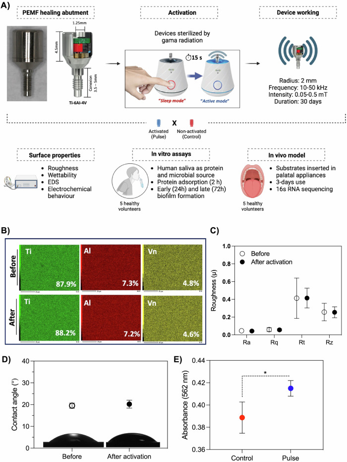
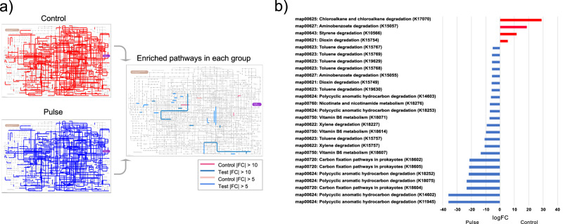
Dental implant-related infections, which lack effective therapeutic strategies, are considered the primary cause for treatment failure. Pulsed electromagnetic field (PEMF) technology has been introduced as a safe and effective modality for enhancing biological responses. However, the PEMF effect on modulating microbial diversity has not been explored. Thus, we tested a miniaturized PEMF biomedical device as a healing component for dental implants. PEMF activation did not alter the chemical composition, surface roughness, wettability, and electrochemical performance. PEMF effectively controlled chronic in vitro polymicrobial microbial accumulation. The in vivo study where devices were inserted in the patients' oral cavities and 16S RNA sequencing analysis evidenced a fivefold or more reduction in 23 bacterial species for PEMF group and the absence of some species for this group, including pathogens associated with implant-related infections. PEMF altered bacterial interactions and promoted specific bacterial pathways. PEMF has emerged as an effective strategy for controlling implant-related infections.


Conflict of interest statement

Competing interests: Shlomo Barak work at Magdent Company. The other authors declare no competing interested.

Figures

Fig. 1
Fig. 1. Pulsed electromagnetic field (PEMF) evaluation.
A Schematic sequence of experimental design. Miniaturized PEMF devices as dental implant healing abutment/component was used. Devices were activated to start pulse emission. Non-activated or device after activation phase (30 days) were used as control. Devices were evaluated in terms of surface properties, in vitro biofilm model and protein adsorption, and in vivo models, where the devices were inserted in the oral cavity of volunteers. B chemical composition of the devices before and after the 30 day activation phase evaluated by energy dispersive spectroscopy. C Surface roughness of the devices before and after 30 day activation phase. D Surface wettability by water contact angle. E Salivary protein adsorption on non-activated (control) and activated (pulse) devices. Pool of stimulated human saliva (5 healthy volunteers) was used for protein adsorption (2 h). Total protein quantification was performed using bicinchoninic acid method and absorbances measured. * Indicates statistical difference (p < 0.05) by Bonferroni t-test.
Fig. 2
Fig. 2. Electrochemical behavior of activated pulsed electromagnetic device (pulse), compared with non-activated (control).
Electrochemical tests were conducted for analyzing the corrosion stability of pulsed and control healing abutments in artificial saliva. A Open circuit potential analysis, performed for 3600 s to evaluate the free corrosion potential of the material. B Nyquist diagram. C Bode plots showing variations in impedance as a function of frequency. D Potentiodynamic polarization curves. E Corrosion rate.
Fig. 3
Fig. 3. Pulsed electromagnetic field (PEMF) evaluated in terms of in vitro polymicrobial biofilm formation.
A Stimulated human saliva (5 healthy volunteers) was used as microbial inoculum/source. Biofilms were incubated for 24 and 72 h and evaluated in terms of live bacterial cells count, structure, pH and composition. B Colony-forming units (CFU) for total live cell counts. Biofilm suspension was serially diluted and plated on Columbia Blood Agar (CBA) for measures. C Biofilm pH medium as bacterial metabolism analysis was evaluated at the beginning and after 72 h, using a pH meter. D Scanning electron microscopy micrographs of both groups after 72 h of biofilm formation. The circles and yellow lines represent microbial (streptococcal) aggregates or mixed-species aggregation on the surface. * Indicates statistical difference (p < 0.05) by t-test.
Fig. 4
Fig. 4. Pulsed electromagnetic field (PEMF) evaluated in terms of in vitro polymicrobial biofilm formation.
Microbiological composition of in vitro biofilms was evaluated by checkerboard DNA–DNA hybridization technique, to assess the presence and levels of 40 bacterial species associated to dental implant-related infections. A Levels (x105) of 40 bacterial species evaluated for both groups, pulse (activated PEMF) and control (non-activated) as average and standard deviation. B Periodontal microbial complexes by bacterial proportion. Bacterial species were grouped as previously described for microbial complexes related to oral infections. C Fold change of bacterial counts from control group divided by the counts in pulse group. Three bacterial species more associated with tissue damage in dental implant-related infections. During biofilm maturation and disease progression, initial colonizers start the process (complexes: Actinomyces – blue, yellow, green, and purple), followed by secondary colonizers (orange complex) that promote biofilm growth and create a suitable environment for the colonization of late colonizers (red complex), which are highly associated with tissue damage. * Indicates statistical difference (p < 0.05) by t-test.
Fig. 5
Fig. 5. Pulsed electromagnetic field (PEMF) evaluated in terms of in vivo polymicrobial biofilm formation.
A For this purpose, 5 healthy volunteers each wore one palatal appliance containing one activated PEMF (pulse) and one non-activated (control) device for 3 days to allow biofilm accumulation. Biofilm composition was evaluated by 16S rRNA sequencing for whole bacterial microbiome. B Alpha diversity analysis by Shannon Index and C Inv Simpson Index of sequenced samples. D Principal coordinates analysis (PCoA) using the Bray-Curtis distance function and ASV abundances. E Stacking bar charts showing dominant bacterial genus by sample and groups.
Fig. 6
Fig. 6. Pulsed electromagnetic field (PEMF) evaluated in terms of in vivo polymicrobial biofilm formation.
For this purpose, 5 healthy volunteers each wore one palatal appliance containing one activated PEMF (pulse) and one non-activated (control) device for 3 days to allow biofilm accumulation. Biofilm composition was evaluated by 16S rRNA sequencing for whole bacterial microbiome. A Heatmap showing the absence or presence of bacterial species for control or pulse group. B Heatmap showing the bacterial species with increased level of abundance of at least 5 times in the control group, compared with the pulse group (ASV - control/pulse). Pulse – activated PEMF; Control – non-activated PEMF.
Fig. 7
Fig. 7. Co-occurrence networks evaluated the bacterial positive correlation.
In all networks, each bacterium was represented by a node, and edges indicated the co-occurrence relationships between them. Bold lines indicated the interactions with significant co-occurrence. A Red - control group; (B) Blue—pulse group.
Fig. 8
Fig. 8. Evaluation of predicted pathways.
a Predicted KEGG pathways were presented in any of the 5 samples for each group (left map) (Pulse - test / control), and only enriched pathways were in each group (right map). b Differential expression pattern of enriched pathways (|FC | > 5). FC – fold change.
Fig. 9
Fig. 9. Hypothesis of mechanisms by which Pulsed Electromagnetic miniaturized device (PEMF) modulates biofilm accumulation.
Non-activated PEMF allows polymicrobial biofilm accumulation with increased pathogens growth. In contrast, activated PEMF showed reduced polymicrobial biofilm formation. Some mechanisms has been raised based on our findings and previous evidence: (1) electroporation effect on microbial cell membranes resulting in bacterial killing; (2) modulation of bacterial-bacterial interactions due electrostatic forces changes; (3) changes in cell-wall surface molecules and surface charges modulating microbial growth.

References

    1. Elani, H. W., Starr, J. R., Da Silva, J. D. & Gallucci, G. O. Trends in dental implant use in the U.S., 1999-2016, and projections to 2026. J. Dent. Res.97, 1424–1430 (2018). - PMC - PubMed
    1. Lybrand, K. E. & Althausen, P. L. The role of value-based implants in orthopedic trauma. Orthop. Clin. North Am.49, 437–443 (2018). - PubMed
    1. Pedrinaci, I. et al. Implant survival in the anterior mandible: a retrospective cohort study. Clin. Oral. Implants Res.34, 463–474 (2023). - PubMed
    1. Pjetursson, B. E. et al. Comparison of survival and complication rates of tooth-supported fixed dental prostheses (FDPs) and implant-supported FDPs and single crowns (SCs). Clin. Oral. Implants Res.18, 97–113 (2007). - PubMed
    1. Albrektsson, T. & Wennerberg, A. On osseointegration in relation to implant surfaces. Clin. Implant Dent. Relat. Res.21, 4–7 (2019). - PubMed