. 2025 Jul;46(7):1864-1876.
doi: 10.1038/s41401-025-01506-w. Epub 2025 Feb 26.

Costunolide normalizes neuroinflammation and improves neurogenesis deficits in a mouse model of depression through inhibiting microglial Akt/mTOR/NF-κB pathway

Affiliations

Costunolide normalizes neuroinflammation and improves neurogenesis deficits in a mouse model of depression through inhibiting microglial Akt/mTOR/NF-κB pathway

Shao-Qi Zhang et al. Acta Pharmacol Sin. 2025 Jul.

Abstract

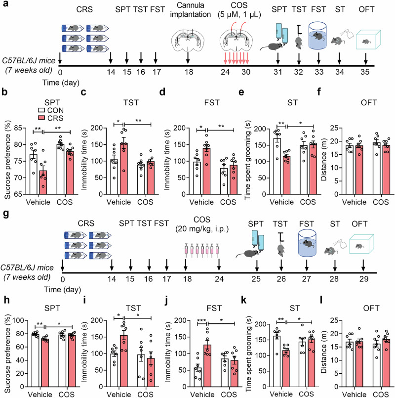
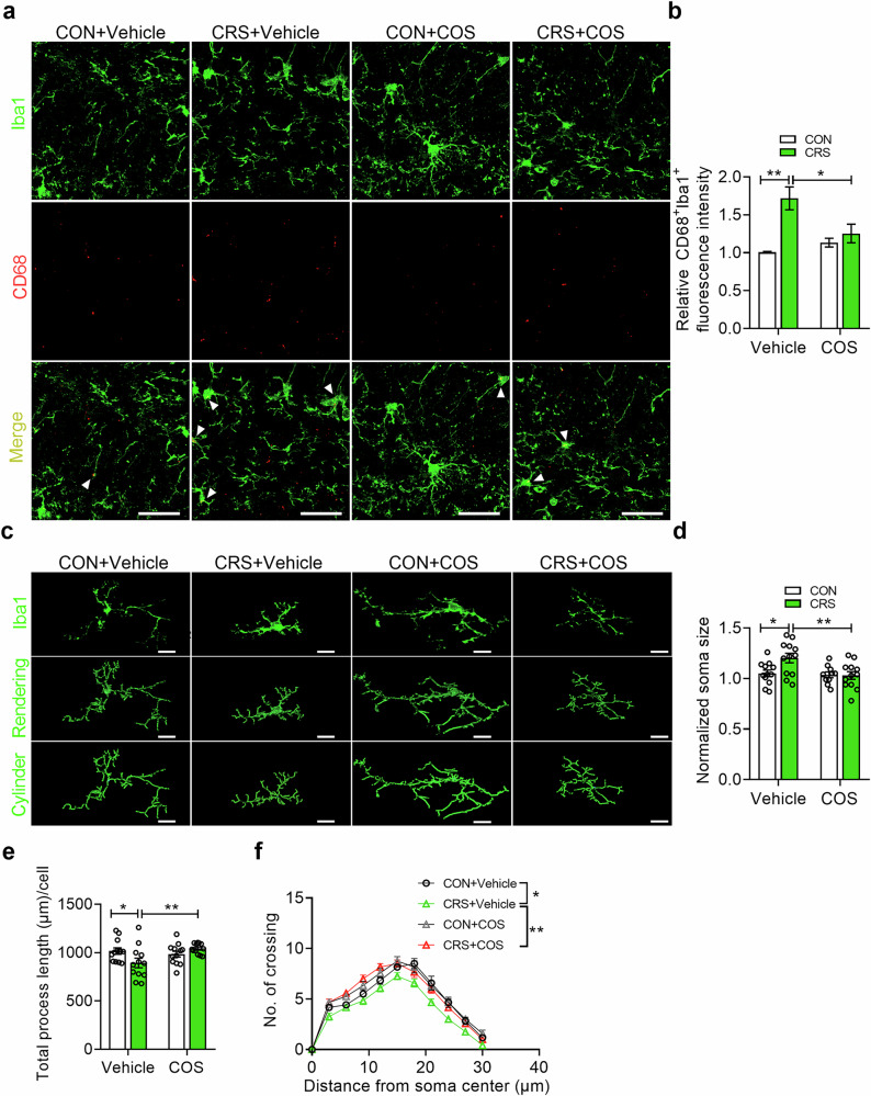
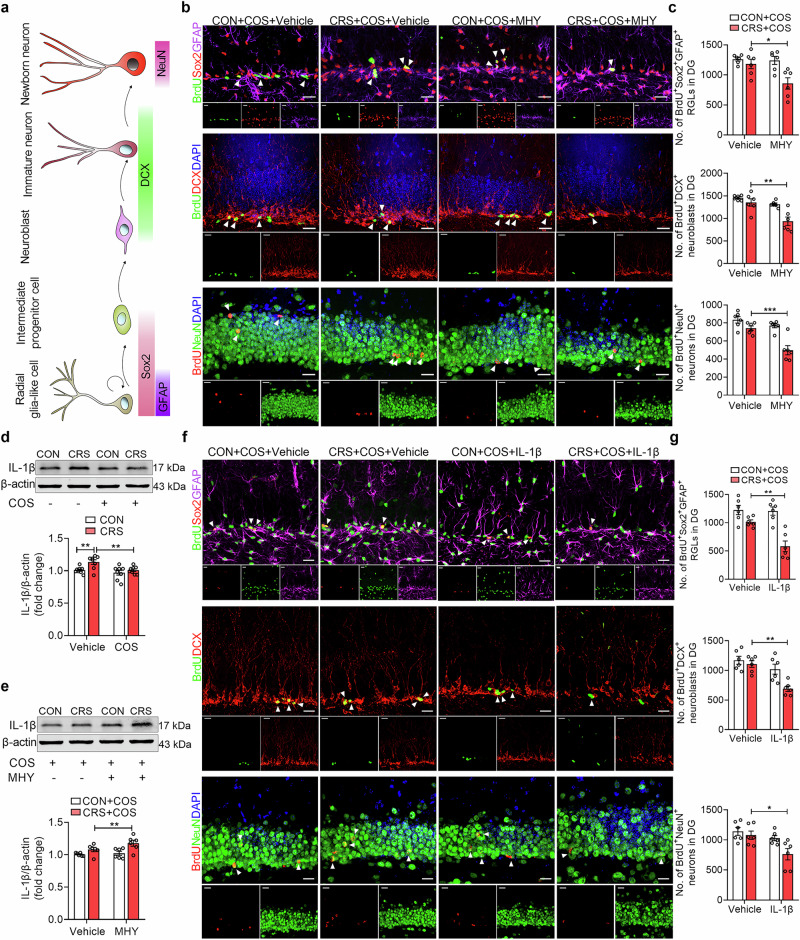
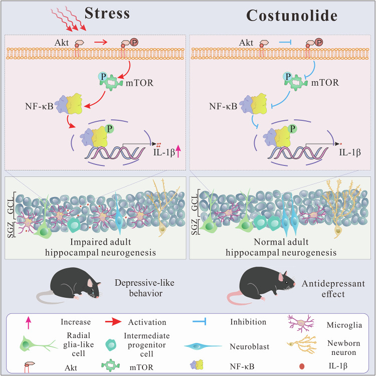
Neuroinflammation is crucial for the pathogenesis of major depression. Preclinical studies have shown the potential of anti-inflammatory agents, specifically costunolide (COS), correlate with antidepressant effects. In this study, we investigated the molecular mechanisms underlying the antidepressant actions of COS. Chronic restraint stress (CRS) was induced in male mice. The mice were treated with either intra-DG injection of COS (5 μM, 1 μL per side) or COS (20 mg/kg, i.p.) for 1 week. We showed that administration of COS through the both routes significantly ameliorated the depressive-like behavior in CRS-exposed mice. Furthermore, administration of COS significantly improved chronic stress-induced adult hippocampal neurogenesis deficits in the mice through attenuating microglia-derived neuroinflammation. We demonstrated that COS (5 μM) exerted anti-neuroinflammatory effects in LPS-treated BV2 cells via inhibiting microglial Akt/mTOR/NF-κB pathway; inactivation of mTOR/NF-κB/IL-1β pathway was required for the pro-neurogenic action of COS in CRS-exposed mice. Our results reveal the antidepressant mechanism of COS that is normalizing neuroinflammation to improve neurogenesis deficits, supporting anti-inflammatory agents as a potential therapeutic strategy for depression.

Keywords: chronic stress; costunolide; mTOR; major depression; microglia; neurogenesis.


Conflict of interest statement

Competing interests: The authors declare no competing interests.

Figures

Fig. 1
Fig. 1. COS treatment ameliorates depressive-like behavior of male mice.
a The experimental timeline. be Behavioral tests showed that intra-DG injection of COS (5 μM, 1 μL per side) ameliorated depressive-like behavior in CRS-exposed mice, including increased sucrose preference in SPT (b), decreased immobility time in TST (c) and FST (d), increased grooming time in ST (e) (n = 7 mice per group). f Open-field test showed that COS did not affect locomotor activity of mice (n = 7 mice per group). g The experimental timeline. h–k Behavioral tests showed that COS (20 mg/kg, i.p.) produced antidepressant action in CRS-exposed mice, including increased sucrose preference in SPT (h), decreased immobility time in TST (i) and FST (j), increased grooming time in ST (k) (n = 7 mice per group). l Open-field test showed that COS did not affect locomotor activity of mice (n = 7 mice per group). Data are presented as mean ± SEM, *P < 0.05, **P < 0.01, ***P < 0.001 by two-way ANOVA (bf, hl) followed by Sidak’s post hoc test. The statistical details can be found in Supplementary Table S1.
Fig. 2
Fig. 2. COS attenuates microglial hyperactivation in the DG induced by CRS.
a Representative images of Iba1 (green) and CD68 (red) positive microglia in DG of control and CRS-exposed mice after vehicle or COS treatment. Scale bars, 30 μm. b Quantification of Iba1 and CD68 immunostaining in DG (n = 3 mice per group). c Representative images of Iba1 (top), 3D reconstruction (middle) and cylinder (bottom) of microglia in DG of control and CRS-exposed mice after vehicle or COS treatment. Scale bars, 10 μm. df Quantification of Iba1 positive cell soma size (d), total process length (e) and number of intersections (f) of microglia in DG (n = 10–12 cells from 3 mice per group). Data are presented as mean ± SEM, *P < 0.05, **P < 0.01 by two-way ANOVA (b, d, e), repeated measures ANOVA (f) followed by Sidak’s post hoc test. The statistical details can be found in Supplementary Table S1.
Fig. 3
Fig. 3. COS improves CRS-induced AHN deficits in DG.
a The experimental timeline. b and c Representative images (b) and quantification (c) of BrdU+Sox2+GFAP+ RGLs and BrdU+DCX+ neuroblasts in DG of control and CRS-exposed mice after vehicle or COS treatment (n = 3 mice per group). White arrows indicate BrdU+ and marker+ cells. Scale bars, 20 μm. d Representative tracers of DCX+ immature neurons in DG of control and CRS-exposed mice after vehicle or COS treatment (n = 3 mice per group). Scale bars, 20 μm. e, and f Quantification of the dendritic length (e) and dendritic complexity (f) of DCX+ immature neurons in DG by Sholl analysis (n = 12 cells of 3 mice per group). g The experimental timeline. h and i Representative images (h) and quantification (i) of BrdU+NeuN+ newborn neurons in DG of control and CRS-exposed mice after vehicle or COS treatment (n = 12 cells of 3 mice per group). White arrows indicate the co-labeling cells. Scale bars, 20 μm. Data are presented as mean ± SEM, *P < 0.05, **P < 0.01, ***P < 0.001 by two-way ANOVA (c, e, i), repeated measures ANOVA (f) followed by Sidak’s post hoc test.
Fig. 4
Fig. 4. COS produces anti-neuroinflammatory effects via inhibiting microglial Akt/mTOR/NF-κB pathway.
ae Representative images (a) of Western blotting analysis and quantification showed that COS treatment prevented the increased phosphorylation protein expression of Akt (b), mTOR (c), S6 (d), NF-κB p65 (e) in BV2 cells induced by LPS exposure (n = 6 independent experiments per group). fj Representative images (f) of Western blotting analysis and quantification showed that COS treatment prevented the increased phosphorylation protein expression of Akt (g), mTOR (h), S6 (i), NF-κB p65 (j) in DG induced by CRS (n = 6 mice per group). k–n Representative images (k) of Western blotting analysis and quantification showed that MHY1458 treatment (10 μM, 1 μL per side) blocked the decreased phosphorylation protein expression of mTOR (l), S6 (m), NF-κB p65 (n) in DG produced by COS treatment of CRS-exposed mice (n = 6 mice per group). o Representative images of Iba1 (top), three-dimensional (3D) reconstruction (middle) and cylinder (bottom) of microglia in DG (n = 12 cells from 3 mice per group). Scale bars, 10 μm. pr Quantification showed that MHY1458 treatment blocked the decreased Iba1+ cell soma size (p) and the increased total process length (q) and number of intersections (r) of microglia in DG by COS treatment of CRS-exposed mice (n = 14 cells from 3 mice per group). Data are presented as mean ± SEM, *P < 0.05, **P < 0.01, ***P < 0.001 by two-way ANOVA (be, gj, ln, p, q), repeated measures ANOVA (r) followed by Sidak’s post hoc test.
Fig. 5
Fig. 5. Inactivation of mTOR/NF-κB/IL-1β pathway is required for the pro-neurogenic action of COS.
a Schematic paradigms of cell-lineage-specific markers in DG during AHN. b, c Representative images (b) and quantification (c) of BrdU+Sox2+GFAP+ RGLs, BrdU+DCX+ neuroblasts, and BrdU+NeuN+ newborn neurons in DG (n = 6 mice per group). White arrows indicate BrdU+ and Marker+ cells. Scale bars, 20 μm. d Treatment with COS (5 μM, 1 μL per side) reversed the increased IL-1β protein expression in DG of CRS-exposed mice (n = 8 mice per group). e Intra-DG injection of MHY1485 (10 μM, 1 μL per side) abolished the decreased IL-1β protein expression of CRS-exposed mice produced by COS (n = 6 mice per group). f, g Representative images (f) and quantification (g) of BrdU+Sox2+GFAP+ RGLs, BrdU+DCX+ neuroblasts, and BrdU+NeuN+ newborn neurons in DG (n = 6 mice per group). White arrows indicate BrdU+ and Marker+ cells. Scale bars, 20 μm. Data are presented as mean ± SEM, *P < 0.05, **P < 0.01, ***P < 0.001 by two-way ANOVA (c, d, e, g) followed by Sidak’s post hoc test.
Fig. 6
Fig. 6. Inhibition mTOR/NF-κB/IL-1β pathway confers to the antidepressant effects of COS.
a The experimental timeline. be Behavioral tests showed that MHY1485 (10 μM, 1 μL per side) reversed the antidepressant effects produced by COS (20 mg/kg, i.p.) in CRS-exposed mice, including decreased sucrose preference in SPT (b), increased immobile time in TST (c) and FST (d), and decreased grooming time in ST (e) (n = 7 mice per group). f The experimental timeline. g-j Behavioral test showed that IL-1β (5 μg/kg, 1 μL per side) abolished antidepressant effects induced by COS in CRS-exposed mice, including decreased sucrose preference in SPT (g), increased immobile time in TST (h) and FST (i), and decreased grooming time in ST (j) (n = 7 mice per group). Data are presented as mean ± SEM, *P < 0.05, ***P < 0.001 by two-way ANOVA (be, gj) followed by Sidak’s post hoc test.
Fig. 7
Fig. 7. Schematic representation of the microglia activation-specific mechanism of COS in mediating the impairment of AHN in chronic stress-induced depression.
COS ameliorates IL-1β-mediated neurogenesis deficits of DG via inhibiting microglial Akt/mTOR/NF-κB-dependent neuroinflammation, further improving chronic stress-induced depressive-like behavior.

References

    1. Smith K. Mental health: a world of depression. Nature. 2014;515:181. - PubMed
    1. Collaborators GBDIBD. The global, regional, and national burden of inflammatory bowel disease in 195 countries and territories, 1990-2017: a systematic analysis for the Global Burden of Disease Study 2017. Lancet Gastroenterol Hepatol. 2020; 5:17–30. - PMC - PubMed
    1. First MB. Diagnostic and statistical manual of mental disorders, 5th edition, and clinical utility. J Nerv Ment Dis. 2013;201:727–9. - PubMed
    1. Setiawan E, Attwells S, Wilson AA, Mizrahi R, Rusjan PM, Miler L, et al. Association of translocator protein total distribution volume with duration of untreated major depressive disorder: a cross-sectional study. Lancet Psychiatry. 2018;5:339–47. - PubMed
    1. Kettenmann H, Hanisch UK, Noda M, Verkhratsky A. Physiology of microglia. Physiol Rev. 2011;91:461–553. - PubMed

MeSH terms