. 2025 Feb 26;8(1):316.
doi: 10.1038/s42003-025-07751-3.

Thrombin cleaves membrane-bound endoglin potentially contributing to the heterogeneity of circulating endoglin in preeclampsia

Affiliations

Thrombin cleaves membrane-bound endoglin potentially contributing to the heterogeneity of circulating endoglin in preeclampsia

Divina El Hamaoui et al. Commun Biol. .

Abstract

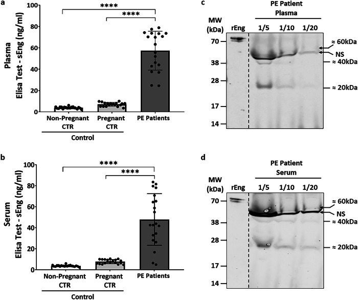
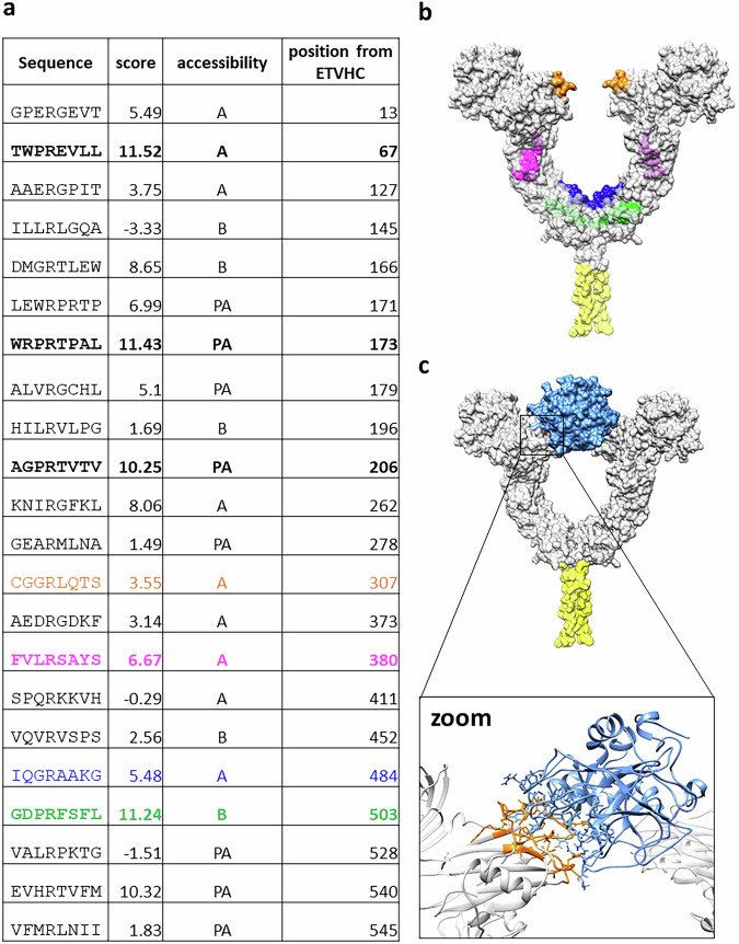
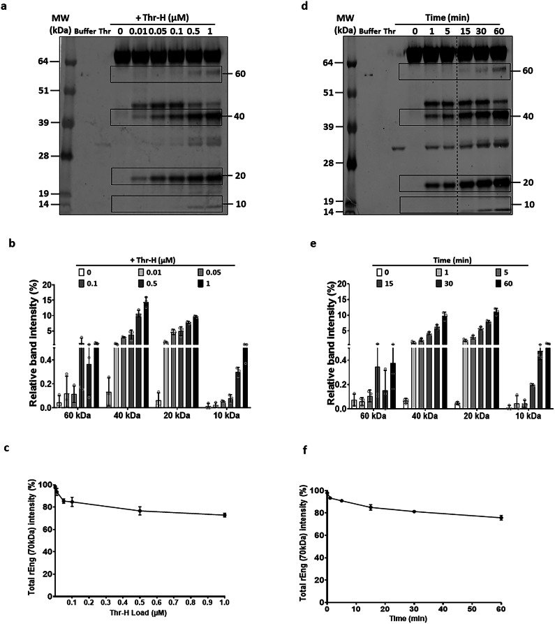
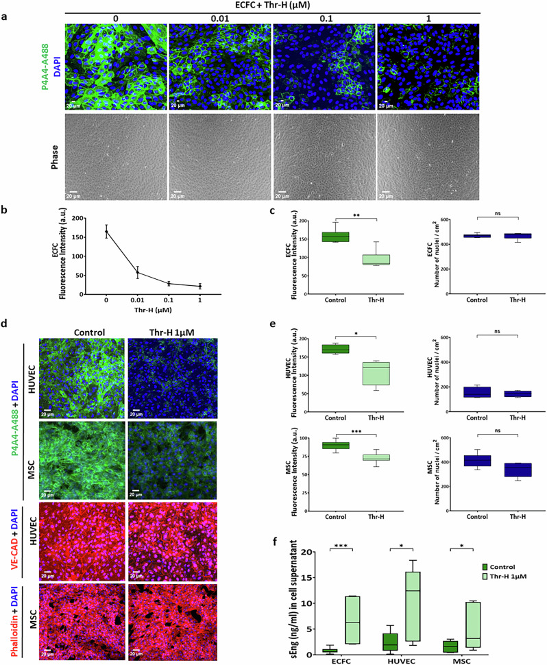
Increased levels of soluble endoglin (sEng) are found in serum, plasma, and urine of preeclampsia patients. sEng is released from membrane-bound endoglin through the proteolytic activity of metalloproteases, but its structural heterogeneity suggests the involvement of additional proteases. Considering the roles of thrombin and sEng in preeclampsia pathogenesis, we investigated whether thrombin cleaves endoglin. Sequence analysis revealed a conserved peptide in endoglin similar to the α-thrombin cleavage site of protease-activated receptor-1. Western blot analysis of plasma from preeclamptic women showed endoglin fragments consistent with thrombin-mediated cleavage. Incubation of purified endoglin with thrombin generated specific fragments, whose N- and C-terminal sequencing confirmed the predicted cleavage sites. Furthermore, thrombin treatment of endoglin-expressing cells released sEng and reduced cell surface endoglin. These findings suggest that multiple protease-targeted cleavage sites lead to the generation of sEng fragments, which may reflect endothelial dysfunction and preeclampsia progression.


Conflict of interest statement

Competing interests: A patent (No. EP24307100) entitled ‘Peptide derived from endoglin for treating bleeding disorders’ was filed by inventor Elisa Rossi and supported by INSERM Transfert (Institut National de la Santé et de la Recherche Médicale) in France.

Figures

Fig. 1
Fig. 1. Plasma and serum analysis of patients with preeclampsia.
The levels of sEng in (a) plasma and in (b) serum were quantified using an ELISA kit assay in a cohort of 60 patients. This cohort comprised 20 non-pregnant women controls, 20 pregnant women controls, and 20 cases diagnosed with preeclampsia. c Plasma and (d) serum samples from preeclampsia patients diluted to ratios of 1:5, 1:10, or 1:20 and reduced with DDT, were subjected to analysis through SDS-PAGE and Western blot with Endoglin/CD105 polyclonal rabbit antibody (ProteinTech). rEng at 100 ng/mL was used as control. The data is presented as mean ± S.D. Statistical significance was determined at *P < 0.05, **P < 0.01, ***P < 0.001, and ****P < 0.0001. NS indicates a non-specific band.
Fig. 2
Fig. 2. Predictive study to identify possible cleavage motifs.
a Possible cleavage motifs found by the Profile Specific Scoring Matrix analysis (left column) and their associated score (right column) combined with accessibility (A: highly accessible; PA: partially accessible; B: buried) giving the extent to which each sequence corresponds to the profile extracted from known thrombin cleavage motifs. The scale of the score is arbitrary and the sequences highlighted in bold have significantly higher scores (score > 6) than the others and are accessible on the surface of Eng within the context of its open homodimeric structure. Colored sequences correspond to those later determined experimentally as cleavage sites. b Theoretical model of endoglin showing possible thrombin cleavage sites which corresponds to location of the experimentally determined cleavage sites on the structure of the open Eng. c The top panel shows a theoretical model of thrombin (blue) in complex with the CGGRLQTS sequence of endoglin, obtained by docking. A detailed view of the corresponding putative interaction site is shown in the bottom panel (“zoom”).
Fig. 3
Fig. 3. Dose and time dependent cleavage of human rEng by thrombin.
rEng at a concentration of 20 µg/mL was treated at 37 °C a with increasing concentrations of Thr-H : 0, 0.01, 0.05, 0.1, 0.5, or 1 µM for 1 h. Following treatment, the samples were resolved using SDS-PAGE and subsequently analyzed by Western blotting with Endoglin/CD105 polyclonal rabbit antibody (ProteinTech). b The percentage of all cleaved bands obtained in a by the action of Thr-H on Eng was quantified relatively to the total rEng (70 kDa). c Percentage of the 70 kDa band reduction, specific to the total rEng band from a. d The same procedure described in a was repeated (Eng 20 µg/mL, treatment at 37 °C), but using a fixed concentration of 1 µM of Thr-H for varying durations: 0, 1, 5, 15, 30 or 60 min. e Graph of percentage of all cleaved bands resulting from the action of Thr-H on rEng at various time intervals. f Percentage of the 70 kDa band reduction, specific to the total rEng band from d. left. In a and d the band above 40 kDa (of approximately 50 kDa) was not reproducibly observed in the repeated experiments and was not further studied. The data is presented as mean ± S.D. from three conducted experiments. All images analysis were performed using Image Studio™ Lite software. The dashed line in panel d indicates a cut made in the gel where a blank column had been.
Fig. 4
Fig. 4. Sequencing of thrombin-cleaved endoglin bands.
a rEng at a concentration of 20 µg/mL was treated at 37 °C for 1 h with 1 µM of Thr-H. The samples were then resolved by SDS-PAGE and subjected to Coomassie Brilliant Blue staining to identify protein spots for subsequent sequencing. Bands at molecular weights of 60, 40, 20, 10, and 8 kDa were excised, with a minimum of 30 bands at each molecular weight selected for further analysis. Plots showing b the decrease in the percentage of the 70 kDa band, specific to the total rEng band and c the percentage of all cleaved bands obtained by the action of Thr-H on Eng, quantified relatively to the total rEng (70 kDa). The optical densities of each Coomassie blue-stained protein band were measured using Image Studio™ Lite software. The data is presented as mean ± S.D. from a minimum of 12 conducted experiments. Statistical significance was considered at *P < 0.05. d Table showing the N-terminal and C-terminal parts of each sequenced band, along with the cleavage position and the cleaved amino acids. The C-terminal information was not determined (ND) for the 8, 40 and 60 kDa bands, and the cleavage position and cleaved amino acids for the last two bands were also unidentified. However, based on the docking data and the Western blot analysis with the monoclonal antibody MAB1097 (R&D), that specifically labels the endoglin orphan region, for we can postulate that the 40 KDa band corresponds to the fragment 329–330. e Schematic diagram illustrating the N-terminal cleavage positions and sequences of each band, including known (solid line) and hypothetical (dashed line) C-terminal cleavages. Because the contribution of glycosylation to the molecular weight of each band is unknown, the correlation between molecular weight and primary structure is not to scale. f Schematic diagram of mEng, indicating cleavage sites. mEng is composed of an orphan region followed by a juxtamembrane zona pellucida (ZP) module and by transmembrane and short cytoplasmic domains. JM: Juxtamembrane; TM: Transmembrane; CP: Cytoplasmic.
Fig. 5
Fig. 5. In vitro cleavage of cellular Endoglin by thrombin.
a Immunofluorescence staining of Endoglin was conducted using the Endoglin/CD105 P4A4-Alexa 488 antibody (green) and vectashield mounting medium with DAPI (blue) after treating ECFC with increasing concentrations of Thr-H (0.01, 0.1 and 1 µM). Observations were carried out using confocal microscopy at 20x magnification. Phase photos at 10x magnification were taken for each condition to confirm the presence of the cell monolayer. b Endoglin fluorescence intensity following ECFC treatment with increasing concentrations of Thr-H (0.01, 0.1 and 1 µM) was quantified using ImageJ software (n = 3). c Plots showing the quantification of Eng fluorescence intensity in ECFC following treatment with 1 µM of Thr-H, alongside the number of nuclei relative to the surface. d Immunofluorescence staining of Eng using the Endoglin/CD105 P4A4-Alexa 488 antibody in different cell types, including HUVECs and MSCs, mounted with Vectashield containing DAPI. HUVECs were additionally stained with VE-Cadherin (VE-CAD) using a Texas Red-conjugated antibody, and MSCs were stained with Phalloidin-Alexa 555 to assess cell integrity after treatment with Thr. e Plots showing the quantification of Eng fluorescence intensity in HUVEC and MSC following treatment with 1 µM of Thr-H, alongside the number of nuclei relative to the surface. f ELISA kit assay results for sEng in supernatants of ECFC, HUVEC and MSC treated or not with 1 µM of Thr-H. The data is presented as mean ± S.D. for a minimum of n = 4. Statistical significance was considered at *P < 0.05, **P < 0.01, ***P < 0.001, and ****P < 0.0001.

References

    1. López-Novoa, J. M. & Bernabeu, C. The physiological role of endoglin in the cardiovascular system. Am. J. Physiol. Heart Circ. Physiol.299, H959–H974 (2010). - PubMed
    1. Rossi, E., Bernabeu, C. & Smadja, D. M. Endoglin as an Adhesion Molecule in Mature and Progenitor Endothelial Cells: A Function Beyond TGF-β. Front. Med. (Lausanne)6, 10 (2019). - PMC - PubMed
    1. Rossi, E. et al. Co-injection of mesenchymal stem cells with endothelial progenitor cells accelerates muscle recovery in hind limb ischemia through an endoglin-dependent mechanism. Thromb. Haemost.117, 1908–1918 (2017). - PubMed
    1. Shovlin, C. L. Hereditary haemorrhagic telangiectasia: Pathophysiology, diagnosis and treatment. Blood Rev.24, 203–219 (2010). - PubMed
    1. Rossi, E., Lopez-Novoa, J. M. & Bernabeu, C. Endoglin involvement in integrin-mediated cell adhesion as a putative pathogenic mechanism in hereditary hemorrhagic telangiectasia type 1 (HHT1). Front. Genet.5, 457 (2014). - PMC - PubMed

LinkOut - more resources