

Using deep learning to differentiate among histology renal tumor types in computed tomography scans
- PMID: 40011809
- PMCID: PMC11866614
- DOI: 10.1186/s12880-025-01606-3
Using deep learning to differentiate among histology renal tumor types in computed tomography scans
Abstract
Background: This study employed a convolutional neural network (CNN) to analyze computed tomography (CT) scans with the aim of differentiating among renal tumors according to histologic sub-type.
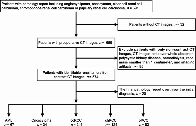
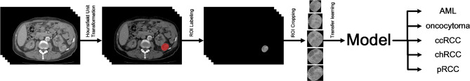
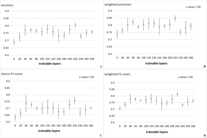
Methods: Contrast-enhanced CT images were collected from patients with renal tumors. The patient cohort was randomly split to create a training dataset (90%) and a testing dataset (10%). Following image dataset augmentation, Inception V3 and Resnet50 models were used to differentiate between renal tumors subtypes, including angiomyolipoma (AML), oncocytoma, clear cell renal cell carcinoma (ccRCC), chromophobe renal cell carcinoma (chRCC), and papillary renal cell carcinoma (pRCC). 5-fold cross validation was then used to evaluate the models in terms of classification performance.
Results: The study cohort comprised 554 patients, including those with angiomyolipoma (n = 67), oncocytoma (n = 34), clear cell renal cell carcinoma (n = 246), chromophobe renal cell carcinoma (n = 124), and papillary renal cell carcinoma (n = 83). Dataset augmentation of the training dataset included this to 4238 CT images for analysis. The accuracy of the models was as follows: Inception V3 (0.830) and Resnet 50 (0.849).
Conclusion: This study demonstrated the efficacy of using deep learning models for the classification of renal tumor subtypes from contrast-enhanced CT images. While the models showed promising accuracy, further development is necessary to improve their clinical applicability.
Keywords: Artificial intelligence; Deep learning; Histology; Renal tumor.
© 2025. The Author(s).
Conflict of interest statement
Declarations. Ethics approval and consent to participate: This study was approved by the ethics committee at Chang Gung Medical Foundation, Taiwan. After reviewing the project application, due to the retrospective nature of this study, the necessity of individual patient informed consent was waived by the ethics committee at Chang Gung Medical Foundation. All methods and procedures in this study were conducted in accordance with relevant guidelines and regulations. Consent for publication: Not applicable. Competing interests: The authors declare no competing interests. Clinical trial number: Not applicable.
Figures

References
-
- Fateh SM, Arkawazi LA, Tahir SH, Rashid RJ, Rahman DH, Aghaways I, Kakamad FH, Salih AM, Bapir R, Fakhralddin SS, Fattah FH, Abdalla BA, Mohammed SH. Renal cell carcinoma T staging: diagnostic accuracy of preoperative contrast-enhanced computed tomography. Mol Clin Oncol. 2023;18(2):11. - PMC - PubMed
-
- Johnson DC, Vukina J, Smith AB, et al. Preoperatively misclassified, surgically removed benign renal masses: a systematic review of surgical series and United States population level burden estimate. J Urol. 2015;193:30. - PubMed
-
- Kutikov A, Fossett LK, Ramchandani P, et al. Incidence of benign pathologic findings at partial nephrectomy for solitary renal mass presumed to be renal cell carcinoma on preoperative imaging. Urology. 2006;68:737. - PubMed
MeSH terms
LinkOut - more resources
Full Text Sources
Medical
