. 2025 Feb 12:15:1525083.
doi: 10.3389/fimmu.2024.1525083. eCollection 2024.

IFNλ1 is a STING-dependent mediator of DNA damage and induces immune activation in lung cancer

Affiliations

IFNλ1 is a STING-dependent mediator of DNA damage and induces immune activation in lung cancer

Stine Høvring Godsk et al. Front Immunol. .

Abstract

Introduction: The importance of the cGAS-STING pathway and type I interferon (IFN) in anti-tumor immunity has been widely studied. However, there is limited knowledge about the role of type III IFNs in cancer settings. Type III IFNs, comprising IFNλ1-4, are opposite to type I IFN only expressed by a few cell types, including epithelial cells, and the receptor subunit IFNLR1, is equally only expressed on limited types of cells.

Methods: Gene and protein expression of the cGAS-STING signaling pathway was characterized in a series of non-small cell lung cancer (NSCLC) cell lines. Herring-testis DNA stimulation and chemotherapy drugs (doxorubicin and cisplatin) were used to activate the cGAS-STING pathway, and the level of activation was determined by measuring changes in the transcriptomic profile as well as type I and III IFNs by ELISA. Re-expression of IFNLR1 on cancer cell lines was achieved using CRISPR activation (CRISPRa) followed by evaluating chemotherapy-induced apoptosis using flow cytometry assays.

Results: STING was not broadly expressed across the NSCLC cell lines. Those cancer cell lines expressing all relevant factors supporting the cGAS-STING pathway secreted IFNλ following STING activation whereas only few of them expressed IFNβ. Treatment with chemotherapy drugs likewise preferentially induced IFNλ, which was abrogated in CRISPR-Cas9 STING knock-out cells. Expression of IFNLR1 was found downregulated in the cancer cell lines compared to the benign epithelial cell line Nuli-1. Rescuing IFNLR1 expression by CRISPRa in multiple cancer cell lines sensitization them to IFNλ-stimulation and resulted in significant reduction in cell viability.

Conclusion: Downregulation of IFNLR1 can be an immune evasion mechanism developed by cancer cells to avoid responding to endogenous type III IFNs. Thus, rescuing IFNLR1 expression in NSCLC in conjunction to chemotherapy may potentially be harnessed to elevate the anti-tumoral responses.

Keywords: CRISPR/Cas9; NSCLC; STING; cancer immunology; interferon lambda.


Conflict of interest statement

The authors declare that the research was conducted in the absence of any commercial or financial relationships that could be construed as a potential conflict of interest.

Figures
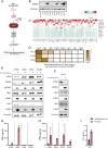
Figure 1
Figure 1
IFNλ is a broad marker of STING pathway activation in NSCLC. (A) An Illustration of the various factors essential for STING pathway activation. (B) Basal STING expression in 11 NSCLC cell lines detected using western blot. Vinculin was used as a loading control. (C) RNA sequencing data from six NSCLC cell lines demonstrating the expression levels of the 50 genes with the average highest log2 fold change, six hours after transfection with HT-DNA (2μg/mL). Data presented as heatmap with Z-scores. (D) A heatmap of IFNL1, IFNL2, and IFNB1 gene expression six hours post stimulation with HT-DNA (2ug/ml), measured by RT-qPCR, in eight NSCLC cell lines. (E, F) Evaluation of STING pathway activation in four NSCLC cell lines and the epithelial control cell line, Nuli-1, following transfection with HT-DNA (2μg/mL) or control for six hours. The expression of total STING, phospho(Ser366)STING, total TBK1, phospho(Ser172)TBK1, total IRF3, and phospho(Ser386)IRF3 were evaluated by western blot using vinculin as loading control. (G-I) NSCLC cell lines and Nuli-1 were stimulated with HT-DNA (2μg/mL) formulated with lipofectamine 2000. Supernatants were harvested after 20 hours and analyzed for IFNβ and IFNλ expression by ELISA. In (G, H) data is shown as mean +/- standard deviation of three independent experiments conducted in triplicates. In (I) mean of one experiment performed in duplicates is shown with +/- standard deviation indicated.
Figure 2
Figure 2
IFNλ and IFNβ share similar cell type dependent kinetic patterns. NSCLC cell lines and Nuli-1 cells were treated with HT-DNA (2μg/mL) formulated with lipofectamine 2000, for the indicated number of hours (hrs), where after gene (A-E) and protein (F-J) expression levels of IFNB1, IFNL1, and IFNL2 were determined. Data is shown as mean +/- standard deviation of duplicates from one experiment.
Figure 3
Figure 3
Doxorubicin and cisplatin induce IFNL1 and IFNB1 transcription. (A) An illustration of chemotherapy-induced STING pathway activation. (B-E) Gene expression of IFNL1 and IFNB1 was determined in NSCLC cell lines after a single IC50-value treatment with doxorubicin or cisplatin for 48 hours. Data is shown as mean +/- standard deviation of triplicates. Two-way ANOVA analysis using Šídák’s multiple comparisons test is performed. *P <.05, **P <.01, ***P value <.001, ****P <.0001.
Figure 4
Figure 4
Doxorubicin and cisplatin induce IFNL1 and IFNB1 transcription in a STING-dependent manner. (A) Western blot of STING expression measured in wild type (WT) or STING knockout (KO) of four NSCLC cell lines. Vinculin is used as loading control. (B-I) Mock and STING KO NSCLC cell lines were stimulated with doxorubicin (B-E) or cisplatin (F-I) for 48 hours. Cells were then harvested and gene expression of IFNL1 and IFNB1 determined by RT-qPCR. Data is shown as mean +/- standard deviation of triplicates. Two-way ANOVA analysis using Šídák’s multiple comparisons test is performed. *P <.05, **P <.01, ***P value <.001, ****P <.0001, ns = none significant.
Figure 5
Figure 5
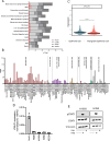
IFNLR1 expression is cell type dependent and downregulated in tumor cells. (A) Gene expression as normalized transcripts per million (nTMP) of IFNLR1, IL10RB, IFNAR1, and IFNAR2 expression in different human tissues generated from the Human Protein Atlas (proteinatlas.org) (59). (B) IFNLR1 gene expression as nTMP in different tissues generated from the Human Protein Atlas (proteinatlas.org) (65). (C) The expression of IFNLR1 in healthy and malignant epithelial cells obtained from 316 patients (healthy controls or lung cancer patients) using single cell RNA sequencing (42). The difference in expression between the groups was evaluated by a Wilcoxon Rank Sum test. ****P <.0001. (D) The basal gene expression of IFNLR1 in four NSCLC cell lines and Nuli-1 cells measured by RT-qPCR. Values are shown as mean +/- standard deviation from 3-5 replicates from one or two experiments. (E) Two NSCLC cell lines were treated for 15 min. with either IFNλ (100ng/mL), IFNβ (100ng/mL) or left untreated prior to cell lysis. The protein expression of STAT1 and pSTAT1 was detected by western blot. Vinculin was used as loading control.
Figure 6
Figure 6
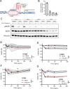
Transcriptional upregulation of IFNLR1 increases IFNλ sensitivity and leads to decreased viability. (A) Illustration of CRISPR activation (CRISPRa) of IFNLR1. Created using Biorender.com. (B) The H1650 cell line was subjected to CRISPRa of IFNLR1, and gene expression evaluated at day 1-4 after treatment. The IFNLR1 expression was normalized to H1650 mock control cells in which transcriptional activation was not performed. The data is shown as mean +/- standard deviation of two replicates from a single experiment. (C) The H1650 cell line was subjected to CRISPRa of IFNLR1 (IFNLR1+). At the indicated days, cells were treated with IFNλ (100ng/mL) or left untreated for 15 min. prior to harvest. The level of STAT1 and pSTAT1 expression was determined by western blot. H1650 mock control cells in which transcriptional activation was not performed were included for each time point. Vinculin was used as a loading control. (D-G) Viability measurement of H1650 (D, E) and H358 (F, G) cell lines. One day after CRISPRa of IFNLR1 and seeding, cells were treated with the indicated concentrations of IFNλ or IFNβ for 48 hours. Viability was compared to an untreated control for both IFNLR1+ and mock cells. Data is shown as mean +/- standard deviation (shaded area) of the average of four replicates from two-three experiments in (D-E, G) In (F) mean +/- standard deviation of four replicates are shown. Two-way ANOVA analysis using Šídák’s multiple comparisons test is performed comparing mock and IFNLR1+ in samples treated with 100ng/mL of IFNβ or IFNλ. *P <.05, **P <.01, ns = none significant.
Figure 7
Figure 7
Transcriptional upregulation of IFNLR1 supports chemotherapy-induced apoptosis. Two NSCLC cell lines were treated with cisplatin for 48 hours with (IFNLR1+) or without (Mock) CRISPRa for IFNLR1. (A, E) Viability was measured by cell count and normalized as percentage of an untreated mock. (B, C, F, G) Caspase 3/7 activation as an early marker of apoptosis was determined by flow cytometry. (B, F) show representative gating strategies. For data in (A, C, E, G), mean +/- standard deviation of 3-4 individual experiments are shown. Two-way ANOVA analysis using Šídák’s multiple comparisons test is performed. *P <.05, **P <.01. (D, H) A score of total caspase 3/7 activation combining both cells dead prior to flow cytometry analysis and cells analyzed by flow cytometry was calculated. Mean +/- standard deviation of 3-4 individual experiments are shown. Multiple paired T tests using Holm-Šídák’s method for multiple testing was performed. *P <.05, **P <.01, ns = none significant.

References

    1. Gresser I, Bourali C. Exogenous interferon and inducers of interferon in the treatment Balb-c mice inoculated with RC19 tumour cells. Nature. (1969) 223:844–5. doi: 10.1038/223844a0 - DOI - PubMed
    1. Yu R, Zhu B, Chen D. Type I interferon-mediated tumor immunity and its role in immunotherapy. Cell Mol Life Sci. (2022) 79:191. doi: 10.1007/s00018-022-04219-z - DOI - PMC - PubMed
    1. Kumar A, Taghi Khani A, Duault C, Aramburo S, Sanchez Ortiz A, Lee SJ, et al. . Intrinsic suppression of type I interferon production underlies the therapeutic efficacy of IL-15-producing natural killer cells in B-cell acute lymphoblastic leukemia. J Immunother Cancer. (2023) 11(5). doi: 10.1136/jitc-2022-006649 - DOI - PMC - PubMed
    1. Ishikawa H, Barber GN. STING is an endoplasmic reticulum adaptor that facilitates innate immune signalling. Nature. (2008) 455:674–8. doi: 10.1038/nature07317 - DOI - PMC - PubMed
    1. Jin L, Waterman PM, Jonscher KR, Short CM, Reisdorph NA, Cambier JC. MPYS, a novel membrane tetraspanner, is associated with major histocompatibility complex class II and mediates transduction of apoptotic signals. Mol Cell Biol. (2008) 28:5014–26. doi: 10.1128/MCB.00640-08 - DOI - PMC - PubMed

MeSH terms