. 2025 Feb 27;82(1):97.
doi: 10.1007/s00018-025-05598-9.

PPP1R13L drives cervical cancer progression by suppressing p63-mediated PTEN transcription

Affiliations

PPP1R13L drives cervical cancer progression by suppressing p63-mediated PTEN transcription

Anjin Wang et al. Cell Mol Life Sci. .

Abstract

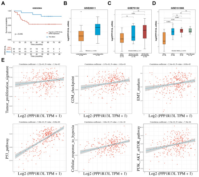
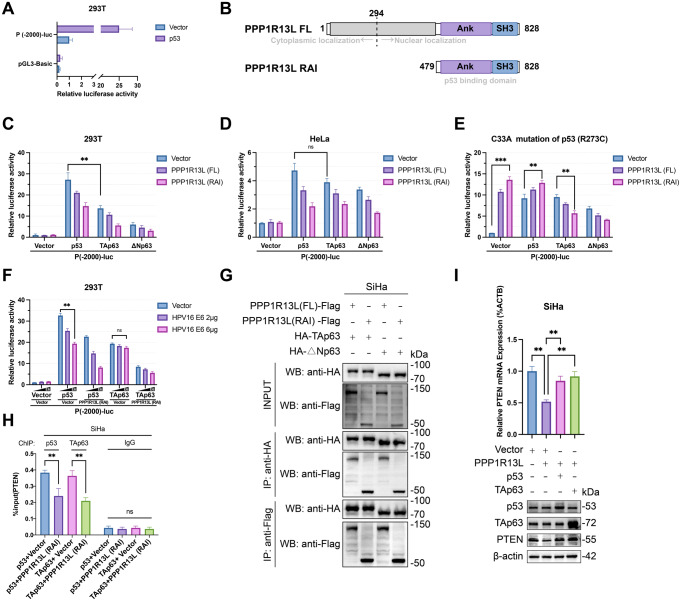
PPP1R13L is a conserved inhibitor of p53, selectively regulating a subset of p53 target genes. Previous studies have reported that PPP1R13L promotes cervical cancer progression, yet its precise mechanism remains unclear and warrants further investigation. In this study, we utilized public databases to reveal the correlation between PPP1R13L and tumor progression pathways. Subsequently, we performed functional assays both in vitro and in xenograft models to assess the impact of PPP1R13L on cervical cancer. Our results demonstrate that PPP1R13L promotes cervical cancer cell proliferation, epithelial-mesenchymal transition, cycle progression, and glycolysis via the PTEN/AKT/mTOR pathway. Mechanistically, PPP1R13L regulates the transcription of PTEN through its Ank-SH3 domain interaction with p53 family, p53 and p63. In 293T cells, p53 originally exhibits significantly higher transcriptional activity than p63. However, in cervical cancer-where E6 continuously degrades p53 and p63 is highly expressed-p63 demonstrates a transcriptional activity for PTEN that is comparable to, or even surpasses, that of p53, depending on E6 expression levels. Additionally, in C33A, an HPV-negative cervical cancer cell line, the p53 R273C mutation causes PPP1R13L to exert an opposite effect, and p63 is shown to be inhibited by PPP1R13L independently of p53. Finally, the response elements of PPP1R13L-regulated p53 family target genes were experimentally validated on p63 for the first time. This provides a sequence basis for the selective regulation of p53 family target genes by PPP1R13L. In summary, we underscore the specificity of the PPP1R13L/p63/PTEN axis in cervical cancer and propose that PPP1R13L holds potential as a therapeutic target for cervical cancer treatment.

Keywords: Cervical cancer; Glycolysis; PPP1R13L; PTEN; p63.


Conflict of interest statement

Declarations. Ethical approval: This study was performed in line with the principles of the Declaration of Helsinki. Approval was granted by the Medical Ethics Committee of Zhongnan Hospital of Wuhan University (Ethics Approval No. 2020029). Consent to participate: Informed consent was obtained from all individual participants included in the study. Consent to publish: The authors affirm that human research participants provided informed consent for publication of the images in Fig. 5G. Competing interests: The authors have no relevant financial or non-financial interests to disclose. Cell lines: The human cervical cancer cell lines HeLa (CL-0101), SiHa (CL-0210), C33A (CL-0045), and the human embryonic kidney cell line 293T (CL-0005) used in this study were purchased from Procell, Wuhan, China, and were verified by STR profiling.

Figures

Fig. 1
Fig. 1
PPP1R13L impacts the prognosis of cervical cancer and reveals the correlation with tumor progression pathways. (A) Kaplan-Meier analysis of overall survival in the GSE52904 cohort revealed that the top 30% of patients with the highest PPP1R13L mRNA expression had significantly lower survival rates compared to the remaining 70% of patients with lower PPP1R13L expression. (B) Analysis of the GSE26511 dataset revealed that cervical cancer patients with lymph node metastasis exhibited higher PPP1R13L mRNA expression compared to those without metastasis. (C) In the GSE75132 dataset, cervical tissues persistently infected with HPV16 (categorized into non-CIN3 and CIN3) were compared to normal tissues without HPV16 infection. The results showed that PPP1R13L mRNA expression progressively increased across these groups. (D) In the GSE151666 dataset, HPV-positive cervical tumor tissues exhibited higher PPP1R13L mRNA expression compared to HPV-negative primary cervical tumor tissues. Elevated PPP1R13L expression was observed in tumors infected with HPV16, HPV18, and other HPV types (HPV33, 59, 58, 52, 45, 31, 56). (E) The ssGSEA analysis of PPP1R13L with tumor proliferation signature, G2M checkpoint, EMT markers, p53 pathway, cellular response to hypoxia, and the PI3K/AKT/mTOR pathway. The data is from TCGA datasets
Fig. 2
Fig. 2
PPP1R13L promotes cervical cancer cell proliferation, cycle progression and facilitates EMT. (A) Western blot showed PPP1R13L overexpression in HeLa cells and PPP1R13L knockdown via siRNA in SiHa cells. (B, C) CCK-8 and colony formation of PPP1R13L-knockdown SiHa cells and PPP1R13L-overexpressing HeLa cells to assess the proliferation capacity. The results indicate that PPP1R13L promotes cervical cancer cell proliferation. (D, E) Transwell assay and wound-healing assay to detect the migration of PPP1R13L-knockdown SiHa cells and PPP1R13L-overexpressing HeLa cells. The results indicate that PPP1R13L promotes cervical cancer cell migration. Scale bar represents 200 μm. (F) The expression of EMT markers (ZEB1, N-cadherin, E‐cadherin, Vimentin, Snail, MMP‐2) in PPP1R13L-knockdown SiHa cells and PPP1R13L-overexpressing HeLa cells. The results indicate that PPP1R13L promotes cervical cancer cell EMT. (G) Flow cytometry results indicate that PPP1R13L knockdown in SiHa cells induces cell cycle arrest in the G0/G1 phase, while PPP1R13L overexpression in HeLa cells reduces the proportion of cells in this phase. (H) Western blot results show that the expression of cell cycle-related proteins is reduced in PPP1R13L-knockdown SiHa cells and increased in PPP1R13L-overexpressing HeLa cells. N = 3 per group. Data are expressed as the mean ± SD. Two-tailed t-tests analyzed two-group differences; one-way ANOVA assessed multiple-group differences. ns, p > 0.05; *p < 0.05; **p < 0.01; ***p < 0.001
Fig. 3
Fig. 3
PPP1R13L downregulates PTEN to activate the PI3K/AKT/mTOR pathway and upregulates glycolysis in cervical cancer cells. (A) Glucose consumption and lactate production in cervical cancer cells were detected after transfection with siRNAs or PPP1R13L overexpression plasmids for 48 h. Glucose consumption and lactate production were decreased by PPP1R13L knockdown in HeLa cells but increased by PPP1R13L overexpression in SiHa cells. (B) Western blot analysis shows that PPP1R13L downregulates PTEN in cervical cancer cells, leading to the activation of the PI3K/AKT/mTOR/HIF1A pathway. Concurrently, several key glycolytic enzymes, including HK2, PGK1, and LDHA, are upregulated. (C) The mRNA expression of PTEN after knockdown and overexpression of PPP1R13L in both SiHa and HeLa cells demonstrates that PPP1R13L inhibits PTEN mRNA levels in cervical cancer cells. (D) The expression of PTEN/AKT/mTOR/HIF1A pathway with key glycolytic enzymes (HK2, PGK1, and LDHA) was detected by western blot. PTEN knockdown reversed the effects of PPP1R13L knockdown in SiHa cells and PTEN overexpression reversed the effects of PPP1R13L overexpression in HeLa cells. (E) Glucose consumption and lactate production in cervical cancer cells were detected after transfection for 48 h. PTEN knockdown reversed the effects of PPP1R13L knockdown in SiHa cells and PTEN overexpression reversed the effects of PPP1R13L overexpression in HeLa cells. (F) Gross images of xenograft tumors in the SiHa-sh-NC and SiHa-sh-PPP1R13L groups indicate that PPP1R13L inhibits cervical cancer cell growth in vivo, as shown by the reduced tumor volume and weight. N = 6 per group. Two-tailed t-tests analyzed two-group differences. p > 0.05; *p < 0.05; **p < 0.01; ***p < 0.001. (G) IHC of the xenograft tumor model showed that the SiHa-sh-PPP1R13L group exhibited lower Ki67 expression and higher PTEN levels. Moreover, the expression of EMT marker Snail and the glucose transporter GLUT1 was also reduced in the SiHa-sh-PPP1R13L group. Scale bar represents 50 μm. N = 3 per group. Data are expressed as the mean ± SD. Two-tailed t-tests analyzed two-group differences; one-way ANOVA assessed multiple-group differences. ns, p > 0.05; *p < 0.05; **p < 0.01; ***p < 0.001
Fig. 4
Fig. 4
PPP1R13L inhibits the transcriptional activity of both p53 and TAp63 on PTEN. (A) 293T cells were transfected with pGL3-basic and P(-2000)-luc, a luciferase reporter containing the PTEN promoter. The results show that P(-2000)-luc has promoter activity and can be enhanced by p53 over 20-fold. (B) PPP1R13L full length (FL) consists of 828 amino acids. PPP1R13L (RAI) contains a C-terminal Ank-SH3 domain (amino acids 479–828). (C) Assess the activation of P(-2000)-luc by p53 family transcription factors and the inhibition by PPP1R13L (FL) and PPP1R13L (RAI) in 293T cells. The results show that p53 has a significantly higher activation capacity than TAp63 in 293T cells. And PPP1R13L (RAI) exhibits stronger inhibitory activity than PPP1R13L (FL). (D) Assess the activation of P(-2000)-luc by p53 family transcription factors and the inhibition by PPP1R13L (FL) and PPP1R13L (RAI) in HeLa cells. In the context of cervical cancer cells where E6 continuously degrades p53, p53 and p63 exhibit comparable activation abilities on P(-2000)-luc. (E) The same experiment was repeated in C33A p53 (R273C) cells. The results showed that p53R273C caused PPP1R13L to have an opposite effect, which could not be reversed even with the overexpression of wild-type p53. However, PPP1R13L still inhibited p63’s activation of P(-2000)-luc independently of p53. (F) Assess the ability of p53 and p63 to activate the P(-2000)-luc reporter under conditions of gradient transfection of the E6 protein. The results demonstrated that, in the context of continuous degradation of p53 by E6, p63 exhibits transcriptional activity comparable to or even surpassing that of p53 in an E6 dose-dependent manner. Additionally, a synergistic effect between E6 and PPP1R13L (RAI) was observed, resulting in the strongest inhibitory effects on transcription. (G) Co-immunoprecipitation and western blot analysis showed that both full-length PPP1R13L and RAI can bind to HA-TAp63 and HA-ΔNp63 in SiHa cells. (H) The ChIP assay demonstrated that PPP1R13L (RAI) reduced the binding of both p53 and TAp63 to the PTEN promoter. (I) Overexpression of p53 or TAp63 reversed the reduction in PTEN caused by PPP1R13L overexpression, indicating that the regulation of PTEN by PPP1R13L is mediated through p53 or TAp63. N = 3 per group. Data are expressed as the mean ± SD. One-way ANOVA assessed multiple-group differences. ns, p > 0.05; *p < 0.05; **p < 0.01; ***p < 0.001
Fig. 5
Fig. 5
PPP1R13L inhibits the transcription of PTEN by p53 and p63, dependent on specific response elements. (A) Two p53 and p63 binding sites were predicted on the PTEN promoter. (B) The p53 family proteins were used to activate P(site1)-luc, P(site2)-luc, P(-2000)-luc-△site1, P(-2000)-luc-△site2, P(-2000)-luc, and pGL3-Basic in 293T cells. The dual-luciferase reporter assay showed that site 1 is the active site. (C) The PPP1R13L-regulated p53 REs (response element) required the 9 C or/and 12G in the basal p53 REs. The p53 RE in the PTEN promoter is similar to the known PPP1R13L-regulated p53 target gene, p21. The black bases represent the conserved characteristics of p53 RE and the red bases represent the features required for PPP1R13L regulation. The C9T mutation in site1 caused overexpression of PPP1R13L to enhance, rather than inhibit, p63’s transcriptional activity on the PTEN promoter. N = 3 per group. Data are expressed as the mean ± SD. One-way ANOVA assessed multiple-group differences. ns, p > 0.05; *p < 0.05; **p < 0.01; ***p < 0.001. (D) RNA expression of PPP1R13L, PTEN, TP53, and TP63 in cervical squamous cell carcinoma, cervical adenocarcinoma, and normal cervical tissue. Data were obtained from TCGA and GTEx datasets. PPP1R13L and p63 are highly expressed in cervical squamous cell carcinoma but not in cervical adenocarcinoma. (E)The positive rate of PPP1R13L, p63, and PTEN in 20 types of tumor slides was ranked, and the RNA expression of them across 55 types of normal tissues were ranked. Data were obtained from The Human Protein Atlas websites. Cervix and cervical cancer have been highlighted in pink. PPP1R13L and p63 are frequently highly expressed in tissues like skin and mucosa, including normal and cancerous cervical tissues. PTEN shows high expression in normal cervical tissues but extremely low positivity in cervical cancer, suggesting it may be suppressed by cervical tissue-specific mechanisms. (F) Expression of PPP1R13L and PTEN in cervical cancer tissue (n = 40) and normal cervical tissue (n = 19). (G) IHC results show that adjacent normal tissues with intact cervical mucosa have high PTEN expression, whereas cancer tissues with a loss of mucosal structure exhibit little to no PTEN expression. PPP1R13L expression is positive in adjacent normal tissues but is even higher in cancer tissues. Scale bar represents 50 μm (left) and 200 μm (right). (H) Our study shows that PPP1R13L promotes cervical cancer progression, EMT, and glycolysis through the PTEN/AKT/mTOR pathway. Mechanistically, PPP1R13L regulates PTEN transcription via its Ank-SH3 domain interaction with p53 family members, including p53 and p63. Given the low levels of p53 and high levels of p63 in cervical cancer, or in cases of p53 mutation, we focus on the role of the PPP1R13L/p63/PTEN axis in cervical cancer progression

Similar articles

References

    1. Wei F, Georges D, Man I, Baussano I, Clifford GM (2024) Causal attribution of human papillomavirus genotypes to invasive cervical cancer worldwide: a systematic analysis of the global literature. Lancet 404:435–444. 10.1016/s0140-6736(24)01097-3 - PubMed
    1. Kusakabe M, Taguchi A, Sone K, Mori M, Osuga Y (2023) Carcinogenesis and management of human papillomavirus-associated cervical cancer. Int J Clin Oncol 28:965–974. 10.1007/s10147-023-02337-7 - PMC - PubMed
    1. Gelbard MK, Munger K (2023) Human papillomaviruses: knowns, mysteries, and unchartered territories. J Med Virol 95:e29191. 10.1002/jmv.29191 - PMC - PubMed
    1. Bergamaschi D, Samuels Y, O’Neil NJ, Trigiante G, Crook T, Hsieh JK, O’Connor DJ, Zhong S, Campargue I, Tomlinson ML, Kuwabara PE, Lu X (2003)iASPP oncoprotein is a key inhibitor of p53 conserved from worm to human. Nat Genet. 33,162–167. 10.1038/ng1070 - PubMed
    1. Bergamaschi D, Samuels Y, Sullivan A, Zvelebil M, Breyssens H, Bisso A, Del Sal G, Syed N, Smith P, Gasco M, Crook T, Lu X (2006) iASPP preferentially binds p53 proline-rich region and modulates apoptotic function of codon 72-polymorphic p53. Nat Genet 38:1133–1141. 10.1038/ng1879 - PubMed

MeSH terms