. 2025 May;388(6746):eadv9789.
doi: 10.1126/science.adv9789. Epub 2025 May 1.

TIGR-Tas: A family of modular RNA-guided DNA-targeting systems in prokaryotes and their viruses

Affiliations

TIGR-Tas: A family of modular RNA-guided DNA-targeting systems in prokaryotes and their viruses

Guilhem Faure et al. Science. 2025 May.

Abstract

RNA-guided systems provide remarkable versatility, enabling diverse biological functions. Through iterative structural and sequence homology-based mining starting with a guide RNA-interaction domain of Cas9, we identified a family of RNA-guided DNA-targeting proteins in phage and parasitic bacteria. Each system consists of a tandem interspaced guide RNA (TIGR) array and a TIGR-associated (Tas) protein containing a nucleolar protein (Nop) domain, sometimes fused to HNH (TasH)- or RuvC (TasR)-nuclease domains. We show that TIGR arrays are processed into 36-nucleotide RNAs (tigRNAs) that direct sequence-specific DNA binding through a tandem-spacer targeting mechanism. TasR can be reprogrammed for precise DNA cleavage, including in human cells. The structure of TasR reveals striking similarities to box C/D small nucleolar ribonucleoproteins and IS110 RNA-guided transposases, providing insights into the evolution of diverse RNA-guided systems.


Figures

Figure 1.
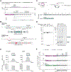
Figure 1.. Discovery and Genomic Organization of TIGR Systems
(A) Structural mining and identification of TIGR systems. The RNA-binding domain (RBD) of SpCas9 (PDB: 8G1I) was used as a seed to identify IS110 as a structural homolog. Further structural mining revealed similarity to the Nop domain-containing family. Genomic mining of Nop domain-containing proteins followed by community detection from embedding of the candidates identified a distinct family of Nop domain-containing proteins associated with Tandem Interspaced Guide RNA (TIGR) systems. Protein structures or models are all colored similarly: RBD, red, blue and green; rest of the Nop domain, wheat. (B) Genomic organization of TIGR systems and architectures of Tas proteins. TIGR-associated (Tas) protein structural models (left) and genomic locus architecture (right) of three representative Tas proteins (TasA, no nuclease; TasR, RuvC; TasH, HNH). Protein amino acid (top) and nucleotide (bottom) coordinates are shown, as are the number of repeat units in the TIGR arrays. (C) Sequence composition of TIGR arrays. Alignment of individual repeat units from the TaTIGR array downstream of the TasR ORF. Conserved regions corresponding to edge and loop repeats are shown in black, and variable regions corresponding to spacers A and B are shown in gray.
Figure 2.
Figure 2.. TIGR arrays are processed into 36-nt tigRNAs
(A) Small RNA-seq of RNA pulled down with Thermoproteota archaeon TasR (TaTasR) mapped to native TaTIGR array expressed in E. coli. Top: All reads mapped to the TIGR array. Bottom: filtered 36-nt long reads. The pre-tigRNA transcript is expressed and processed into distinct 36-nt tigRNA units (see also fig. S5). (B) Top: Schematic representation of pre-tigRNA processing into tigRNA units. Bottom: Schematic representation of pre-crRNA processing (SpCas9 CRISPR system) into crRNA units. (C) Experimental design to test wild-type (WT) or catalytically-inactive TasR (dTasR, D11A) association with native tigRNA arrays or a minimal tigRNA array consisting of three repeats of tigRNA1 (3X tR1) via ribonucleic acid protein (RNP) pulldown. (D) Composition of purified TaTasR RNPs using the experimental design shown in (C). Left: SDS-PAGE protein gel stained with Coomassie blue. Right: 10% denaturing PAGE gel stained with SYBR Gold to show nucleic acids. (E) Small RNA-seq of RNAs present in TaTasR RNPs showing comparison between the processing of the native array (top) and the 3X tR1 array (bottom). (F) Small RNA-seq of total RNA from E. coli. The TIGR-TasR expression plasmid contained an arabinose-inducible promoter (PBAD) before the TasR gene, and cells were grown in the presence of arabinose. Coverage and read starts/ends are shown for reads that mapped to the expression plasmid. From top to bottom, the plasmid expresses the (1) WT system, (2) WT system with a nonsense mutation at Asp200 of TasR, (3) Residues 1-322 of TasR (331 residues total) replaced with GFP, and (4) Residues 1-322 to TasR deleted.
Figure 3.
Figure 3.. Identification of the TasR nuclease target
(A) Experimental scheme to identify TasR cleavage sites in the E. coli genome. (B) Mapping of read starts to the E. coli genome. The annotated peak is at the wcaD gene. Example reads start with a ‘T’ due to the non-templated A added during NGS library preparation with Klenow DNA polymerase and identifies the 5′ end of the input DNA fragment. The pArray variant was either native, minimized to three identical units (3X tR1), or absent (apoprotein). (C) Top: The wcaD gene sequence with potential matches to spacer A and spacer B indicated, and the sequence of a full matching synthetic target. Bottom: denaturing polyacrylamide gel of an in vitro cleavage reaction using purified TasR RNP (coexpressed with a 3X tR1 TIGR array) and strand-specifically labelled substrates. FL, fluorescein label on the strand targeted by spacer A.
Figure 4∣
Figure 4∣. DNA targeting rules for TIGR-TasR
(A) Top: base-pairing scheme of tigRNA1 from the TaTasR TIGR array with the synthetic optimized target sequence. Triangles indicate cleavage sites. Bottom: comparison with the targeting rules for CRISPR Cas9. PAM, protospacer-adjacent motif. (B) In vitro cleavage reactions with TasR. Left: TaTasR (WT) or catalytically inactivated RuvC domain (d, D11A mutation) RNPs were purified and incubated with synthetic target DNA matching the first tigRNA from the TaTasR TIGR array (RNP “–” indicates apoprotein was used). Right: Purified WT TaTasR apoprotein was incubated with the synthesized cognate tigRNA1, no RNA, or a non-targeting (NT) tigRNA, with the target (T) DNA substrate or non-target (NT) substrate. tigRNA2 from the TaTasR TIGR array was used as the non-targeting tigRNA, and PCR amplicon of the SpTasH target was used for the non-target DNA substrate. (C) Sanger traces for sequencing of the TasR in vitro-cleaved optimized DNA target. The polymerase used in Sanger sequencing adds a non-templated A after running off the template, indicated with an asterisk, and this delineates the precise cleavage site. (D) In vitro activity of TasR containing tigRNA1 on labelled single-stranded or double-stranded DNA or RNA substrates containing matches to spacer A and/or spacer B. (E) Effects of transversions on the in vitro cleavage of the optimized DNA target by TasR RNPs containing tigRNA1. All transversions were strictly A→T, T→A, G→C and C→G. Both strands of the target were mutated. The seed and nickase mutations are indicated. For (B, D, and E) reactions were resolved by denaturing PAGE and visualized by fluorescent labels on the DNA 5′ ends.
Figure 5∣
Figure 5∣. Human genome editing with TIGR-TasR
(A) Experimental scheme to test programmable gene editing with TasR in HEK293FT cells. hs, Homo Sapiens; CMV, cytomegalovirus; NLS, nuclear localization signal. (B) Average indel rates (%) generated by TaTasR and a second TasR ortholog from Parcubacteria of the candidate phyla radiation (ParTasR) at six genomic loci in HEK293FT; data are presented as mean ± s.d. (n = 3). (C) Sequence of the CXCR4 target site (highlighted in green) in the human genome and corresponding TasR gRNA with spacer matches shown in green. (D) Indels generated by TaTasR (left) and ParTasR (right) at the CXCR4 target site. (E) Distribution of indel size generated by TaTasR (top) and ParTasR (bottom) at the CXCR4 target site.
Figure 6∣
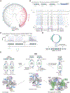
Figure 6∣. Structural basis for RNA-guided DNA cleavage by TIGR-TasR
(A) Cryo-EM structure of TasR containing tigRNA1 and the optimized DNA substrate in a post-reaction product state. The protein is a dimer, and each protomer is labelled ‘A’ or ‘B’ according to whether its RuvC domain has cleaved the DNA strand targeted by spacer A or B. (B) Detail of the RuvC domain of monomer A interacting with the spacer A RNA/DNA heteroduplex. The 5′ phosphate of the nick is still in proximity to the RuvC active site, key residues of which are shown and labelled. (C) Diagram of the secondary structures of the RNA and DNA components of the TasR product complex. (D) Interaction of the coiled-coil dimerization domain of TasR monomer A with the seed region of the spacer B RNA/DNA heteroduplex (see Fig. 4E). CTD, C-terminal domain. (E) Recognition of the pseudosymmetric tigRNA by the symmetric TasR protein dimer. (F) Interactions of the box C and box D motifs of the edge (left) and loop (right) repeats with TasR residues. (G) Consensus sequences for edge and loop repeats from nine different TasR TIGR arrays. Excerpts from the full alignments used to derive these consensuses can be found in fig. S4. Arcs show the Watson/Crick base pairs observed in the structure (which are between different edge repeats on a processed tigRNA but are shown here on the same edge repeat for simplicity) which are either absolutely conserved (box C/D pairing) or show covariance.
Figure 7∣
Figure 7∣. Evolutionary and functional diversity of TIGR systems.
(A) Phylogenetic tree of the Tas Nop domain indicating array association, viral origin, and presence of RuvC (regardless of catalytic activity) or HNH nuclease domain. (B) Example of a TasA TIGR system with a stem-loop array (from the gut microbiome of Peromyscus leucopus). Top: locus architecture with TasA protein domain coordinates (in aa) above (coiled-coil region in light green; RNA binding C-terminal domain in blue) and nucleotide coordinates below. Bottom: Alignment of individual stem units, with hairpin sequences (palindromic sequence) in blue flanking each unit. Conserved C and D motifs are shown in black, spacers are in gray, and linkers between units are indicated. (C) Small RNA-seq mapping of the TIGR array following RNP pulldown. All three stem arrays are actively expressed and processed at linker regions. (D) Schematic of a representative stem-loop repeat unit (tigRNA2 from the TIGR system found in the gut microbiome of Peromyscus leucopus). (E) Comparative schematic of TIGR systems, IS110, and box C/D. tigRNAs are the simplest and shortest ncRNAs, while IS110 bridge RNAs resemble a fusion of two tigRNAs. box C/D snoRNAs are depicted as tigRNA-like molecules stabilizing the stem structure via the L7 k-turn motif. Bottom: comparison of the TasR-product complex with the crystal structure of an archaeal snoRNP (PDB 3PLA) (111) and a dimer excerpted from a cryo-EM structure of the IS110 bridge RNA complex (PDB 8WT6) (32). The RNP structures of all systems harbor the same domain architecture. TasR is structurally closer to IS110, while the box C/D protein (Nop5) harbors an inactive RuvC recruiting the fibrillarin methylase (green) and stabilizing the C and D box via an interaction with L7 (white). A proposed model positions TIGR as an ancestral system to IS110 and box C/D snoRNAs, highlighting its evolutionary relevance.

References

    1. Makarova KS, Wolf YI, Iranzo J, Shmakov SA, Alkhnbashi OS, Brouns SJJ, Charpentier E, Cheng D, Haft DH, Horvath P, Moineau S, Mojica FJM, Scott D, Shah SA, Siksnys V, Terns MP, Venclovas Č, White MF, Yakunin AF, Yan W, Zhang F, Garrett RA, Backofen R, van der Oost J, Barrangou R, Koonin EV, Evolutionary classification of CRISPR-Cas systems: a burst of class 2 and derived variants. Nat Rev Microbiol 18, 67–83 (2020). - PMC - PubMed
    1. Hille F, Richter H, Wong SP, Bratovič M, Ressel S, Charpentier E, The Biology of CRISPR-Cas: Backward and Forward. Cell 172, 1239–1259 (2018). - PubMed
    1. Dexheimer PJ, Cochella L, MicroRNAs: From Mechanism to Organism. Front Cell Dev Biol 8, 409 (2020). - PMC - PubMed
    1. Senti K-A, Brennecke J, The piRNA pathway: a fly’s perspective on the guardian of the genome. Trends Genet 26, 499–509 (2010). - PMC - PubMed
    1. Omer AD, Lowe TM, Russell AG, Ebhardt H, Eddy SR, Dennis PP, Homologs of small nucleolar RNAs in Archaea. Science 288, 517–522 (2000). - PubMed

MeSH terms

Supplementary concepts

LinkOut - more resources