. 2025 Mar 12;147(10):8618-8628.
doi: 10.1021/jacs.4c17726. Epub 2025 Feb 27.

Probing the Dynamics of Yersinia Adhesin A (YadA) in Outer Membranes Hints at Requirements for β-Barrel Membrane Insertion

Affiliations

Probing the Dynamics of Yersinia Adhesin A (YadA) in Outer Membranes Hints at Requirements for β-Barrel Membrane Insertion

Jayasubba Reddy Yarava et al. J Am Chem Soc. .

Abstract

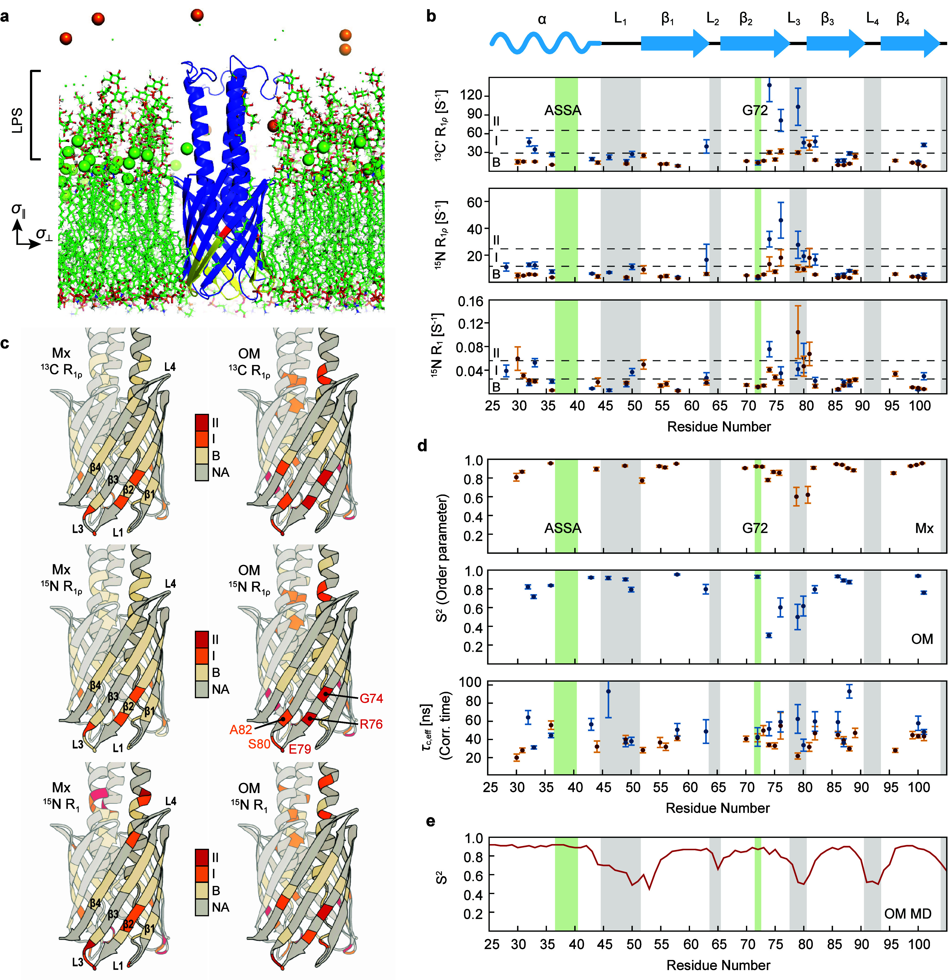
The vast majority of cells are protected and functionalized by a dense surface layer of glycans, proteoglycans, and glycolipids. This surface represents an underexplored space in structural biology that is exceedingly challenging to recreate in vitro. Here, we investigate β-barrel protein dynamics within an asymmetric outer membrane environment, with the trimeric autotransporter Yersinia adhesin A (YadA) as an example. Magic-angle spinning NMR relaxation data and a model-free approach reveal increased mobility in the second half of strand β2 after the conserved G72, which is responsible for membrane insertion and autotransport, and in the subsequent loop toward β3. In contrast, the protomer-protomer interaction sites (β1i-β4i-1) are rigid. Intriguingly, the mobility in the β-strand section following G72 is substantially elevated in the outer membrane and less so in the detergent environment of microcrystals. A possible source is revealed by molecular dynamics simulations that show the formation of a salt bridge involving E79 and R76 in competition with a dynamic interplay of calcium binding by E79 and the phosphate groups of the lipids. An estimation of overall barrel motion in the outer membrane and detergent-containing crystals yields values of around 41 ns for both. The global motion of YadA in the outer membrane has a stronger rotational component orthogonal to the symmetry axis of the trimeric porin than in the detergent-containing crystal. In summary, our investigation shows that the mobility in the second half of β2 and the loop to β3 required for membrane insertion and autotransport is maintained in the final folded form of YadA.


Conflict of interest statement

The authors declare no competing financial interest.

Figures

Figure 1
Figure 1
Dynamics properties of YadAM in OMs and the microcrystalline environment. (a) YadA anchor domain (YadAM) in an asymmetric bacterial OM environment. The outer leaflet contains LPS, whereas the inner leaflet contains mostly phospholipids. Membrane axes as used in this study are shown on the left. G72 is colored red, and the second half of β2 is shown in yellow. Green spheres are calcium ions, and orange spheres are potassium ions. (b) Experimentally determined 15N R1 and 15N as well as 13C′ R relaxation rates of YadAM-Mx (orange) and OM (blue). The gray bars represent loop regions, and green bars represent the residues that are involved in autotransporter (ASSA) and membrane insertion (G72) mechanisms. The black dashed lines define the classes employed as the color code in (c). The three classes of relaxation times are defined as fast (II, red), medium (I, orange), and base (B, ivory). The 15N R1 rates were categorized as 0–0.025 s–1 (B), 0.025–0.056 s–1 (I), and >0.056 s–1 (II). The 15N R rates were categorized as 0–12 s–1 (B), 12–25 s–1 (I), and >25 s–1 (II). The 13C R rates were categorized as 0–29 s–1 (B), 29–66 s–1 (I), and >66 s–1 (II). (c) Visualization of relaxation times sorted into three classes; see the dashed lines in (b). All analyzed residues appear in red, orange, or ivory. Nonanalyzed residues are in gray. One protomer of the trimeric barrel is shown with a slightly darker gray shade. (d) Interpretation of the 15N R1 and R relaxation rates with the SMF formalism. Order parameters for YadAM-Mx and YadAM-OM are shown in the top and center panels, respectively. Correlation times are shown in the lower panel, with values depicted in blue for YadAM-OM and in orange for YadAM-Mx. (e) MD-derived order parameters of YadAM-OM at the NH peptide plane vectors were obtained from 2 μs MD simulations in a realistic OM model. The values represent the average of 3 replicas.
Figure 2
Figure 2
Structural variability at the intracellular face of YadAM. (a) Correlation of OM and Mx S2 values. (b) Calcium ions bound to a highly conserved region (residues 75–80) (see also Figure S10 for ions bound vs time). (c) Alternative interactions between E79 and Y49.
Figure 3
Figure 3
Grid search of the data from the SMF-3D GAF analysis. The grid search results of the SMF-3D GAF analysis for YadAM-Mx and YadAM-OM are shown in (a) and (b), respectively. The grid search analysis was performed on the amplitudes (σ, σ) by stepwise fixing both σ values individually in the range from 1° to 15° in steps of 0.5°. For each pair of fixed amplitudes, we optimized the remaining fit parameters.

References

    1. Gaboriaud F.; Gee M. L.; Strugnell R.; Duval J. F. L. Coupled Electrostatic, Hydrodynamic, and Mechanical Properties of Bacterial Interfaces in Aqueous Media. Langmuir 2008, 24, 10988–10995. 10.1021/la800258n. - DOI - PubMed
    1. Henderson J. C.; Zimmerman S. M.; Crofts A. A.; Boll J. M.; Kuhns L. G.; Herrera C. M.; Trent M. S. The Power of Asymmetry: Architecture and Assembly of the Gram-Negative Outer Membrane Lipid Bilayer. Annu. Rev. Microbiol. 2016, 70, 255–278. 10.1146/annurev-micro-102215-095308. - DOI - PubMed
    1. Horne J. E.; Brockwell D. J.; Radford S. E. Role of the Lipid Bilayer in Outer Membrane Protein Folding in Gram-Negative Bacteria. J. Biol. Chem. 2020, 295, 10340–10367. 10.1074/jbc.REV120.011473. - DOI - PMC - PubMed
    1. Yildiz Ö.; Vinothkumar K. R.; Goswami P.; Kühlbrandt W. Structure of the Monomeric Outer-Membrane Porin OmpG in the Open and Closed Conformation. EMBO J. 2006, 25, 3702–3713. 10.1038/sj.emboj.7601237. - DOI - PMC - PubMed
    1. Retel J. S.; Nieuwkoop A. J.; Hiller M.; Higman V. A.; Barbet-Massin E.; Stanek J.; Andreas L. B.; Franks W. T.; Rossum B.-J.; Vinothkumar K. R.; et al. Structure of Outer Membrane Protein G in Lipid Bilayers. Nat. Commun. 2017, 8, 2073.10.1038/s41467-017-02228-2. - DOI - PMC - PubMed

MeSH terms

LinkOut - more resources