. 2025 May 5;224(5):e202312120.
doi: 10.1083/jcb.202312120. Epub 2025 Feb 27.

The pathway of unconventional protein secretion involves CUPS and a modified trans-Golgi network

Affiliations

The pathway of unconventional protein secretion involves CUPS and a modified trans-Golgi network

Amy J Curwin et al. J Cell Biol. .

Abstract

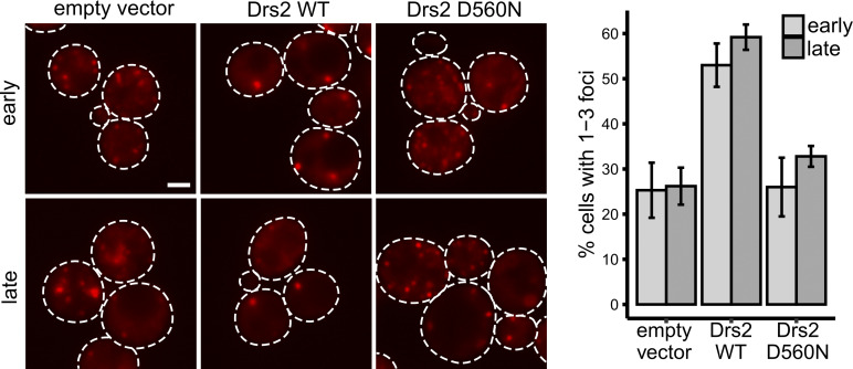
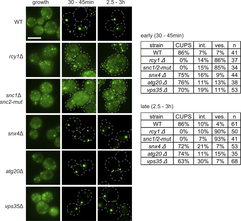
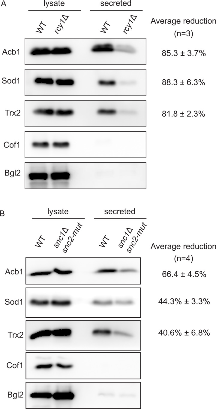
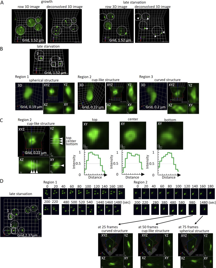
Compartment for unconventional protein secretion (CUPS), a compartment for secretion of signal sequence-lacking proteins, forms through COPI-independent extraction of membranes from early Golgi cisternae, lacks Golgi-specific glycosyltransferases, and requires phosphatidylinositol 4-phosphate (PI4P) for biogenesis, as well as phosphatidylinositol 3-phosphate for stability. Our findings demonstrate that Drs2, a PI4P effector from the trans-Golgi network (TGN), is essential for CUPS formation, specifically through its interaction with Rcy1, and Rcy1 is crucial for the unconventional secretion. Using 4D super-resolution confocal live imaging microscopy, we observed that CUPS interact with a modified TGN that contains Drs2 in addition to proteins Tlg2 and Snc2, which are necessary for membrane fusion. Notably, while CUPS remain stable, the modified TGN undergoes remodeling during the later stages of unconventional secretion. In summary, we suggest that CUPS and the modified TGN, without the function of COPII and COPI, participate in collecting and sorting unconventionally secreted proteins, reflecting the role of Golgi membranes in receiving cargo from the ER during conventional secretion.


Conflict of interest statement

Disclosures: The authors declare no competing interests exist.

Figures

Figure 1.
Figure 1.
Drs2 is required for CUPS biogenesis. (A) Cells genomically expressing Drs2-3xGFP and Grh1-2xmCherry were visualized by confocal spinning-disk microscopy in growth conditions and starvation by incubation in 2% potassium acetate. Short movies were acquired at 10-s intervals to assess the frequency and duration of colocalization. Scale bar = 2 μm. (B) WT and drs2Δ cells expressing Grh1-2xGFP were visualized by epifluorescence microscopy in growth conditions and after incubation in 2% potassium acetate for the indicated times. Cells were classified with normal CUPS (one to three larger foci per cell); intermediate CUPS (“int.”), where a large focus is observed in addition to smaller structures; and vesiculated CUPS (“ves.”), where only small foci of Grh1 are observed. Scale bar = 2 μm.
Figure S1.
Figure S1.
Drs2 flippase activity is required for CUPS biogenesis. Cells genomically expressing pGalS-Drs2-3xGFP and Grh1-2xmCherry were transformed with an empty vector, WT Drs2, or mutant Drs2 (D560N) plasmids in a galactose-containing medium. Cells were switched to glucose medium, in order to repress the expression of Drs2-3xGFP, 72 h prior to starvation in 2% potassium acetate. Cells were visualized by epifluorescence microscopy at the indicated times (early, 30- to 35-min incubation; late, 2-h incubation). (A) Representative images of Grh1-positive foci. Scale bar = 2 μm. (B) Quantification of the percentage of cells showing one to three foci per cell.
Figure S2.
Figure S2.
No CUPS defect in cells lacking Gea2, Arl1, Chc1, Apl6, Aps1, Snc1, or Snc2. The indicated deletion strains expressing Grh1-2xGFP were grown to the log phase and starved for 2.5 h. Scale bar = 2 μm.
Figure 2.
Figure 2.
Drs2-Rcy1 pathway and the v-SNAREs, Snc1 and Snc2, are required for CUPS formation. WT and the indicated deletion or mutant strains expressing Grh1-2xGFP were visualized by epifluorescence microscopy in growth conditions and after incubation in 2% potassium acetate for the indicated times. Cells were classified with normal CUPS (one to three larger foci per cell); intermediate CUPS (int.), where a large focus is observed in addition to smaller structures; and vesiculated CUPS (ves.), where only small foci of Grh1 are observed. Scale bar = 2 μm.
Figure 3.
Figure 3.
Rcy1 and v-SNAREs are required for unconventional secretion. (A and B) WT, rcy1Δ, or snc1Δ snc2-V39A,M42A cells were grown in the logarithmic phase, washed twice, and cultured in 2% potassium acetate for 2.5 h. The cell wall proteins were extracted from an equal number of cells followed by precipitation with TCA (“secreted”). Lysates and secreted proteins were analyzed by western blot, and the ratio of the secreted to lysate for the indicated protein was determined and compared with that of WT in each experiment. Statistical analyses were performed for the indicated unconventional cargo proteins, and the reduction in secretion compared with WT is indicated as ± SD. Source data are available for this figure: SourceData F3.
Figure 4.
Figure 4.
CUPS contain a pool of the v-SNARE Snc2 and the t-SNARE Tlg2. (A and B) Cells genomically expressing Grh1-2xCherry with GFP-Snc1, GFP-Snc2, or GFP-Tlg2 were visualized by confocal spinning-disk microscopy in growth conditions and (B) throughout the time course of culture in 2% potassium acetate. Short movies were acquired at 10-s intervals to assess the frequency and duration of colocalization. Scale bar = 2 μm.
Figure S3.
Figure S3.
Drs2, Tlg2, and Snc2 label the same compartment in starvation—modified TGN. Cells co-expressing GFP-Tlg2 with Drs2-3xmCherry or mCherry-Snc2 were visualized by spinning-disk confocal microscopy in the indicated conditions. In both combinations, average Pearson’s coefficient increased from ∼0.3 in the growth to ∼0.7 in the starvation condition (n = 25–40 cells). Scale bar = 2 μm.
Figure 5.
Figure 5.
SCLIM reveals the dynamic structure of CUPS. (A) Grh1-2xGFP cells were incubated in normal growth conditions or 2% potassium acetate for 3 h and visualized by SCLIM. In growth, Grh1 labeled many small and mobile structures (early Golgi membranes and ER exit sites). In starvation, Grh1 labeled fewer, larger, and less mobile membrane structures (CUPS) (arrowheads). Grid = 1.52 μm. (B and C) Line-scan analysis in 3D of multiple CUPS structures revealed three forms: spherical (3/14), complex curved (8/14), or cup-shaped (3/14). (D) Visualization of CUPS over time showed stable, mature CUPS are still dynamic, able to change morphology between the different forms. Region 1 = moving structures; Region 2 = nonmoving structure. The time-lapse images of Region 2 at 25, 50, and 75 frames are shown in XYZ images. Scale bar = 1 μm.
Figure 6.
Figure 6.
SCLIM analysis of Drs2 and Tlg2 labeled structures in growth (TGN) and starvation (modified TGN). (A) Drs2-3xCherry (magenta) and GFP-Tlg2 (green) cells were visualized in the growth condition. Time-lapse images of the two regions are indicated. Scale bar = 0.5 μm. Relative fluorescence intensities of Drs2-3xCherry and GFP-Tlg2 in the cisterna are shown on the right. (B) Drs2-3xCherry (magenta) and GFP-Tlg2 (green) cells were visualized at 2-h starvation. Time-lapse images of the two regions are indicated. Scale bar = 0.5 μm. Relative fluorescence intensities of Drs2-3xCherry and GFP-Tlg2 in the cisterna are shown on the right.
Figure 7.
Figure 7.
SCLIM analysis of CUPS-modified TGN contacts. (A) Grh1-2xmCherry (magenta) and GFP-Tlg2 (green) cells cultured in the starvation condition for 1.5 h. 3D time-lapse images (10-s intervals) are indicated. Light blue arrows show separated membrane structures labeled with GFP-Tlg2. Yellow arrows indicate where the membrane structures have been cut. White arrows indicate where Grh1 contacts with a Tlg2 protrusive membrane. Scale bar = 0.5 µm. (B) Grh1-2xCherry (magenta) and Drs2-3xGFP (green) cells cultured in the starvation condition for 1.5 h. 3D time-lapse images (20-s intervals) are indicated. Light blue arrows show separated membrane structures labeled with Drs2-3xGFP. Yellow arrows indicate where the membrane structures have been cut. White arrows indicate where Grh1 contacts with a Drs2 protrusive membrane. Scale bar = 0.5 µm.
Figure S4.
Figure S4.
Drs2 and Snc2 also label small vesicles that contact with or are near CUPS. (A and B) Grh1-2xCherry (magenta) cells co-expressing either (A) Drs2-3xGFP (green) or (B) GFP-Snc2 (green) were cultured in the starvation condition for 1 h.
Figure 8.
Figure 8.
Working scheme building CUPS-modified TGN for unconventional secretion. During growth, cells predominantly depend on the conventional ER–Golgi pathway of protein secretion. When cells are cultured in starvation medium, there is a sharp reduction in the use of conventional secretory pathway and the cells switch to a new or an unconventional mode to release essential proteins to the cell’s exterior. A cis-Golgi membrane produces small fragments, which do not contain glycosylation enzymes, in a COPI-independent manner to synthesize CUPS (magenta). The early TGN produces small membranes to generate a compartment that we have called the modified TGN (green). Our data show that tubules emanating from the modified TGN are collared by CUPS, which is followed by severing of the tubule. We suggest that these contacts, over a period, lead to the consumption of the modified TGN to produce smaller elements (vesicles + tubules). These smaller elements are likely used for delivering essential proteins to other compartments of the cell and releasing proteins such as SOD1 and Acb1 to the cell’s exterior. This mode of TGN consumption is common to both the conventional and unconventional protein secretion processes. Upon shifting cells to growing conditions, components of the CUPS are delivered by COPI vesicles to the ER, which then traffic the respective components to the Golgi, thereby restoring the Golgi to restart the conventional mode of protein secretion.

References

    1. Bai, L., Kovach A., You Q., Hsu H.C., Zhao G., and Li H.. 2019. Autoinhibition and activation mechanisms of the eukaryotic lipid flippase Drs2p-Cdc50p. Nat. Commun. 10:4142. 10.1038/s41467-019-12191-9 - DOI - PMC - PubMed
    1. Best, J.T., Xu P., McGuire J.G., Leahy S.N., and Graham T.R.. 2020. Yeast synaptobrevin, Snc1, engages distinct routes of postendocytic recycling mediated by a sorting nexin, Rcy1-COPI, and retromer. Mol. Biol. Cell. 31:944–962. 10.1091/mbc.E19-05-0290 - DOI - PMC - PubMed
    1. Bruns, C., McCaffery J.M., Curwin A.J., Duran J.M., and Malhotra V.. 2011. Biogenesis of a novel compartment for autophagosome-mediated unconventional protein secretion. J. Cell Biol. 195:979–992. 10.1083/jcb.201106098 - DOI - PMC - PubMed
    1. Cabral, M., Anjard C., Malhotra V., Loomis W.F., and Kuspa A.. 2010. Unconventional secretion of AcbA in Dictyostelium discoideum through a vesicular intermediate. Eukaryot. Cell. 9:1009–1017. 10.1128/EC.00337-09 - DOI - PMC - PubMed
    1. Caielli, S., Balasubramanian P., Rodriguez-Alcazar J., Balaji U., Robinson L., Wan Z., Baisch J., Smitherman C., Walters L., Sparagana P., et al. . 2024. Type I IFN drives unconventional IL-1β secretion in lupus monocytes. Immunity. 57:2497–2513.e12. 10.1016/j.immuni.2024.09.004 - DOI - PMC - PubMed

MeSH terms

Substances

LinkOut - more resources