. 2025 Mar 17;8(3):2263-2274.
doi: 10.1021/acsabm.4c01793. Epub 2025 Feb 27.

Mucin Coatings Establish Multifunctional Properties on Commercial Sutures

Affiliations

Mucin Coatings Establish Multifunctional Properties on Commercial Sutures

Ufuk Gürer et al. ACS Appl Bio Mater. .

Abstract

During the wound healing process, complications such as bacterial attachment or inflammation may occur, potentially leading to surgical site infections. To reduce this risk, many commercial sutures contain biocides such as triclosan; however, this chemical has been linked to toxicity and contributes to the occurrence of bacterial resistance. In response to the need for more biocompatible alternatives, we here present an approach inspired by the innate human defense system: utilizing mucin glycoproteins derived from porcine mucus to create more cytocompatible suture coatings with antibiofouling properties. By attaching manually purified mucin to commercially available sutures through a simple and rapid coating process, we obtain sutures with cell-repellent and antibacterial properties toward Gram-positive bacteria. Importantly, our approach preserves the very good mechanical and tribological properties of the sutures while offering options for further modifications: the mucin matrix can either be condensed for controlled localized drug release or covalently functionalized with inorganic nanoparticles for hard tissue applications, which allows for tailoring a commercial suture for specific biomedical use cases.

Keywords: antibacterial; bioactive glass; biopolymer; drug release; surgical site infection.


Conflict of interest statement

The authors declare no competing financial interest.

Figures

Figure 1
Figure 1
Optical and mechanical characterization of mucin coated sutures. Mucin-coated Vicryl (a) Vicryl Plus (b) samples were imaged on a microscope in bright field mode (left) and in fluorescence mode (right). ATTO-594 labeled mucin was used and the exposure time for capturing the fluorescence images was set to 100 ms. Scale bars represent 250 μm. (c) Maximum tensile force determined for different suture variants: Vicryl (V), Vicryl Plus (VP), and their mucin coated variances Vicry mucin (Vm) and Vicryl Plus mucin (VPm). Data shown represents mean values; error bars denote the standard deviation as calculated from n ≥ 6 independent samples. “n.s.” indicates statistically nonsignificant differences (based on a p-value of 0.05).
Figure 2
Figure 2
Interaction of different suture variants (Vicryl (V), Vicryl Plus (VP), and their mucin-coated counterparts Vm and VPm, respectively) with eukaryotic cells. (a) Viability of HeLa cells after conducting an cytotoxicity test with the sutures. Data shown represents mean values; error bars denote the standard deviation as calculated from n = 6 independent samples. (b–f) Suture colonization tests conducted with HeLa and NIH/3T3 cells. The exemplary fluorescence microscopy images (b–e) show live (green) and dead (red) HeLa and NIH/3TC cells, respectively. The scale bar in (b) denotes 250 μm and applies to all images of this figure. Data shown in (f) represents mean values determined from such microscopy images; error bars denote the standard deviation as calculated from n = 4 independent samples. Asterisks and “n.s.” indicate statistically significant and nonsignificant differences, respectively (based on a p-value of 0.05).
Figure 3
Figure 3
Antibacterial properties of different suture variants. Exemplary agar plate images showing the inhibition zones created by uncoated Vicryl sutures (V) and mucin-coated Vicryl (Vm) sutures toward S. aureus (a, b) and E. coli (c, d). Scale bars represent 10 mm. (e) Quantification of the inhibition zones created by suture pieces of 2 cm length. Data shown in the table represents mean values together with the standard deviation calculated from n = 4 independent samples. Asterisks and “n.s.” indicate statistically significant and nonsignificant differences, respectively (based on a p-value of 0.05).
Figure 4
Figure 4
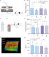
Friction and wear tests performed with different suture variants. (a) A first set of friction tests was conducted on perforated porcine skin samples, and the corresponding friction energy values are shown in (b). (c) By employing a modified suture coating procedure (in which only half of each suture is coated), the same chicken stomach sample could be used for up to four consecutive friction tests (d). The corresponding friction energy values are shown in (e); here, identical colors represent data obtained from the same suture/tissue combination. (f) Exemplary profilometry image depicting wear scars on a chicken stomach sample as inflicted by the sliding process of a mucin coated Vicryl Plus (VPm) suture and its uncoated counterpart (VP), respectively. (g) Sdr values were calculated from profilometric images to compare the local surface roughness of the damaged tissue samples after the sliding tests. Data points obtained on the same tissue sample are represented by identical colors. In all graphs, the data shown represents mean values, and error bars depict the standard deviation as calculated from n ≥ 5 independent samples. “n.s.” indicates statistically nonsignificant differences based on a p-value of 0.05. The schematic drawings were created using BioRender: https://BioRender.com/c83j150.
Figure 5
Figure 5
Additional suture modification using aCu-MBGNs. (a, b) SEM images of mucin coated Vicryl sutures carrying aCu-MBGNs (VmBG); the scale bars represent (a) 4 and (b) 1 μm, respectively. Exemplary agar plate images showing the inhibition zones created by VmBG sutures toward S. aureus (c) and E. coli (d). Scale bars represent 10 mm. (e) Quantification of the inhibition zones created by suture pieces of 2 cm length. Data shown in the table represents mean values together with the standard deviation calculated from n ≥ 3 independent samples.
Figure 6
Figure 6
Triggerable drug release from condensed mucin coatings on Vicryl sutures: (a–c) Schematic representation of the process used to (a) load the mucin surface with the target drug TCL; (b) trap the drug through mucin layer condensation and stabilization with cations; (c) release TCL upon contact of the coating with a physiological NaCl concentration. (d) TCL release profiles from uncoated and mucin coated Vicryl sutures obtained in a 154 mM NaCl solution (dark blue symbols) and in UPW (light blue symbols). Data shown in gray represents the control group (uncoated Vicryl sutures). The concentration of released TCL is calculated with the help of a standard curve (Figure S8, Supporting Information). Data shown represents mean values; error bars denote the standard deviation as calculated from n ≥ 4 independent samples. Asterisks and “n.s.” indicate statistically significant and nonsignificant differences, respectively (based on a p-value of 0.05). The schematic drawings were created using BioRender: https://BioRender.com/v75v402.

References

    1. Mahmoud N. N.; Hamad K.; Al Shibitini A.; Juma S.; Sharifi S.; Gould L.; Mahmoudi M. Investigating Inflammatory Markers in Wound Healing: Understanding Implications and Identifying Artifacts. ACS Pharmacol. Transl. Sci. 2024, 7 (1), 18–27. 10.1021/acsptsci.3c00336. - DOI - PMC - PubMed
    1. Mehrjou B.; Wu Y.; Liu P.; Wang G.; Chu P. K. Design and Properties of Antimicrobial Biomaterials Surfaces. Adv. Healthcare Mater. 2023, 12 (16), 220207310.1002/adhm.202202073. - DOI - PubMed
    1. Wang C.; Su Y.; Shahriar S. M. S.; Li Y.; Xie J. Emerging strategies for treating medical device and wound-associated biofilm infections. Microb. Biotechnol. 2024, 17 (10), e7003510.1111/1751-7915.70035. - DOI - PMC - PubMed
    1. Schmitz N. D.; Ovington L.; Berlin J.; Zhang S.; Collier J. Optimal usage of antibacterial sutures for wound closure in clinical trials addressing SSI. Lancet 2023, 401 (10387), 1497–1498. 10.1016/S0140-6736(23)00668-2. - DOI - PubMed
    1. Lovric V.; Goldberg M. J.; Heuberer P. R.; Oliver R. A.; Stone D.; Laky B.; Page R. S.; Walsh W. R. Suture wear particles cause a significant inflammatory response in a murine synovial airpouch model. J. Orthop. Surg. Res. 2018, 13 (1), 311.10.1186/s13018-018-1026-4. - DOI - PMC - PubMed

LinkOut - more resources