. 2025 Feb 27;16(1):140.
doi: 10.1038/s41419-025-07430-5.

MicroRNA-379-5p attenuates cancer stem cells and reduces cisplatin resistance in ovarian cancer by regulating RAD18/Polη axis

Affiliations

MicroRNA-379-5p attenuates cancer stem cells and reduces cisplatin resistance in ovarian cancer by regulating RAD18/Polη axis

Devendra Shukla et al. Cell Death Dis. .

Abstract

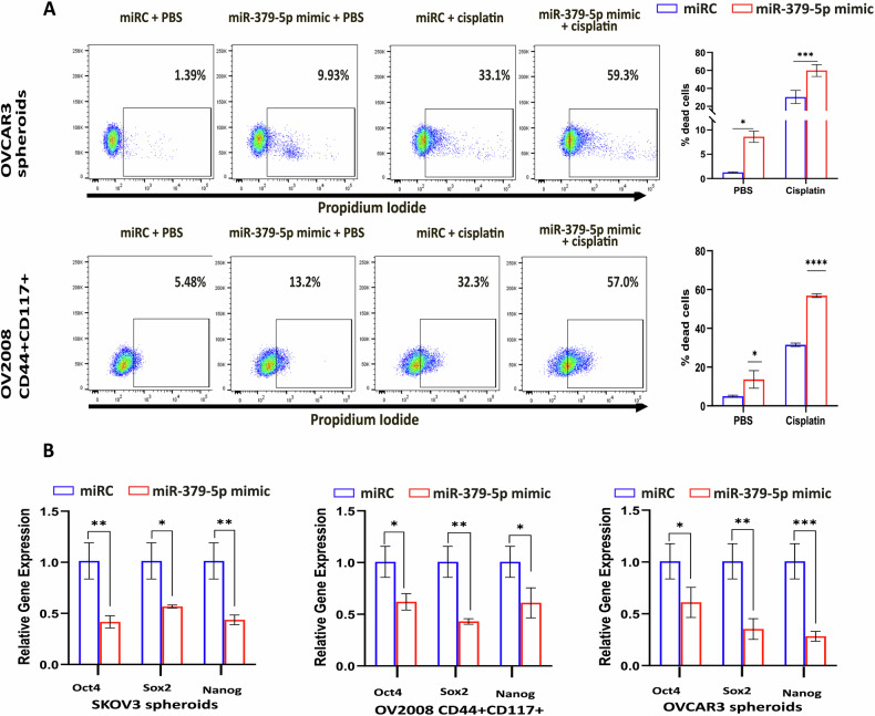
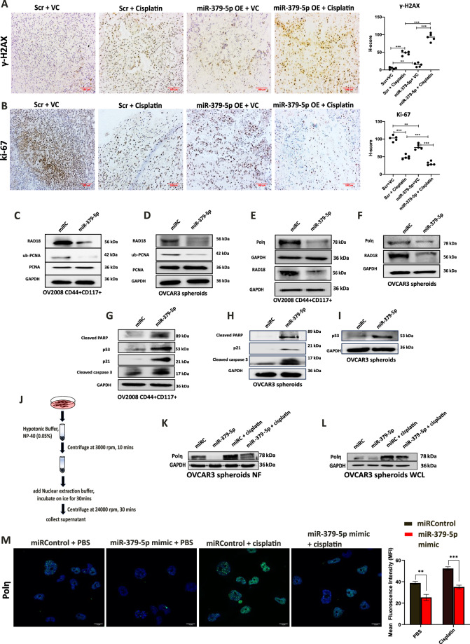
Ovarian cancer (OC) is an aggressive malignancy of the female reproductive organs, associated with a low 5-year survival rate. Emerging evidence suggests the pivotal role of microRNAs (miRNAs) in regulating chemoresistance and metastasis in OC, primarily through cancer stem cells (CSCs), also known as cancer stem-like cells (CSLCs). Herein, we demonstrate that miR-379-5p is downregulated in several OC cell populations including both cell lines and patient tumor samples. Furthermore, overexpression of miR-379-5p effectively inhibits CSCs and counteracts cisplatin-induced expansion of CSCs. Further mechanistic investigations identify RAD18, a DNA repair protein involved in translesion DNA synthesis (TLS), as a direct target of miR-379-5p. Moreover, a negative correlation between miR-379-5p and RAD18 expression is observed in ovarian CSCs isolated from OC patients. The downregulation of RAD18 inhibits stem-like phenotypes and enhances the sensitivity of ovarian CSCs to cisplatin treatment. Importantly, miR-379-5p-mediated inhibition of RAD18 prevents the repair synthesis in CSCs by promoting the accumulation of DNA damage. In vivo studies further reveal that miR-379-5p enhances DNA damage, which, in turn, inhibits tumor cell proliferation in athymic nude mice. Remarkably, targeting of RAD18 by miR-379-5p prevents monoubiquitination of proliferating cell nuclear antigen (PCNA), resulting in reduced DNA Polymerase η (a TLS polymerase that helps to bypass DNA lesions) recruitment to lesion sites. In the absence of Polη, the persisting DNA lesions cause activation of cell cycle arrest and apoptosis pathway in CSCs. Therefore, our findings unveil a novel mechanism whereby miR-379-5p overexpression curtails CSCs by modulating the RAD18/Polη axis.


Conflict of interest statement

Competing interests: The author declares no competing interests. Ethics approval and consent to participate: All experiments related to patients were performed at CNCI, Kolkata. Before starting the study, the mandatory institutional human ethical clearance was taken (Ref No. CNCI-IEC-40104). Patients were made aware of the study’s objectives, and their written consent was also received. Animal care and related experiments were conducted following institutional guidelines and with the approval of Ohio State University’s Institutional Animal Care and Use Committee.

Figures

Fig. 1
Fig. 1. miR-379-5p is downregulated in ovarian CSCs.
A The GEO dataset GSE107155 was reanalyzed to identify a list of differentially expressed microRNAs in ovarian cancer cell line SKOV3 grown in adherent and spheroid cultures. B qPCR analysis showed decreased miR-379-5p expression in spheroids derived from ovarian cancer cell lines and HGSOC patients. C Representative H&E of normal tissue and tumor tissue derived from ovarian cancer patients. D Pan-cancer expression profiling of miR-379-5p in normal vs tumor samples using CancerMIRNome (Wilcoxon rank-sum test, ***: P < 0.001; **: P < 0.01; *: P < 0.05; ns: P > 0.05). E Colony formation assay was conducted on control and miR-379-5p-overexpressing OC cells. F Representative images show spheroid formation ability in control and miR-379-5p-overexpressing ovarian CSCs (n = 3, Bar, SD; Significance levels denoted as ns > 0.05, *P < 0.05, **P < 0.01, ***P < 0.001 and ****P < 0.0001).
Fig. 2
Fig. 2. miR-379-5p downregulation is required for CSCs maintenance.
A Overexpression of miR-379-5p reduces the enrichment of CD44 + CD117+ cell populations within SKOV3 and OVCAR3 bulk cells following cisplatin treatment. CD44 + CD117+ cells were identified using flow cytometry, and the accompanying graph shows the percentage of CD44 + CD117+ cell enrichment. B Athymic nude mice (n = 5) were injected with either miRControl or miR-379-5p-proficient OVCAR3 cells. Once the tumor diameter reached around 0.5 cm, both groups of mice received weekly treatments with PBS or cisplatin (5 mg/Kg body weight) for 3 weeks. C Representative images of the tumors 3 weeks after the initial drug treatment. D Quantification of the time-dependent progression in tumor volume, measured with calipers. E After the mice were sacrificed, tumor weights were determined in grams. F The graph shows that xenograft cells with miR-379-5p overexpression exhibit a reduced enrichment of CD44 + CD117+ cell populations in response to cisplatin treatment (n = 3, Bar, SD; Significance levels indicated as *P < 0.05, **P < 0.01, ***P < 0.001 and ****P < 0.0001).
Fig. 3
Fig. 3. miR-379-5p enhances the sensitivity of CSCs to cisplatin while decreasing the expression of genes associated with stemness.
A Propidium iodide (PI) live-dead assay on OVCAR3 spheroid cells and OV2008 CD44 + CD117+ cells exhibit higher cytotoxicity in cells overexpressing miR-379-5p upon treatment with cisplatin. The accompanying chart illustrates the percentage of cell death. B In SKOV3 spheroids, OV2008 CD44 + CD117+ cells, and OVCAR3 spheroids, the overexpression of miR-379-5p significantly diminishes the expression of stem cell marker genes such as oct4, sox2, and nanog (n = 3, Bar, SD; Significance levels are denoted as *P < 0.05, **P < 0.01, ***P < 0.001, and ****P < 0.0001).
Fig. 4
Fig. 4. RAD18, a direct target for miR-379-5p.
A The Venn diagram illustrates three common targets (KIF5A, RAD18, and CFTR) of miR-379-5p identified from various databases. B Pan-cancer expression analysis of RAD18 in tumor vs. normal samples using GEPIA. C Schematic diagram illustrating the alignment of miR-379-5p with its wild-type binding site in the Homo sapiens RAD18 3′UTR sequence, along with the corresponding mutated binding site. D Results from the dual-luciferase reporter assay reveal the direct interaction between miR-379-5p and RAD18. OVCAR3 cells were transfected with either wild-type RAD18 3′-UTR-psiCHECK2/miR-379-5p or mutated RAD18 3′-UTR-psiCHECK2/miR-379-5p alongside a wild-type 3′-UTR-psiCHECK2/scramble miRControl. Renilla luciferase activity was measured, and normalized to firefly activity. E The expression levels of RAD18 in HGSOC patients were determined via qPCR. F, G RAD18 levels were analyzed in SKOV3 adherent and spheroid cells and OV2008 CD44-CD117- and CD44 + CD117+ cells through qPCR and western blotting. H Single-cell RNA analysis demonstrates the variation in RAD18 expression between metastatic and non-metastatic conditions in different cell types (n = 3, Bar, SD; Significance levels indicated as *P < 0.05, **P < 0.01, and ***P < 0.001).
Fig. 5
Fig. 5. RAD18 expression is associated with CSCs progression.
A Downregulation of RAD18 reduces the enrichment of CD44 + CD117+ cell populations within SKOV3 and OVCAR3 bulk cells following cisplatin treatment. CD44 + CD117+ cells were identified using flow cytometry, and the accompanying graph shows the percentage of CD44 + CD117+ cell enrichment. B The propidium iodide (PI) live-dead assay on SKOV3 and OVCAR3 spheroids showed enhanced cytotoxicity in cells with RAD18 downregulation after cisplatin treatment. C Downregulation of RAD18 was confirmed through western blotting in OV2008 and OVCAR3. D The downregulation of RAD18 in xenograft transplants resulted in reduced tumor size in athymic nude mice treated with either PBS or cisplatin (5 mg/kg body weight). E Tumor weights were measured in grams (gm) at the conclusion of the experiments (Bar, SD; Significance levels indicated as *P < 0.05, **P < 0.01, ***P < 0.001 and ****P < 0.0001).
Fig. 6
Fig. 6. miR-379-5p impairs DNA repair and reduces cisplatin-induced mutagenesis.
A, B Flow cytometry analysis of γ-H2AX, a marker for DNA damage, in SKOV3 spheroids and OVCAR3 spheroids, reveals increased γ-H2AX levels in cells having miR-379-5p overexpression (OE: overexpression); The DNA repair kinetics assay revealed insignificant DNA damage repair in cells with miR-379-5p OE group, compared to the significant repair observed in both the miRControl and miR-379-5p OE + siRAD18 OE groups after 3 h and 6 h of recovery. C, D Immunofluorescence study of γ-H2AX accumulation in SKOV3 and OVCAR3 adherent cells overexpressing miR-379-5p. The corresponding graph indicates the mean fluorescence intensity (MFI) of γ-H2AX staining. E Mutagenesis analysis in OVCAR3 adherent cells with miR-379-5p overexpression revealed reduced mutagenesis in response to cisplatin treatment compared to control cells. The corresponding graph shows the mutation frequency observed in each group (n = 3, Bar, SD; Significance levels indicated as *P < 0.05, **P < 0.01, and ***P < 0.001).
Fig. 7
Fig. 7. miR-379-5p induces DNA damage, suppresses proliferation in xenografts, and modulates the TLS pathway via targeting RAD18, leading to cell cycle arrest and apoptosis in CSCs.
A Immunohistochemistry (IHC) staining using γ-H2AX, a marker of DNA damage, in tumor tissues obtained from xenografts. B IHC staining employing Ki-67 in tumor tissues derived from xenografts. The corresponding graphs depict the H-scores for each staining. C, D miR-379-5p overexpression in CSCs inhibits RAD18-mediated monoubiquitination of PCNA in OV2008 CD44 + CD117+ and OVCAR3 spheroids. E, F The nuclear fraction isolated from miR-379-5p mimic-transfected cells displays reduced Polη and RAD18 expression in OV2008 CD44 + CD117+ and OVCAR3 spheroids. GI miR-379-5p overexpression activates apoptotic genes in OV2008 CD44 + CD117+ and OVCAR3 spheroids. J The schematic diagram outlines the nuclear fraction isolation method utilized in this study. K Polη expression levels in response to PBS or cisplatin treatment in nuclear fractions (NF) isolated from miRC or miR-379-5p-overexpressing OVCAR3 spheroids. L Polη expression levels in response to PBS or cisplatin treatment in whole cell lysate (WCL) isolated from miRC or miR-379-5p-overexpressing OVCAR3 spheroids. M miR-379-5p mimic prevents the increased Polη expression in response to cisplatin treatment. The corresponding graph indicates the mean fluorescence intensity (MFI) values of Polη immunostaining (n = 3, Bar, SD; Significance levels denoted as **P < 0.01, and ***P < 0.001).
Fig. 8
Fig. 8. RAD18 facilitates the recruitment of Polη to the DNA damage sites within the nucleus.
A The ribbon diagram of RAD18, Polη, and ub-PCNA reproduced from PDB. B Protein-protein docking for RAD18-Polη, RAD18- ub-PCNA, and Polη-ub-PCNA was performed using HDOCK. C A positive correlation between RAD18 and Polη expression in female gynecological malignancies was obtained from the CorrelationAnalyzeR tool. D Co-immunoprecipitation of RAD18, ub-PCNA, and Polη was performed using Polη antibody. A representative western blot image validates the interaction between RAD18, ub-PCNA, and Polη. E Immunofluorescence staining of OVCAR3 adherent cells with RAD18 and Polη showed that Polη had reduced nuclear localization in the absence of RAD18 following cisplatin treatment.

References

    1. Arora T, Mullangi S, Vadakekut ES, Lekkala MR. Ovarian cancer. 2023 [cited 2023 Oct 24]. https://pubmed.ncbi.nlm.nih.gov/33620837.
    1. Ip CKM, Li SS, Tang MYH, Sy SKH, Ren Y, Shum HC, et al. Stemness and chemoresistance in epithelial ovarian carcinoma cells under shear stress. Sci Rep. 2016;6:26788. 10.1038/srep26788. - PMC - PubMed
    1. Walcher L, Kistenmacher AK, Suo H, Kitte R, Dluczek S, Strauß A, et al. Cancer stem cells—origins and biomarkers: perspectives for targeted personalized therapies. Front Immunol. 2020;11. 10.3389/fimmu.2020.01280. - PMC - PubMed
    1. Nör C, Zhang Z, Warner KA, Bernardi L, Visioli F, Helman JI, et al. Cisplatin induces Bmi-1 and enhances the stem cell fraction in head and neck cancer. Neoplasia. 2014;16:137–46. - PMC - PubMed
    1. Louka M, Boutou E, Bakou V, Pappa V, Georgoulis A, Stürzbecher HW, et al. DNA damage response/repair in cancer stem cells—potential vs. controversies. In: Chen CC, editor. Advances in DNA repair. Rijeka: IntechOpen; 2015. p. 15. 10.5772/61355.

MeSH terms