. 2025 Jul;46(7):2056-2072.
doi: 10.1038/s41401-025-01507-9. Epub 2025 Feb 27.

Tetrandrine augments melanoma cell immunogenicity via dual inhibition of autophagic flux and proteasomal activity enhancing MHC-I presentation

Affiliations

Tetrandrine augments melanoma cell immunogenicity via dual inhibition of autophagic flux and proteasomal activity enhancing MHC-I presentation

Li-Na He et al. Acta Pharmacol Sin. 2025 Jul.

Abstract

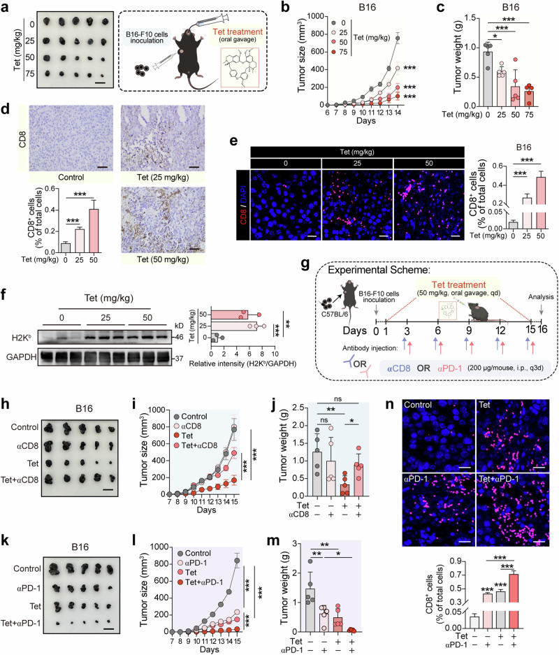
MHC-I-mediated antigen presentation is pivotal in antitumor immunity, enabling the recognition and destruction of tumor cells by CD8+ T cells. Both the proteasome and autophagy serve as essential cellular degradation mechanisms that regulate the stability and functionality of MHC-I molecules. In melanoma, modulating the pathways that affect MHC-I antigen presentation is pivotal and can profoundly influence the therapeutic outcomes of immunotherapy. Our initial effort of this study was a screening process to identify natural compounds capable of amplifying MHC-I surface expression on B16 melanoma cells. Utilizing flow cytometry with fluorescently tagged antibodies, we identified tetrandrine (Tet), a bisbenzylisoquinoline alkaloid derived from the root of Stephania tetrandra, as a potent enhancer of MHC-I-mediated antigen presentation in B16 melanoma cells. We demonstrate that tetrandrine (2.5, 5, 7.5 μM) dose-dependently upregulates both surface and total MHC-I protein levels in B16 or A375 melanoma cells by simultaneously inhibiting autophagy and proteasomal activity, two key pathways involved in MHC-I degradation. This dual inhibition stabilizes MHC-I molecules, leading to enhanced tumor antigen presentation and improved recognition by CD8+ T cells. In co-culture systems, tetrandrine treatment increased CD8+ T cell activation and cytotoxicity against melanoma cells, evidenced by elevated IFN-γ secretion and increased tumor cell apoptosis. Administration of tetrandrine (50 mg·kg-1·d-1, i.g., for 15 days) significantly suppressed melanoma growth in mouse models accompanied by increased CD8+ T cell infiltration and activation within the tumor microenvironment. Notably, tetrandrine synergized with anti-PD-1 immune checkpoint therapy, leading to enhanced tumor growth inhibition compared to either treatment alone. We revealed that tetrandrine (7.5 μM) blocked the lysosomal calcium efflux channel TPC2, disrupting lysosomal calcium homeostasis, thus impairing lysosomal acidification and proteasomal activity, thereby stabilizing MHC-I molecules and promoting antigen presentation. These results highlight tetrandrine's unique mechanism of action in enhancing MHC-I-mediated antigen presentation through dual inhibition of autophagic flux and proteasomal degradation. This study underscores tetrandrine's potential as a novel immunomodulatory agent to boost CD8+ T cell-mediated tumor cell eradication and enhance the efficacy of immune checkpoint therapies.

Keywords: MHC-I; TPC2 channel; autophagy; melanoma; proteasome; tetrandrine.


Conflict of interest statement

Competing interests: The authors declare no conflict of interest.

Figures

Fig. 1
Fig. 1. Tetrandrine enhances MHC-I-mediated antigen presentation in melanoma cells.
a Schematic representation of the screening for natural product small molecules that increase MHC-I on the surface of melanoma cells. Using flow cytometry with fluorescently labeled antibodies, we screened for natural products from the TargetMol natural product library that enhance the expression of H2Kb (MHC-I) on the surface of B16 melanoma cells. b Chemical structure of tetrandrine. c Concentration-dependent increase of MHC-I on the surface of melanoma cells by tetrandrine. B16 or A375 melanoma cells were treated with a gradient of tetrandrine concentrations (0, 2.5, 5, 7.5 μM) for 24 h, followed by flow cytometric analysis using an H2Kb (HLA-A/B) fluorescent antibody to assess the effect of tetrandrine on the surface expression of MHC-I. d Concentration-dependent increase of MHC-I on the surface of melanoma cells by tetrandrine. B16 or A375 melanoma cells were treated with a gradient of tetrandrine concentrations (0, 2.5, 5, 7.5 μM) for 24 h, followed by Western blot analysis using an H2Kb (HLA-A/B) antibody to assess the effect of tetrandrine on the total expression of MHC-I. e Concentration-dependent enhancement of MHC-I-mediated antigen presentation in melanoma cells by tetrandrine. B16-OVA cells (B16 melanoma cells stably expressing OVA protein) were treated with a gradient of tetrandrine concentrations (0, 2.5, 5, 7.5 μM) for 24 h. Subsequently, flow cytometry was used to quantify the cell surface H2Kb/SIINFEKL complexes. f Concentration-dependent enhancement of MHC-I-mediated antigen presentation in melanoma cells by tetrandrine. B16 cells were incubated with the SIINFEKL peptide and treated with a range of tetrandrine concentrations (0, 2.5, 5, 7.5 μM) for 24 h. Following treatment, flow cytometry was performed to quantify the cell surface H2Kb/SIINFEKL complexes. Tet, tetrandrine. **P < 0.05, ***P < 0.001 indicate levels of statistical significance.
Fig. 2
Fig. 2. Tetrandrine treatment enhances CD8+ T cell recognition and killing of melanoma cells.
a Schematic of the in vitro immune cytotoxicity assay. B16-OVA cells pretreated with a gradient of tetrandrine concentrations (0, 2.5, 5, 7.5 μM) for 24 h were co-cultured with CD8+ T cells from OT-I mice at a 1:10 ratio. After 24 h, CD8+ T cell activation and their cytotoxic effect on melanoma cells were assessed by crystal violet staining, ELISA for IFN-γ release, and flow cytometry analysis of B16-OVA cell apoptosis. b Tetrandrine enhances the cytotoxicity of CD8+ T cells against B16-OVA melanoma cells (crystal violet staining). Results indicate that tetrandrine alone does not have a significant cytotoxic effect on melanoma cells. However, in the presence of CD8+ T cells, tetrandrine enhances the cytotoxic effect in a concentration-dependent manner. c Tetrandrine treatment increases IFN-γ release by CD8+ T cells. Following treatment as depicted, IFN-γ release was measured by ELISA. d Tetrandrine enhances the killing of melanoma cells by CD8+ T cells (flow cytometry - proportion of APC-CD8(-) FVS620(+) cells). After treatment as shown, the death of B16-OVA cells was assessed by flow cytometry, following co-staining with CD8 antibody and FVS620. Cells negative for CD8 and positive for FVS620 were identified as dead B16-OVA cells. Tet tetrandrine. ***P < 0.001 indicate levels of statistical significance.
Fig. 3
Fig. 3. Tetrandrine inhibits the late stage of autophagic flux in melanoma cells.
a Treatment with tetrandrine increases the number of autophagosomes in melanoma cells. A375-GFP-LC3 cells were treated with vehicle, tetrandrine (5 μM) for 24 h, HBSS (to induce autophagy) for 6 h, or Baf (20 nM) for 24 h, followed by confocal microscopy to count green fluorescent puncta. Scale bar, 10 μm. b Tetrandrine induces a concentration-dependent increase in P62 and LC3-II in melanoma cells. A375 and B16 cells were exposed to varying concentrations of tetrandrine (0, 2.5, 5, 7.5 μM) for 24 h. Subsequently, proteins were harvested for Western blot analysis. c Time-dependent elevation of P62 and LC3-II is observed with tetrandrine treatment. A375 or B16 melanoma cells were treated with tetrandrine (5 μM) for intervals up to 24 h, followed by protein isolation and Western blot assessment, with CQ (chloroquine, 20 μM) serving as a positive control. d Combined HBSS and tetrandrine treatment exacerbates autophagosome accumulation. A375-GFP-LC3 cells underwent treatments with vehicle, HBSS for 6 h, tetrandrine (5 μM) for 24 h, or tetrandrine for 18 h followed by HBSS for the remaining 6 h with continuous presence of tetrandrine. Subsequent analysis of green fluorescent puncta was performed via confocal microscopy. Scale bar, 10 μm. e Tetrandrine treatment enhances the colocalization of red and green fluorescence in mCherry-GFP-LC3-transfected melanoma cells, indicating a blockade at the late stage of autophagic flux. After transfection with mCherry-GFP-LC3 plasmids, A375 cells were treated with vehicle, HBSS for 6 h, tetrandrine (5 μM) for 24 h, or CQ (20 μM) for 24 h, followed by confocal microscopy to assess the presence of yellow puncta, signifying the impaired late stage of autophagic flux. Scale bar, 10 μm. Tet tetrandrine, CQ chloroquine, Baf bafilomycin A1, HBSS Hanks’ Balanced Salt Solution. **P < 0.01, ***P < 0.001 indicate levels of statistical significance.
Fig. 4
Fig. 4. Tetrandrine increases MHC-I levels by simultaneously inhibiting autophagy and proteasomal activity.
a, b Mutation of Atg4b blocks autophagy and increases the levels of both total and surface MHC-I (H2Kb) in melanoma cells. B16-Atg4bC74A cells, induced by Dox, express a mutant form of Atg4b that blocks autophagy. Cells were treated with varying concentrations of Dox (0, 2, 4, 8 μg/mL) for 24 h. Total MHC-I levels were assessed by Western blot, and surface MHC-I levels were measured using H2Kb fluorescent antibody staining and flow cytometry to determine mean fluorescence intensity. c Blocking autophagy with mutant Atg4b increases the level of MHC-I presentation. B16-Atg4bC74A cells were incubated with SIINFEKL peptide and treated with varying concentrations of Dox (0, 2, 4, 8 μg/mL) for 24 h. Surface H2Kb/SIINFEKL complex was assessed using fluorescent antibody staining and flow cytometry to measure mean fluorescence intensity. d, e Combined treatment with tetrandrine and Atg4b mutation, which blocks autophagy, leads to a further increase in both total and surface MHC-I in melanoma cells compared to individual treatments. B16-Atg4bC74A cells were treated with vehicle, Dox (8 μg/mL), Tet (7.5 μM), or Dox+Tet for 24 h. Total MHC-I levels were analyzed by Western blot, and surface MHC-I levels were measured using H2Kb fluorescent antibody staining and flow cytometry. f Blocking proteasomal activity with MG132 leads to an increase in MHC-I levels, and combined treatment with tetrandrine and MG132 results in a further increase in MHC-I levels. B16 cells were treated with vehicle, MG132 (1 μM), Tet (7.5 μM), or MG132+Tet for 24 h. Surface MHC-I was assessed using FITC-H2Kb fluorescent antibody staining and flow cytometry. g Combined treatment with tetrandrine, MG132, and Dox does not lead to a further increase in MHC-I levels compared to MG132 and Dox combined. B16-Atg4bC74A cells were treated with vehicle, MG132 (1 μM) combined with Dox (8 μg/mL), Tet (7.5 μM), or Tet+MG132+Dox for 24 h. Surface MHC-I was assessed using FITC-H2Kb fluorescent antibody staining and flow cytometry. h Combined treatment with tetrandrine, MG132, and CQ does not lead to a further increase in MHC-I levels compared to MG132 and CQ combined. B16 cells were treated with vehicle, MG132 (1 μM) combined with CQ (20 μM), Tet (7.5 μM), or Tet+MG132 + CQ for 24 h. Surface MHC-I was assessed using FITC-H2Kb fluorescent antibody staining and flow cytometry. i Tetrandrine treatment decreases proteasomal activity in melanoma cells. B16 cells were treated with vehicle, Tet (5, 7.5 μM), or MG132 (1 μM) for 24 h. Proteasomal activity was measured using a 20S proteasome activity assay kit. Tet tetrandrine, CQ chloroquine, Dox doxycycline. *P < 0.05, **P < 0.01, ***P < 0.001 indicate levels of statistical significance.
Fig. 5
Fig. 5. Tetrandrine inhibits the late stage of autophagic flux by impeding lysosomal acidification rather than inhibiting the fusion of lysosomes and autophagosomes.
a A375 cells transfected with the hLAMP1-mCherry plasmid were exposed to tetrandrine (5 μM) or chloroquine (CQ, 20 μM) for 24 h. Following immunostaining for LC3-I/II (green), images were acquired using confocal microscopy, and colocalization between hLAMP1-mCherry (red) and LC3 (green) was analyzed using ImageJ software. Yellow fluorescence indicates the overlap of lysosomes and autophagosomes. The red and green lines in the figure correspond to the arbitrary units (a.u.) of red and green intensity within the rectangular region shown in the zoomed-in image. Scale bar: 5 μm. b Tetrandrine inhibits lysosomal acidification. A375 cells were treated with vehicle, tetrandrine (5 μM), or Baf (20 nM) for 24 h and subsequently stained with Lysotracker to evaluate lysosomal acidification. A decrease in red fluorescence intensity indicates suppressed lysosomal acidification, with both tetrandrine and Baf treatments showing a significant reduction in Lysotracker red fluorescence. Scale bar, 10 μm. c, d Tetrandrine inhibits the maturation of Cathepsins. A375 or B16 cells were treated with various concentrations of tetrandrine (0, 2.5, 5, 7.5 μM) for 24 h or with a fixed concentration of tetrandrine (5 μM) over different time points (0, 2, 6, 12, 24 h). Proteins were then extracted, and Western blot analysis was performed to assess Cathepsin maturation. Baf (20 nM) was used as a positive control. e Analysis of autophagosome-lysosome fusion by electron microscopy. A375 cells were treated with vehicle or tetrandrine (7.5 μM) for 24 h, and samples were collected for electron microscopy. As indicated by the red arrows, a higher number of autolysosomes were observed in the tetrandrine-treated group, suggesting that tetrandrine does not inhibit the fusion of autophagosomes and lysosomes. Tet tetrandrine, CQ chloroquine, Baf bafilomycin A1. ***P < 0.001 indicate levels of statistical significance.
Fig. 6
Fig. 6. Tetrandrine inhibits lysosomal acidification and cytosolic proteasomal activity by blocking the lysosomal calcium efflux channel TPC2.
a Tet reduces cytosolic calcium levels. A375 melanoma cells were treated with vehicle, Tet (7.5 μM), TPC2-A1-N (30 μM, TPC2 activator), or Ned-19 (50 μM, an antagonist of the endogenous TPC2 activator NAADP) for 6 h, followed by Fluo-4/AM staining (3 μM, 37°C, 30 min). Cytosolic calcium levels were measured using confocal microscopy based on green fluorescence intensity. Scale bar, 5 μm. b Tet increases lysosomal calcium levels. A375 melanoma cells were treated with vehicle, Tet (7.5 μM), or BAPTA-AM (5 μM, calcium chelator) for 6 h, followed by Fluo-4/AM staining (3 μM, 37°C, 30 min). GPN (400 μM) was then used to permeabilize lysosomes, and lysosomal calcium content was inferred from changes in cytosolic calcium measured by confocal microscopy. c Tet inhibits TPC2-mediated calcium release. A375 melanoma cells stably expressing TPC2-GCaMP6m, a calcium-sensitive reporter that emits green fluorescence upon TPC2 activation, were treated with vehicle, Tet (7.5 μM), or BAPTA-AM (5 μM) for 6 h. After Fluo-4/AM staining (3 μM, 30 min), TPC2-A1-N (30 μM) was added to stimulate TPC2, and calcium release was measured by confocal microscopy based on green fluorescence intensity. d, e TPC2 activation reverses Tet-induced autophagic flux blockade and Cathepsin B maturation inhibition. A375 melanoma cells were treated with vehicle, Tet (7.5 μM), TPC2-A1-N (30 μM), or Tet combined with TPC2-A1-N for 24 h. Western blot analysis showed that TPC2 activation reversed Tet-induced accumulation of LC3-II and P62, as well as the reduction in Cathepsin B maturation. Densitometric quantification of protein bands is presented in the bar graphs. f TPC2 activation restores lysosomal acidification inhibited by Tet. A375 melanoma cells were treated with vehicle, Tet (7.5 μM), TPC2-A1-N (30 μM), Tet + TPC2-A1-N, Ned-19 (50 μM), or Tet + Ned-19 for 6 h. Lysosomal acidification was assessed using LysoTracker staining, with red fluorescence intensity quantified by confocal microscopy in at least 15 cells per group. Scale bar, 10 μm. g TPC2 activation restores proteasomal activity inhibited by Tet. A375 melanoma cells were treated with vehicle, Tet (7.5 μM), TPC2-A1-N (30 μM), or Tet + TPC2-A1-N for 6 h. Proteasomal activity was measured using the Amplite® Fluorometric 20S Proteasome Assay Kit, with MG132 as a positive control. h, i TPC2 activation reverses Tet-induced upregulation of HLA expression in human melanoma cells. A375 melanoma cells were treated with vehicle, Tet (7.5 μM), TPC2-A1-N (30 μM), or Tet + TPC2-A1-N for 24 h. Surface HLA expression was measured by flow cytometry after PE-conjugated anti-HLA antibody staining, while total HLA-A/B protein levels were assessed by Western blot analysis. j TPC2 activation reverses Tet-induced upregulation of surface H2Kb in murine melanoma cells. B16 murine melanoma cells were treated with vehicle, Tet (7.5 μM), TPC2-A1-N (30 μM), or Tet + TPC2-A1-N for 24 h. Surface H2Kb expression was measured by flow cytometry following PE-conjugated anti-H2Kb antibody staining (20 min). BAPTA BAPTA-AM, Tet tetrandrine, TPC2 two-pore calcium channel 2. *P < 0.05, **P < 0.01, ***P < 0.001 indicate levels of statistical significance.
Fig. 7
Fig. 7. Tetrandrine inhibits melanoma growth through CD8+ T cell-mediated immune responses and enhances anti-PD-1 therapy efficacy.
ac Tumor growth curves a and tumor weight analysis b showing the inhibitory effect of tetrandrine on melanoma growth in vivo. C57BL/6 mice (n = 5 per group) were subcutaneously injected with 2 × 105 B16 melanoma cells and were treated with varying concentrations of tetrandrine (0, 25, 50, 75 mg/kg). Tumor growth was monitored every day, at the end of the experiment, tumors were harvested and weighed. Scale bar, 2 cm. d, e Immunohistochemical and immunofluorescence analysis of CD8+ T cell infiltration in paraffin-embedded tumor tissues, indicating the percentage of CD8+ T cells. Scale bar, 50 μm d and 20 μm e. f Western blot analysis of H2Kb expression in tumor tissues from mice (n = 3 per group) treated with tetrandrine. g Schematic representation of CD8 depletion and anti-PD-1 combination therapy in animal experiments. hj Tumor images h, tumor growth curves i and tumor weight analysis j showing the reversal of tetrandrine’s tumor inhibitory effect upon CD8 depletion. C57BL/6 mice were divided into groups: control, αCD8 (200 μg/mouse, i.p., every 3 days), Tet (50 mg/kg), and αCD8+Tet. Scale bar, 2 cm. km Tumor images k, tumor growth curves l and tumor weight analysis m showing the enhanced tumor inhibitory effect of tetrandrine in combination with anti-PD-1 therapy. C57BL/6 mice were divided into groups: control, αPD1 (200 μg/mouse, i.p., every 3 days), Tet (50 mg/kg), and αPD1+Tet. Scale bar, 2 cm. n CD8+ T cell infiltration in tumor tissues. Immunofluorescence analysis of tumor tissues from B16-F10 tumor-bearing C57BL/6 mice. The bar graph quantifies the percentage of CD8+ T cells among total cells, showing significantly increased infiltration in the Tet+αPD-1 group. Scale bar, 20 μm. Tet, tetrandrine. *P < 0.05, **P < 0.01, ***P < 0.001 indicate levels of statistical significance.
Fig. 8
Fig. 8. Mechanistic illustration of tetrandrine-mediated enhancement of melanoma cell recognition and killing by CD8+ T cells through the inhibition of autophagy and proteasomal activity.
The diagram illustrates how MHC-I molecules in melanoma cells can be degraded through both autophagy and proteasomal pathways, leading to a reduction in surface MHC-I molecules and facilitating immune escape of the melanoma cells. Tetrandrine disrupts this degradation process by concurrently inhibiting late-stage autophagic flux, through lysosomal acidification disruption, and suppressing proteasomal activity. This dual inhibition prevents MHC-I degradation, thereby increasing MHC-I-mediated antigen presentation on the surface of melanoma cells. The elevated antigen presentation enhances CD8+ T cell recognition and cytotoxicity against melanoma cells. Further mechanistic exploration revealed that tetrandrine exerts its effects by blocking the lysosomal calcium efflux channel TPC2, leading to elevated lysosomal calcium levels and reduced cytosolic calcium concentrations. This calcium imbalance inhibits lysosomal acidification and suppresses cytoplasmic proteasomal activity, collectively contributing to reduced MHC-I degradation. The schematic emphasizes tetrandrine’s pivotal role in modulating both autophagic and proteasomal pathways, ultimately enhancing the immunogenicity of melanoma cells and increasing their susceptibility to CD8+ T cell-mediated cytotoxicity. Tet tetrandrine.

References

    1. Ribas A, Wolchok JD. Cancer immunotherapy using checkpoint blockade. Science. 2018;359:1350–5. - PMC - PubMed
    1. Hiam-Galvez KJ, Allen BM, Spitzer MH. Systemic immunity in cancer. Nat Rev Cancer. 2021;21:345–59. - PMC - PubMed
    1. Wu X, Li T, Jiang R, Yang X, Guo H, Yang R. Targeting MHC-I molecules for cancer: function, mechanism, and therapeutic prospects. Mol Cancer. 2023;22:194. - PMC - PubMed
    1. Mellman I, Chen DS, Powles T, Turley SJ. The cancer-immunity cycle: Indication, genotype, and immunotype. Immunity. 2023;56:2188–205. - PubMed
    1. Oliveira G, Stromhaug K, Klaeger S, Kula T, Frederick DT, Le PM, et al. Phenotype, specificity and avidity of antitumour CD8+ T cells in melanoma. Nature. 2021;596:119–25. - PMC - PubMed

MeSH terms

Substances

LinkOut - more resources