Review
. 2025 Feb 5:31:101503.
doi: 10.1016/j.mtbio.2025.101503. eCollection 2025 Apr.

Potentially commercializable nerve guidance conduits for peripheral nerve injury: Past, present, and future

Affiliations
Review

Potentially commercializable nerve guidance conduits for peripheral nerve injury: Past, present, and future

Chundi Liu et al. Mater Today Bio. .

Abstract

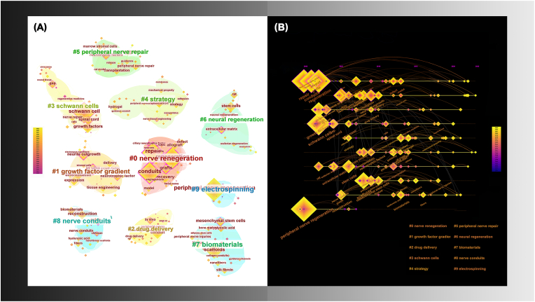
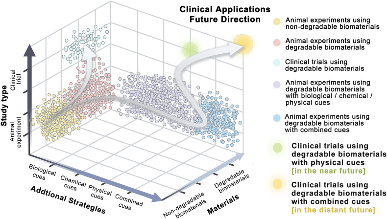
Peripheral nerve injuries are a prevalent global issue that has garnered great concern. Although autografts remain the preferred clinical approach to repair, their efficacy is hampered by factors like donor scarcity. The emergence of nerve guidance conduits as novel tissue engineering tools offers a promising alternative strategy. This review aims to interpret nerve guidance conduits and their commercialization from both clinical and laboratory perspectives. To enhance comprehension of clinical situations, this article provides a comprehensive analysis of the clinical efficacy of nerve conduits approved by the United States Food and Drug Administration. It proposes that the initial six months post-transplantation is a critical window period for evaluating their efficacy. Additionally, this study conducts a systematic discussion on the research progress of laboratory conduits, focusing on biomaterials and add-on strategies as pivotal factors for nerve regeneration, as supported by the literature analysis. The clinical conduit materials and prospective optimal materials are thoroughly discussed. The add-on strategies, together with their distinct obstacles and potentials are deeply analyzed. Based on the above evaluations, the development path and manufacturing strategy for the commercialization of nerve guidance conduits are envisioned. The critical conclusion promoting commercialization is summarized as follows: 1) The optimization of biomaterials is the fundamental means; 2) The phased application of additional strategies is the emphasized direction; 3) The additive manufacturing techniques are the necessary tools. As a result, the findings of this research provide academic and clinical practitioners with valuable insights that may facilitate future commercialization endeavors of nerve guidance conduits.

Keywords: Add-on strategy; Biomaterial; Clinical efficacy; Nerve guidance conduit; Peripheral nerve.


Conflict of interest statement

The authors declare that they have no known competing financial interest or personal relationships that could have appeared to influence the work reported in this manuscript.

Figures

Image 1
Graphical abstract
Fig. 1
Fig. 1
Diagram peripheral nerve injury repair (A) with artificial nerve guide conduit (B). (C–E) Nerve regeneration processes after injury. (C) After the injury, Waller degeneration occurs, axons are disintegrated, myelin is shed, and Schwann cells (SCs) transform into a repair phenotype, recruiting macrophages M1 (Mø-1) to enter the site of injury, followed by joint phagocytosis and removal of axon and myelin debris. (D) Macrophages are polarized into M2 type (Mø-2) in response to a hypoxic environment, which stimulates endothelial cell (EC) proliferation, migration, and budding to form new blood vessels. The new vasculature system acts as a physical track to guide SC migration. The reprogrammed SCs extend lengthwise to form Büngner bands. The axon tip forms a new growth cone and is directed to regenerate under the guidance of SC. (E) The microvascular network in the nerve is reconstructed, SCs are redifferentiated to form myelin, and the regenerated axons are remyelinated, representing that the repair was completed [38].
Fig. 2
Fig. 2
(A) Basic information on the 13 included studies. (B–D) Forest plot. (B) Effect of FDA-approved NGCs on s2PD. (C) Subgroup analysis of the effect of FDA-approved conduits on the recovery rate of s2PD based on the follow-up duration. There were four subgroups: 3 months (3m); 6 months (6m); 12 months (12m); and longer than 12 months (>12m) according to the duration of follow-up. (D) Comparative analysis of the efficacy of FDA-approved artificial conduits versus standard repairs on s2PD. N, number of participants; IG, intervention group; CG, control group.
Fig. 3
Fig. 3
Keywords cluster (A) related to peripheral nerve guidance conduit and time diagram (B), including 10 clusters from #0 to #9. Keyword nodes can be observed under each cluster; the size of the nodes represents the keyword frequency, while the node color reflects the time development of the keyword. (For interpretation of the references to color in this figure legend, the reader is referred to the Web version of this article.)
Fig. 4
Fig. 4
Summary of ideal NGC properties: biocompatibility; biodegradability; mechanical properties; and porosity.
Fig. 5
Fig. 5
Biological modification of NGCs prepared from FDA-approved materials in animal experiments. (A–C) Stem cell transplantation for nerve repair. (A) Schematic of inductively differentiated SC-like ADSCs implanted into a collagen sponge scaffold for nerve repair (reproduced with permission) [149]. Copyright 2019, Elsevier. (B) The schematic of inductively differentiated SC-like BMMSCs implanted into a collagen scaffold for nerve repair (reproduced with permission) [147]. Copyright 2011, Elsevier. (C) Schematic of BMMSCs anchored via DABLs in a PGA conduit (reproduced under the terms of the Creative Commons Attribution 4.0 International License (CC BY 4.0)) [148]. Copyright 2023, Springer. (D) Pre-vascularization strategy for nerve repair. Schematic of subcutaneous construction of a pre-vascularized scaffold of SKP-SCs and its combined use in chitosan conduits. The expression condition of NGF after subcutaneous implantation and bridging of defects with different scaffolds is also important (reproduced with permission) [151]. Copyright 2023, Wiley.
Fig. 6
Fig. 6
Chemical modification of NGCs prepared from FDA-approved materials in animal experiments. (A) Load and release of the chemical factors. Schematic of PLCL conduits loaded with different amounts of MeCbl (reproduced with permission) [158]. Copyright 2020, Wiley. (B) On-demand release of chemical factors. Schematic of a biologically orthogonal functionalized scaffold with esterase activation release (reproduced with permission) [162]. Copyright 2023, Elsevier. (C–D) Sustainable release of chemical factors. (C) Schematic of a collagen conduit for continuous delivery of GDNF (30 d) (reproduced with permission) [163]. Copyright 2018, Elsevier. (D) Schematic of a chitosan scaffold with long-term controlled release of NGF (8 weeks) (reproduced under the terms of the Creative Commons Attribution-NonCommercial-ShareAlike License (CC BY-NC-SA)) [164]. Copyright 2022, Wolters Kluwer.
Fig. 7
Fig. 7
Physical modification of NGCs prepared from FDA-approved materials in animal experiments. (A–C) 2D topological cues. (A) Schematic of the PLCL conduit modified with ridge/groove micropatterns and surface-anchored GO nanosheet promoting nerve regeneration (reproduced with permission) [172]. Copyright 2020, American Chemical Society. (B) Fabrication flow chart of the PLCL conduit containing 3D printed longitudinally oriented collagen hydrogels on the inner surface (reproduced with permission) [170]. Copyright 2020, Royal Society of Chemistry. (C) Fabrication of the chitosan conduit with ridge/groove micropatterned inner wall and seamless side wall, with optical and scanning electron microscopy images of the micropatterned structures (reproduced with permission) [171]. Copyright 2018, Elsevier. (D–G) 3D topological cues. (D) Schematic of the multi-channel nerve conduit based on chitosan and chitosan derivatives (reproduced with permission) [173]. Copyright 2023, Elsevier. (E) Fabrication schematic and cross-sectional images of multi-channel collagen conduits with different numbers of channels (reproduced with permission) [171]. Copyright 2010, Elsevier. (F) Schematic of the chitosan conduit with longitudinal chitosan film introduced into the lumen (reproduced under the terms of the Creative Commons Attribution-NonCommercial-NoDerivatives 4.0 International License (CC BY-NC-ND 4.0)) [174]. Copyright 2016, Elsevier. (G) Schematic of the collagen conduit with unidirectional and multidirectional multilayered pore structure promoting nerve repair (reproduced with permission) [175]. Copyright 2021, Elsevier. (H–I) Electromagnetic signal cues. (H) Schematic of the chitosan/GO conduit fabricated by electrodeposition to repair a nerve defect and macroscopic diagram of the material surface morphology (reproduced under the terms of the Creative Commons Attribution-NonCommercial-ShareAlike License (CC BY-NC-SA)) [176]. Copyright 2023, Wolters Kluwer. (I) Schematic of PLCL conduits combined with direct current or charge-balanced pulse stimulation (pulse) to repair facial nerve injury (reproduced with permission) [177]. Copyright 2023, American Chemical Society. (J) Schematic of PVDF/PLCL/PEDOT self-powering conduits that facilitated the repair of injured peripheral nerves (reproduced with permission) [178]. Copyright 2024, Wiley.
Fig. 8
Fig. 8
Biomanufacturing technology for physically modified structures of NGCs prepared from FDA-approved materials. (A) 3D printing technique. Schematic illustration and electron microscopy characterization of a collagen conduit with a high-precision microchannel structure and NGF gradient. (reproduced with permission) [67]. Copyright 2023, Wiley. (B) Compression molding technique. Fabrication routes and electron microscopy images of regular groove/ridged micropattern structures using a template thermo-pressing method (reproduced under the terms of the Creative Commons Attribution-NonCommercial-NoDerivatives 4.0 International License (CC BY-NC-ND 4.0)) [186]. Copyright 2022, Elsevier. (C–D) Electrospinning. (C) General images and electron microscopic images of suitable pore structures at nanometer/micron resolution in the inner wall of PLCL nerve conduit fabricated by electrospinning (reproduced with permission) [187]. Copyright 2022, American Chemical Society. (D) Schematic and electron microscopy of the nanopore structures with appropriate permeability of PLCL fiber scaffolds manufactured by electrospinning (reproduced with permission) [170]. Copyright 2020, Royal Society of Chemistry. (E) Blow-spinning. Schematic diagram and electron microscopic images of a double-layer collagen nanofiber conduit with internal fiber orientation and external fiber random arrangement prepared by multi-needle blow-spinning technique (reproduced under the terms of the Creative Commons Attribution-NonCommercial-NoDerivatives 4.0 International License (CC BY-NC-ND 4.0)) [188]. Copyright 2024, Elsevier.
Fig. 9
Fig. 9
Combined modification of NGCs prepared from FDA-approved materials in animal experiments. (A–C) Combination of physical and chemical cues on NGCs. (A) Schematic of the fabrication of ridge/groove micropatterned PLCL conduit with gradient-density CQAASIKVAV peptide modification (reproduced under the terms of the Creative Commons Attribution-NonCommercial-NoDerivatives 4.0 International License (CC BY-NC-ND 4.0)) [186]. Copyright 2022, Elsevier. (B) Schematic representation and conductivity properties of the PLCL/graphene scaffold incorporating ridge/grooved micropatterns, polydopamine modification, and ES (reproduced with permission) [201]. Copyright 2023, Wiley. (C) Schematic of magnetic nanoparticles modified with NGF (MNP-NGF) and collagen conduits (Neuragen) containing magnetically aligned gels with anisotropic MNP-NGF distribution (reproduced with permission) [202]. Copyright 2021, Wiley. (D) Combination of physical and biological cues in NGCs. The fabrication flow diagram of a 3D collagen hydrogel scaffold containing highly aligned SCs (reproduced Creative Commons Attribution 3.0 Unported (CC BY 3.0)) [203]. Copyright 2013, Elsevier. (E) Combination of biological and chemical cues in NGCs. The schematic of the collagen conduit with anchored bFGF and transplanted neural stem/progenitor cells (NS/PCs) (reproduced with permission) [199]. Copyright 2017, Elsevier.
Fig. 10
Fig. 10
Schematic representation of future advanced additive manufacturing technologies adapted for commercial development of NGC. (A) Inanimate 3D/4D fabrication techniques are suitable for the construction of NGCs modified with physical strategies in the short term. (B) 3D/4D biofabrication techniques are suitable for the construction of multifunctional NGCs modified with combined strategies in the long term.
Fig. 11
Fig. 11
The historical process and development trend of nerve guidance conduits. Potential strategies to achieve future clinical translation are highlighted with glowing dots.

Similar articles

Cited by

References

    1. Zheng T., Gao H., Liu Y., Sun S., Guan W., Wu L., Yang Y., Li G. Development of ovalbumin implants with different spatial configurations for treatment of peripheral nerve injury. Bioact. Mater. 2024;35:401–415. doi: 10.1016/j.bioactmat.2024.01.025. - DOI - PMC - PubMed
    1. Soto P.A., Vence M., Piñero G.M., Coral D.F., Usach V., Muraca D., Cueto A., Roig A., van Raap M.B.F., Setton-Avruj C.P. Sciatic nerve regeneration after traumatic injury using magnetic targeted adipose-derived mesenchymal stem cells. Acta Biomater. 2021;130:234–247. doi: 10.1016/j.actbio.2021.05.050. - DOI - PubMed
    1. Mahar M., Cavalli V. Intrinsic mechanisms of neuronal axon regeneration. Nat. Rev. Neurosci. 2018;19(6):323–337. doi: 10.1038/s41583-018-0001-8. - DOI - PMC - PubMed
    1. Li J., Yao Y., Wang Y., Xu J., Zhao D., Liu M., Shi S., Lin Y. Modulation of the crosstalk between schwann cells and macrophages for nerve regeneration: a therapeutic strategy based on a multifunctional tetrahedral framework nucleic acids system. Adv. Mater. 2022;34(46) doi: 10.1002/adma.202202513. - DOI - PubMed
    1. Sun Y., Zhang H., Zhang Y., Liu Z., He D., Xu W., Li S., Zhang C., Zhang Z. Li-Mg-Si bioceramics provide a dynamic immuno-modulatory and repair-supportive microenvironment for peripheral nerve regeneration. Bioact. Mater. 2023;28:227–242. doi: 10.1016/j.bioactmat.2023.05.013. - DOI - PMC - PubMed

LinkOut - more resources