. 2025 Oct 15;152(20):dev204244.
doi: 10.1242/dev.204244. Epub 2025 Mar 27.

A single-cell transcriptomic atlas of sensory-dependent gene expression in developing mouse visual cortex

Affiliations

A single-cell transcriptomic atlas of sensory-dependent gene expression in developing mouse visual cortex

Andre M Xavier et al. Development. .

Abstract

Sensory experience drives the maturation of neural circuits during postnatal brain development through molecular mechanisms that remain to be fully elucidated. One likely mechanism involves the sensory-dependent expression of genes that encode direct mediators of circuit remodeling within developing cells. To identify potential drivers of sensory-dependent synaptic development, we generated a single-nucleus RNA sequencing dataset describing the transcriptional responses of cells in the mouse visual cortex to sensory deprivation or to stimulation during a developmental window when visual input is necessary for circuit refinement. We sequenced 118,529 nuclei across 16 neuronal and non-neuronal cell types isolated from control, sensory deprived and sensory stimulated mice, identifying 1268 sensory-induced genes within the developing brain. While experience elicited transcriptomic changes in all cell types, excitatory neurons in layer 2/3 exhibited the most robust changes, and the sensory-induced genes in these cells are poised to strengthen synapse-to-nucleus crosstalk and to promote cell type-specific axon guidance pathways. Altogether, we expect this dataset to significantly broaden our understanding of the molecular mechanisms through which sensory experience shapes neural circuit wiring in the developing brain.

Keywords: Cell signaling; Circuit development; Neuron; Sensory experience; Synapse; Transcription; Visual cortex.


Conflict of interest statement

Competing interests The authors declare no competing or financial interests.

Figures

Fig. 1.
Fig. 1.
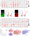
Experimental design and introduction to the single-nucleus RNA sequencing dataset. (A) Schematic illustrating the late dark-rearing (LDR) paradigm and the workflow of the single-nucleus RNA sequencing (snRNAseq) experiments. (B) Quantification of Fos mRNA expression in sensory deprived (LDR) mice and in mice acutely exposed to light for between 15 min and 2 h, with stimulation timepoints labeled as follows: LDR15m (15 min of light), LDR30m (30 min), LDR1h (1 h) and LDR2h (2 h). Fos expression assessed by qPCR and normalized to Gapdh. Values plotted are additionally normalized to the LDR condition. (C) qPCR quantification of Jun mRNA expression (normalized to Gapdh) in V1 across all timepoints. Data obtained by qPCR and values plotted are normalized to LDR. In B and C, data are mean±s.e.m. n=3 mice per condition. One-way ANOVA followed by Tukey's post hoc test: *P<0.05; **P<0.01; ***P<0.001; ****P<0.0001. (D) Example confocal images of V1 in sections from a sensory deprived mouse (LDR) and a mouse re-exposed to light for 30 min (LDR30m). Fos mRNA (red), Jun mRNA (green) and DAPI (blue). Scale bar: 44 µm. (E) UMAP plot illustrating the 118,529 nuclei in the dataset categorized by general cell class: excitatory neurons (blue), inhibitory neurons (pink) and glia (green). (F) UMAP plot with all 16 clusters colored and labeled by cell type. (G) UMAP plot with cells colored by condition according to the legend on the left. (H) Numbers of cells of each type included in the final dataset across all conditions. See also Table S1. (I) Violin plot demonstrating the enrichment of markers used to assign nuclei in the dataset to distinct cell types. The top enriched gene per cluster is listed on the y-axis on the right; normalized FPKM expression given on the y-axis on the left; cluster identity shown on the x-axis.
Fig. 2.
Fig. 2.
Excitatory and inhibitory neurons mount shared and distinct responses to sensory stimulation. (A) Bubble plot illustrating the induction of canonical immediate-early genes (IEGs) across timepoints and cell types. Color indicates relative expression level according to the scale on the right. Size of circle represents the percentage of cells expressing the gene. (B) Confocal images of V1 in late dark-reared (LDR) mice and in mice re-exposed to light for 30 min (LDR30m) subjected to single molecule fluorescence in situ hybridization (smFISH) to label Fos mRNA. Scale bar: 100 µm. (C) Quantification of Fos expression (arbitrary units, A.U.) in L2/3 and L4 of V1 in LDR and LDR30m mice. Data are mean±s.e.m. Two-way ANOVA with Tukey's post-hoc test: **P<0.01, ***P<0.001; n=3 mice/condition. (D) Confocal images of V1 in LDR and LDR30m mice subjected to smFISH to label Nr4a1 mRNA. Scale bar: 100 µm. (E) Quantification of Nr4a1 expression in L2/3 and L4 in LDR and LDR30m mice. Data are mean±s.e.m. Two-way ANOVA with Tukey's post-hoc test: **P<0.01, ***P<0.001; n=3 mice/condition. (F) Bubble plot demonstrating late-response gene (LRG) expression across cell types and conditions. Scaled expression is indicated on the right. (G) Graph displaying the numbers of genes significantly upregulated at each stimulation timepoint (compared to LDR control) across conditions for aggregated excitatory (pink) and inhibitory (purple) neurons. (H,I) Venn diagrams demonstrating overlap between sensory-dependent gene programs in excitatory (pink) versus inhibitory (purple) neurons at LDR30m (H) and LDR6h (I). (J,K) Gene ontology (GO) categories enriched among genes upregulated in excitatory (J) and inhibitory (K) neurons at LDR30m.
Fig. 3.
Fig. 3.
Comparison of sensory-driven gene expression in L2/3 and L4 excitatory neurons reveals a shared protein kinase signature and divergent axon guidance pathways. (A) Schematic of the pathway from the retina to the primary visual cortex (V1) in the mouse. L2/3 neurons principally receive ‘top-down’ input from other regions of cortex (blue), while L4 neurons receive ‘bottom-up’ inputs from visual thalamus (magenta). (B) Graph displaying the numbers of genes significantly upregulated at each stimulation timepoint [compared to late dark-reared (LDR) control] across conditions for L2/3 (pink) and L4 (purple) neurons. (C,D) Volcano plots illustrating genes that were significantly upregulated (red) or downregulated (blue) in L2/3 (C) and L4 (D) neurons after 30 min of light re-exposure following LDR. Genes of particular interest are in bold. (E,F) Volcano plots illustrating genes that were significantly upregulated (red) or downregulated (blue) in L2/3 (E) and L4 (F) neurons after 4 h of light re-exposure following LDR. (G-J) Venn diagrams displaying overlap between upregulated genes identified in L2/3 (pink) versus L4 neurons (purple) at the LDR30m (G), LDR2h (H), LDR4h (I) and LDR6h (J) timepoints. (K,L) Gene ontology (GO) analyses of genes upregulated by light in L2/3 neurons at LDR30m (K) and LDR4h (L). (M,N) GO analysis of genes upregulated by light in L4 neurons at LDR30m (M) and LDR4h (N).
Fig. 4.
Fig. 4.
Sensory-dependent gene expression in inhibitory neurons and glia. (A-J) Volcano plots demonstrating transcripts that were significantly differentially expressed (differentially expressed genes, DEGs) in inhibitory neurons (A-F) and glia (G-J) following light re-exposure after dark rearing. Y-axis, negative Log(10) adjusted P value (threshold of P.adj<0.05 indicated by dashed horizontal line); x-axis, Log(2) fold change [threshold of log2(1.5) indicated by dashed vertical lines]. Red, genes that are upregulated by experience; blue, genes that are downregulated by experience; gray, genes that are unchanged by experience. Genes of particular interest (i.e. mentioned in the text) are in bold.
Fig. 5.
Fig. 5.
In situ validation of the cell type-specific induction of Bdnf, Crh and Dlx6os1. (A-J) Example confocal images of coronal sections of visual cortex subjected to fluorescence in situ hybridization and probed for Bdnf, Crh and Dlx6os1 as follows: Bdnf (magenta) expression in Rorb+ excitatory L4 neurons (white) and VIP+ inhibitory neurons (green) at LDR0 (A) and LDR30m (B); Crh (magenta) expression in L4 neurons (white) and VIP+ neurons (green) at LDR0 (C) and LDR30m (D); Crh expression (magenta) in Pde11a+ NPY inhibitory neurons (white) and Cemip+ PV inhibitory neurons (green) at LDR0(E) and LDR30m (F); Dlx6os1 (magenta) expression in NPY+ neurons (white) and VIP+ neurons (green) at LDR0 (G) and LDR30m (H); and Dlx6os1 (magenta) expression in L4 neurons (white) and PV neurons (green) at LDR0 (I) and LDR30m (J). Scale bars: 20 µm (left); 5 µm (right). (K) Quantification of Bdnf expression in L4 versus VIP neurons plotted as fluorescence intensity averaged across at least six cells per biological replicate at LDR30m divided by intensity at LDR0; n=4 where each biological replicate is one mouse. Data are mean±s.e.m. Unpaired Student's t-test: ****P<0.0001. (L,M) Similar quantifications of Crh expression (L) and Dlx6os1 expression (M) in L4, VIP, NPY and PV neurons. Data are mean±s.e.m. One-way ANOVAs followed by Tukey's post-test: *P<0.01, ***P<0.001.
Fig. 6.
Fig. 6.
Transcriptional induction and repression events in L2/3 and L4 neurons revealed by RNA velocity. (A) UMAP plots generated based upon RNA velocity displaying transcriptional dynamics across each cell-state transition. L2/3 neurons, top row, L4 neurons, bottom row. (B,C) Bar graphs displaying the total numbers of induced (red) and repressed (blue) genes across each cell-state transition in L2/3 (B) and L4 (C) neurons. (D,E) Venn diagrams displaying overlap between the genes induced at LDR30m (red) and the genes that are repressed between LDR30m and LDR2h (blue) in L2/3 (D) and L4 (E) neurons. (F,G) Venn diagrams displaying overlap between the genes induced between LDR2h and LDR4h (red) and the genes that are repressed between LDR4h and LDR6h (blue) in L2/3 (F) and L4 (G) neurons. (H,I) Overlap between upregulated DEGs and induced genes in L2/3 (H) and L4 (I) neurons at LDR30m.
Fig. 7.
Fig. 7.
Inference of putative cell:cell interactions in developing V1 using CellChat. (A) Cellular communication plot demonstrating the predicted numbers of intercellular ligand-receptor interactions between all cell types in the dataset. (B) Comparative weights/strengths of the predicted cell:cell interactions plotted in A. (C,D) Heatmaps displaying distinct cell signaling modules (y-axis, pathways of interest in bold) predicted by CellChat across all cell types (x-axis) in the dataset. Top: bars representing the contributions of each cell type to outgoing (C) or incoming (D) signals aggregated across signaling modules. Bar graphs on the right of each heatmap demonstrate the contribution of each individual signaling pathway to the overall interaction score generated in CellChat. Heatmap colors indicate the relative strength of signaling activity of a given pathway, as predicted by CellChat, according to the scale on the right.

Update of

References

    1. Adewale, Q., Khan, A. F., Bennett, D. A. and Iturria-Medina, Y. (2024). Single-nucleus RNA velocity reveals critical synaptic and cell-cycle dysregulations in neuropathologically confirmed Alzheimer's disease. Sci. Rep. 14, 7269. 10.1038/s41598-024-57918-x - DOI - PMC - PubMed
    1. Akkermans, O., Delloye-Bourgeois, C., Peregrina, C., Carrasquero-Ordaz, M., Kokolaki, M., Berbeira-Santana, M., Chavent, M., Reynaud, F., Raj, R., Agirre, J.et al. (2022). GPC3-Unc5 receptor complex structure and role in cell migration. Cell 185, 3931-3949.e26. 10.1016/j.cell.2022.09.025 - DOI - PMC - PubMed
    1. Allen, N. J. and Eroglu, C. (2017). Cell biology of astrocyte-synapse interactions. Neuron 96, 697-708. 10.1016/j.neuron.2017.09.056 - DOI - PMC - PubMed
    1. Bading, H., Ginty, D. D. and Greenberg, M. E. (1993). Regulation of gene expression in hippocampal neurons by distinct calcium signaling pathways. Science 260, 181-186. 10.1126/science.8097060 - DOI - PubMed
    1. Cheadle, L., Tzeng, C. P., Kalish, B. T., Harmin, D. A., Rivera, S., Ling, E., Nagy, M. A., Hrvatin, S., Hu, L., Stroud, H.et al. (2018). Visual experience-dependent expression of Fn14 is required for retinogeniculate refinement. Neuron 99, 525-539.e10. 10.1016/j.neuron.2018.06.036 - DOI - PMC - PubMed

LinkOut - more resources