. 2025 Feb 28;11(9):eadr1938.
doi: 10.1126/sciadv.adr1938. Epub 2025 Feb 28.

A long-lived pool of PINK1 imparts a molecular memory of depolarization-induced activity

Affiliations

A long-lived pool of PINK1 imparts a molecular memory of depolarization-induced activity

Liam Pollock et al. Sci Adv. .

Abstract

The Parkinson's disease-linked kinase, PINK1, is a short-lived protein that undergoes cleavage upon mitochondrial import leading to its proteasomal degradation. Under depolarizing conditions, it accumulates on mitochondria where it becomes activated, phosphorylating both ubiquitin and the ubiquitin E3 ligase Parkin, at Ser65. Our experiments reveal that in retinal pigment epithelial cells, only a fraction of PINK1 becomes stabilized after depolarization by electron transport chain inhibitors. Furthermore, the observed accrual of PINK1 cannot be completely accounted for without an accompanying increase in biosynthesis. We have used a ubiquitylation inhibitor TAK-243 to accumulate cleaved PINK1. Under these conditions, generation of unconjugated "free" phospho-ubiquitin serves as a proxy readout for PINK1 activity. This has enabled us to find a preconditioning phenomenon, whereby an initial depolarizing treatment leaves a residual pool of active PINK1 that remains competent to seed the activation of nascent cleaved PINK1 following a 16-hour recovery period.


Figures
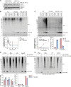
Fig. 1.
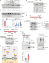
Fig. 1.. The UBA1 inhibitor TAK-243 reveals de novo phosphorylation of free ubiquitin in the cytosol.
(A) hTERT-RPE1 and HCT116 cells were treated ± TAK-243 (1 μM) for the indicated times, lysed, and proteins analyzed by nonreducing SDS-PAGE and Western blot. Blue and red arrowheads indicate the unloaded forms and the Ub-loaded forms, respectively, of UBA1 and UBC13. Representative Western blot of two independent experiments. (B and C) hTERT-RPE1 cells were treated ± TAK-243 (1 μM), antimycin A (1 μM) and oligomycin A (1 μM) (AO), or both TAK-243 and AO. Representative Western blot of three independent experiments and quantification showing the decay of conjugated ubiquitin (B) and accumulation of free and conjugated pSer65-Ubiquitin [pUb (C)]. Brackets indicate the molecular weight (MW) range used for the quantification. Error bars show SD. (D and E) hTERT-RPE1 cells were treated for 4 hours ± TAK-243 (1 μM), AO (1 μM), or TAK-243 and AO and subjected to subcellular fractionation. PNS, post-nuclear supernatant; Mito, mitochondria-enriched fraction; LM, light membrane fraction. SDS-PAGE was performed under reducing (D) or nonreducing (E) conditions to assess levels of free pUb. Bracket indicates the MW range of signal used for conjugated pUb quantification. Quantification of data shown in (D) shows the enrichment of conjugated and free pUb in mitochondrial, LM, and cytosolic fractions (normalized to PNS) for two independent experiments.
Fig. 2.
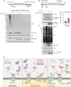
Fig. 2.. TAK-243 reveals continued processing of PINK1 and generation of cleaved active PINK1 at depolarized mitochondria.
(A and B) hTERT-RPE1 cells were treated ± TAK-243 (1 μM), antimycin A (1 μM) and oligomycin A (1 μM) (AO), or ± epoxomicin (100 nM) as indicated. Representative Western blots of three independent experiments. Colored arrowheads correspond to full-length (FL) and cleaved PINK1 (cl). (C) hTERT-RPE1 cells were treated ± AO, ±TAK-243 for 4 hours before subcellular fractionation (the same samples as in Fig. 1D). Representative Western blot and associated quantification. (D) hTERT-RPE1 cells were pretreated ± AO for 22 hours before addition of TAK-243 for a further 2 hours. Representative Western blot and quantification of cleaved PINK1. Error bars, SD; n = 3 independent experiments. (E) hTERT-RPE1 YFP-Parkin cells were pretreated ± AO for 24 hours before addition of TAK-243 for a further 4 hours. Representative Western blot of four independent experiments. (F) hTERT-RPE1 PINK1 KO cells inducibly expressing PINK1-Flag were treated with doxycycline (Dox, 0.1 μg/ml) for 1 hour ± AO, ±TAK-243, and lysates subjected to anti-Flag immunoprecipitation (IP). Representative Western blot and quantification. Error bars, SD; n = 3 independent experiments. (G) Working model proposing two distinct pools of PINK1 on depolarized mitochondria: (i) A labile pool is imported, processed, and released as cPINK1 from mitochondria and can be visualized by TAK-243 treatment; (ii) a separate pool of PINK1 remains intact and accumulates upon depolarization. Both pools are active in this model.
Fig. 3.
Fig. 3.. PINK1 stability measurements.
(A) hTERT-RPE1 cells were treated for 4 hours with AO (1 μM) followed by addition of cycloheximide (CHX; 100 μg/ml, ± AO) for 0.5 to 4 hours. Experimental configuration and representative Western blots are shown. Protein loading was adjusted to obtain a similar PINK1 signal at T = 0 (3× protein loaded for the first seven lanes in the PINK1 blot; 2× for the first seven lanes of the pUb blot). An untreated PINK1 KO cell–derived sample was loaded alongside for background subtraction. (B) Quantification of the rate of PINK1 decay assessed as in (A). Error bars show SD, n = 4 independent experiments. (C) Graph showing the fold increase in PINK1 levels after 4 hours of AO treatment [T = 0 hours in (A)] and the fraction of the accrued PINK1 that is stable [PINK1 (+AO at T = 3 hours)/PINK1 (+AO at T = 0 hours)], leaving an unaccounted for pool that may reflect increased translation. Error bars (fold increase and stable fraction) show SD of four independent experiments. (D) Quantitative RT-PCR reactions of PINK1 (normalized to actin) were performed with cDNA derived from cells treated ± AO (1 μM) for 4 hours. Error bars show SD, n = 3 independent experiments.
Fig. 4.
Fig. 4.. Correlation between cleaved PINK1 and free pUb generation upon E1 inhibition.
(A) Schematic diagram showing experimental overview and representative Western blot for ubiquitin. hTERT-RPE1 cells were treated ± AO (1 μM) for 4 hours, followed by lysis (0 hour) or replacement of media (washout) and a 0.5 to 2 hours chase ± TAK-243 (1 μM). (B) Representative Western blots for pUb and PINK1 from experiments depicted schematically in (A). The bracket indicates the range used for conjugated pUb quantification. * indicates a nonspecific band. (C to E) Quantification of data represented in (B) showing conjugated pUb (conj. pUb) decay (C), accumulation of free pUb (D), and cleaved PINK1 (cPINK1) (E). Error bars show SD for five independent experiments.
Fig. 5.
Fig. 5.. PINK1 exhibits sustained activity after mitochondria have recovered from depolarization.
(A) hTERT-RPE1 cells were treated for 4 hours ± AO (1 μM) and lysed or incubated in media without AO (washout; w/o). After 16 hours of recovery, the cells were either lysed or treated with TAK-243 (1 μM) for indicated times. Representative of three independent experiments. (B and C) hTERT-RPE1 PINK1 KO cells inducibly expressing PINK1-FLAG were cotreated with doxycycline (Dox) ± AO for 1 hour, washed, and incubated ± AO for further 3 hours. AO was washed out and cells left to recover for 16 hours before treatment ± TAK-243 for 4 hours. Lysates were subjected to anti-Flag IP. Bound proteins (B) were analyzed alongside lysates (C) by Western blot. Graph shows pSer228-cPINK1 normalized to total cPINK1 generated upon TAK-243 treatment after recovery from a prior depolarization insult (AO). Error bars, SD; n = 3 independent experiments. (D) Working model of PINK1 behavior under basal, depolarizing, and recovery conditions. Under resting conditions (basal state), PINK1 is rapidly turned over through cleavage in mitochondria and subsequent ubiquitin-dependent degradation. This labile pool persists under depolarizing conditions alongside the emergence of a stable pool of active PINK1, which is proposed to promote the mobilization of PINK1 mRNA resulting in an increase in PINK1 protein synthesis. A small pool of PINK1 remains stable and active long after mitochondria have recovered from depolarization and can facilitate activation of nascent, labile PINK1, which can be visualized and stabilized by inhibiting ubiquitylation.

References

    1. Jin S. M., Lazarou M., Wang C., Kane L. A., Narendra D. P., Youle R. J., Mitochondrial membrane potential regulates PINK1 import and proteolytic destabilization by PARL. J. Cell Biol. 191, 933–942 (2010). - PMC - PubMed
    1. Matsuda N., Sato S., Shiba K., Okatsu K., Saisho K., Gautier C. A., Sou Y. S., Saiki S., Kawajiri S., Sato F., Kimura M., Komatsu M., Hattori N., Tanaka K., PINK1 stabilized by mitochondrial depolarization recruits Parkin to damaged mitochondria and activates latent Parkin for mitophagy. J. Cell Biol. 189, 211–221 (2010). - PMC - PubMed
    1. Narendra D. P., Jin S. M., Tanaka A., Suen D. F., Gautier C. A., Shen J., Cookson M. R., Youle R. J., PINK1 is selectively stabilized on impaired mitochondria to activate Parkin. PLOS Biol. 8, e1000298 (2010). - PMC - PubMed
    1. Kondapalli C., Kazlauskaite A., Zhang N., Woodroof H. I., Campbell D. G., Gourlay R., Burchell L., Walden H., Macartney T. J., Deak M., Knebel A., Alessi D. R., Muqit M. M., PINK1 is activated by mitochondrial membrane potential depolarization and stimulates Parkin E3 ligase activity by phosphorylating Serine 65. Open Biol. 2, 120080 (2012). - PMC - PubMed
    1. Kane L. A., Lazarou M., Fogel A. I., Li Y., Yamano K., Sarraf S. A., Banerjee S., Youle R. J., PINK1 phosphorylates ubiquitin to activate Parkin E3 ubiquitin ligase activity. J. Cell Biol. 205, 143–153 (2014). - PMC - PubMed

LinkOut - more resources