Review
. 2025 Feb;4(2):101528.
doi: 10.1016/j.jacadv.2024.101528.

Reperfused Myocardial Infarction: The Road to CCS Classification of Acute MI and Beyond

Affiliations
Review

Reperfused Myocardial Infarction: The Road to CCS Classification of Acute MI and Beyond

Rohan Dharmakumar et al. JACC Adv. 2025 Feb.

Abstract

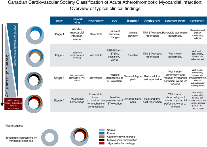
The Canadian Cardiovascular Society recently put forth a new classification of acute reperfused myocardial infarction (MI) based on stages of myocardial injury. Backed by more than 5 decades of intense investigation in the field, the key message of this new classification is that not all MIs are the same and that the type and extent of myocardial injury should be considered in diagnosing and treating MI. We review the literature with the goal of highlighting the progressive advances that enabled the synthesis of the Canadian Cardiovascular Society classification into 4 distinct stages of tissue injury. We emphasize the major breakthroughs from insights gained from experimental, translational, and clinical studies to date. We also identify current gaps in knowledge and critical research directions that need to be pursued to improve patient care and reduce post-MI complications such as chronic heart failure and malignant arrhythmias, whose risk is linked to stage and extent of myocardial injury.

Keywords: CCS stages of myocardial tissue injury; intramyocardial hemorrhage; myocardial infarction; reperfusion injury; reperfusion therapy.


Conflict of interest statement

Funding support and author disclosures This work was funded in part by National Institutes of Health (HL133407, HL136578, and HL147133) to Dr Dharmakumar. Dr Dharmakumar has an ownership interest in Cardio-theranostics, LLC. Dr Kalra is the founding director of makeadent.org Aavishqaar Fund. Dr Kovacs is on the Clinical Events Adjudication Committee for Cook Inc; is on the ECG Adjudication for Eli Lilly; is on the data safety monitoring board for Immunovant and GSK; is a consultant for Jazz and GenInCode; and receives research grant from Pfizer. Dr Henry is a consultant for Corflow. Dr Kumar is an advisor to Cardio-Theranostics, LLC. All other authors have reported that they have no relationships relevant to the contents of this paper to disclose.

Figures

None
Graphical abstract
Figure 1
Figure 1
Ultrastructural Changes During Ischemia and Infarction (A) swollen mitochondria containing amorphous densities (arrows) and wide I bands in dyskinetic fibers and (B) lipid droplets in myocytes (arrows from heart of experimental animal sacrificed after 2 hours of coronary occlusion.
Figure 2
Figure 2
Mechanisms of Cell Death in Reperfused Myocardial Infarction A schematic illustration of mechanisms contributing to multiple cell death pathways attributable to necrosis, necroptosis, apoptosis, autophagy, ferroptosis and pyroptosis. Necrosis is the cell death which is morphologically characterized by rupture of mitochondria, rupture of the sarcolemma and an inflammatory response.,, Traditionally, all IS which was quantified by lack of triphenyl tetrazolium chloride (TTC) staining or gadolinium contrast enhancement in CMR was considered as necrosis. It is currently unclear to which extent the novel, more regulated modes of cell death are reflected in lack of TTC staining or contrast-enhanced CMR. Apoptosis is, in strong contrast to necrosis, morphologically characterized by lack of sarcolemmal rupture and lack of an inflammatory reaction; the nucleus displays chromatin condensation and DNA fragmentation, and mitochondria are small and rounded. Necroptosis shares features with both necrosis and apoptosis, is characterized by sarcolemmal rupture but still a regulated mode of cell death which is initiated by sarcolemmal receptors, resulting in a characteristic necrosome complex of receptor-interacting protein kinase, death domain proteins and caspases which induce cytoplasmic and organelle disintegration., Pyroptosis is an "inflammatory" mode of cell death in response to damage-associated molecular patterns (DAMPs) such as mitochondrial DNA and pathogen-associated molecular patterns (PAMPs) such as endotoxins., Ferroptosis was originally related to iron overload and the generation of reactive oxygen species but is now more precisely related to a deficiency or reduced activity of glutathione peroxidase 4 (GPX4). This reduction of GPX4 activity increases the formation reactive oxygen species (ROS), which cause lipid peroxidation and damage to cellular organelles and sarcolemma., Autophagy differs from the other regulated modes of cell death in that it serves primarily a physiological function, that is, the removal and re-circulation of damaged cellular organelle material, notably mitochondria, through a signaling pathway which centers on the inhibition of the mammalian target of rapamycin (mTOR) and induces the formation of double-membrane autophagosome vesicles., , Adapted with permission from Heusch.
Figure 3
Figure 3
Multimodality Cardiac Imaging Demarcates Reversible from Irreversible Myocardial Injury This composite figure highlights imaging techniques to differentiate reversible from irreversible myocardial injury. In the ECG panel, the top row demonstrates ST-segment elevation (red arrows) indicative of acute ischemia, which may be reversible, while the bottom row shows pathological Q waves (red arrows), suggesting irreversible infarction. In the echocardiography panel, the top row highlights hypokinesis (white arrows), representing potentially viable myocardium, whereas the bottom row shows nonviable thinned akinetic IVS (white arrows), a hallmark of chronic infarction with remodeling. The cardiac MRI panel includes T2-weighted imaging in the top row, where white arrows indicate myocardial edema, a reversible injury marker, while the bottom row demonstrates late gadolinium enhancement (LGE, dashed arrows), reflecting irreversible myocardial scarring, and an inset T2∗ image showing intramyocardial hemorrhage (dotted line), which indicates severe microvascular injury. In the cardiac CT panel, the top row presents a transient perfusion defect (white arrow), which suggests reversible ischemia, while the bottom row displays permanent intramyocardial fat (black arrows), indicating chronic infarction. The SPECT/PET panel shows areas of reduced tracer uptake (top row, black arrows), indicative of ischemia with salvageable myocardium, whereas the bottom row highlights regions with no tracer uptake, confirming nonviable infarcted myocardium. These imaging modalities play a crucial role in differentiating reversible ischemia from infarcted myocardium, guiding revascularization decisions and prognosis assessment. ECG = electrocardiogram; LGE = late gadolinium enhancement; MVO = microvascular obstruction; PET = positron emission tomography; SPECT = single-photon emission computed tomography.
Figure 4
Figure 4
Schematic Illustration of the Initial Pro- and Anti-Inflammatory Reparative Response in Acute Myocardial Infarction During acute MI, there is an induction of the pro-inflammatory response characterized by the production of DAMPs, ROS, and complement activation. These processes lead to the release of pro-inflammatory cytokines and chemokines including IL-1a, IL1-b, IL6, IL18, IFN-g, TNF-a, CCL2, CCL5, and MCP-1, promoting the accumulation of various immune cells such as neutrophils, monocytes, macrophages, and lymphocytes such as CD8+CD28+ T-cells within the infarct zone. Subsequently, the anti-inflammatory reparative response ensues, allowing the resolution of inflammatory response through the activation of anti-inflammatory factors such as IL-2, IL-10, and TGF-bb. This also involves changes in the polarization of monocytes and macrophages, as well as the recruitment of Tregs and dendritic cells. Dysregulated or excessive inflammation response can aggravate myocardial injury and contribute to adverse cardiac remodeling and recurrent MACE. CCL2/5 = chemokine (C-C motif) ligand 2/5; DAMP = damage-associated molecular pattern; IFN = interferon; IL = interleukin; MACE = major adverse cardiovascular events; MCP = monocyte chemoattractant protein; ROS = reactive oxygen species; TGF = transforming growth factor; TNF = tumor necrosis factor.
Figure 5
Figure 5
Overview of Typical Clinical Findings at Each Stage of the Canadian Cardiovascular Society Classification of Acute Myocardial Infarction Typical changes are shown; precise diagnostic criteria will require further research and expert consensus (see manuscript). The stages build on each other, reflecting progression of severity of tissue injury. Timely reperfusion can halt tissue injury at an earlier stage and prevent progression to a more severe stage of injury. Hemorrhagic infarction is the worst stage and a cause of infarct expansion and risk factor for mechanical complications. Wall motion abnormalities and ECG changes also depend on the size of the affected myocardium. CCS = Canadian Cardiovascular Society; LV = left ventricle; MI = myocardial infarction; other abbreviations as in Figure 3. Adapted from Kumar et al.
Central Illustration
Reperfused Myocardial Infarction: The Road to CCS-AMI Classification Abbreviation as in Figure 5.

References

    1. Parikh N.I., Gona P., Larson M.G., et al. Long-term trends in myocardial infarction incidence and case fatality in the National Heart, Lung, and Blood Institute's Framingham Heart study. Circulation. 2009;119:1203–1210. - PMC - PubMed
    1. Martin S.S., Aday A.W., Almarzooq Z.I., et al. 2024 heart disease and stroke statistics: a report of US and global data from the American heart association. Circulation. 2024;149:e347–e913. - PubMed
    1. Salari N., Morddarvanjoghi F., Abdolmaleki A., et al. The global prevalence of myocardial infarction: a systematic review and meta-analysis. BMC Cardiovasc Disord. 2023;23:206. - PMC - PubMed
    1. Kalra A., Jose A.P., Prabhakaran P., et al. The burgeoning cardiovascular disease epidemic in Indians - perspectives on contextual factors and potential solutions. Lancet Reg Health Southeast Asia. 2023;12 - PMC - PubMed
    1. Murugiah K., Wang Y., Nuti S.V., et al. Are non-ST-segment elevation myocardial infarctions missing in China? Eur Heart J Qual Care Clin Outcomes. 2017;3:319–327. - PubMed

LinkOut - more resources